Physicochemical Properties
| Molecular Formula | C25H41NO9 |
| Molecular Weight | 499.5943 |
| Exact Mass | 499.278 |
| CAS # | 509-20-6 |
| Related CAS # | 509-20-6 |
| PubChem CID | 417761 |
| Appearance | White to off-white solid |
| Density | 1.4±0.1 g/cm3 |
| Boiling Point | 626.1±55.0 °C at 760 mmHg |
| Melting Point | 129-131ºC |
| Flash Point | 332.4±31.5 °C |
| Vapour Pressure | 0.0±4.1 mmHg at 25°C |
| Index of Refraction | 1.624 |
| LogP | -1.63 |
| Hydrogen Bond Donor Count | 5 |
| Hydrogen Bond Acceptor Count | 10 |
| Rotatable Bond Count | 6 |
| Heavy Atom Count | 35 |
| Complexity | 878 |
| Defined Atom Stereocenter Count | 0 |
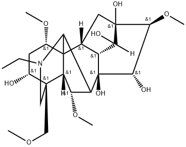
| SMILES | O([H])[C@]12[C@]([H])([C@@]([H])([C@@]3([C@@]([H])([C@@]1([H])[C@@]([H])(C3([H])[H])[C@@]13[C@]([H])(C([H])([H])[C@]([H])([C@@]4(C([H])([H])OC([H])([H])[H])C([H])([H])N(C([H])([H])C([H])([H])[H])C1([H])[C@]2([H])[C@@]([H])([C@@]34[H])OC([H])([H])[H])O[H])OC([H])([H])[H])O[H])O[H])OC([H])([H])[H])O[H] |
| InChi Key | SQMGCPHFHQGPIF-JIOYIOPFSA-N |
| InChi Code | InChI=1S/C25H41NO9/c1-6-26-9-22(10-32-2)12(27)7-13(33-3)24-11-8-23(30)19(28)14(11)25(31,20(29)21(23)35-5)15(18(24)26)16(34-4)17(22)24/h11-21,27-31H,6-10H2,1-5H3/t11-,12-,13+,14-,15+,16+,17-,18?,19-,20+,21+,22+,23-,24+,25-/m1/s1 |
| Chemical Name | (1S,2R,3R,4R,5R,6S,7S,8R,9R,13R,14R,16S,17S,18R)-11-ethyl-6,16,18-trimethoxy-13-(methoxymethyl)-11-azahexacyclo[7.7.2.12,5.01,10.03,8.013,17]nonadecane-4,5,7,8,14-pentol |
| Synonyms | Jesaconine; Aconine |
| HS Tariff Code | 2934.99.9001 |
| Storage |
Powder-20°C 3 years 4°C 2 years In solvent -80°C 6 months -20°C 1 month |
| Shipping Condition | Room temperature (This product is stable at ambient temperature for a few days during ordinary shipping and time spent in Customs) |
Biological Activity
| Targets |
NF-κB - Aconine exerts inhibitory effects by targeting nuclear factor-kappa B (NF-κB), nuclear factor of activated T-cells cytoplasmic 1 (NFATc1), and dendritic cell-specific transmembrane protein (DC-STAMP)[1] |
| ln Vitro |
Treatment with aconine dose-dependently reduces NF-κB'stranscriptionalactivity that is induced by RANKL. By reducing the expression of the cell-cell fusion molecule DC-STAMP and the activation of NF-B and NFATc1, aconine prevents RANKL from inducing osteoclast differentiation in RAW264.7 cells. RAW264.7 cells are unaffected by aconine (0.125, 0.25 μM), but it dose-dependently reduces the activity of osteoclasts and bone resorption caused by RANKL. Aconine reduces the expression of osteoclast-specific genes (c-Src, β3-Integrin, cathepsin K and MMP-9) and dendritic cell-specific transmembrane protein (DC-STAMP), which is crucial for cell-cell fusion[1]. It also inhibits the RANKL-induced activation of κB and NFATc1 in RAW264.7 cells. - Inhibition of osteoclast differentiation: In RANKL-induced RAW264.7 cells (a murine macrophage cell line used as an osteoclast precursor model), Aconine exhibited concentration-dependent inhibition of osteoclast differentiation. At concentrations of 10 μM, 20 μM, and 40 μM, the number of tartrate-resistant acid phosphatase (TRAP)-positive multinucleated osteoclasts was reduced by 35%, 62%, and 85%, respectively, compared to the RANKL-only group [1] - Suppression of bone resorption activity: Aconine (20 μM, 40 μM) significantly reduced the area of bone resorption pits formed by RANKL-induced RAW264.7 cells on bone slices. The resorption pit area in the 40 μM Aconine group was 78% smaller than that in the RANKL-only group [1] - Inhibition of NF-κB and NFATc1 activation: Aconine (10-40 μM) suppressed RANKL-induced phosphorylation of NF-κB p65 and IκBα, and reduced the nuclear translocation of NF-κB p65 (observed via immunofluorescence staining). It also downregulated RANKL-induced NFATc1 expression at both mRNA and protein levels; at 40 μM, NFATc1 mRNA and protein levels were reduced by 72% and 68%, respectively [1] - Downregulation of DC-STAMP expression: Aconine (10-40 μM) concentration-dependently decreased RANKL-induced DC-STAMP mRNA and protein expression. At 40 μM, DC-STAMP mRNA and protein levels were reduced by 65% and 60%, respectively, compared to the RANKL-only group [1] - Cell viability assessment: Aconine at concentrations up to 40 μM did not affect the viability of RAW264.7 cells (cell viability >90% compared to the untreated control group), indicating that its inhibitory effect on osteoclast differentiation was not due to cytotoxicity [1] |
| Cell Assay |
- Osteoclast differentiation induction and TRAP staining: RAW264.7 cells were seeded in 24-well plates (5×10³ cells/well) and cultured in medium containing 10% fetal bovine serum. Cells were divided into control group (no RANKL), RANKL group (50 ng/mL RANKL), and Aconine + RANKL groups (10 μM, 20 μM, 40 μM Aconine + 50 ng/mL RANKL). After 5 days of culture, cells were fixed with 4% paraformaldehyde, stained with TRAP staining kit, and TRAP-positive multinucleated cells (≥3 nuclei) were counted under a light microscope [1] - Bone resorption pit assay: RAW264.7 cells were seeded on bone slices in 24-well plates (1×10⁴ cells/slice) and treated with RANKL (50 ng/mL) and Aconine (20 μM, 40 μM) for 7 days. Bone slices were then treated with 10% sodium hypochlorite to remove cells, stained with 1% toluidine blue, and the area of resorption pits was analyzed using image analysis software [1] - Western blot analysis: RAW264.7 cells were treated with RANKL (50 ng/mL) and Aconine (10-40 μM) for 24-48 hours. Total protein was extracted, and nuclear protein was isolated for NF-κB p65 detection. Protein samples were separated by SDS-PAGE, transferred to PVDF membranes, and probed with primary antibodies against NF-κB p65, phospho-NF-κB p65, IκBα, phospho-IκBα, NFATc1, DC-STAMP, and β-actin (loading control). Secondary antibodies were added, and bands were visualized using an enhanced chemiluminescence system; band intensity was quantified with image software [1] - RT-PCR analysis: RAW264.7 cells were treated with RANKL (50 ng/mL) and Aconine (10-40 μM) for 24 hours. Total RNA was extracted, reverse-transcribed into cDNA, and PCR amplification was performed using specific primers for NFATc1, DC-STAMP, and GAPDH (internal control). The relative mRNA expression levels were calculated using the 2⁻ΔΔCt method [1] - Cell viability assay: RAW264.7 cells were seeded in 96-well plates (1×10⁴ cells/well) and treated with Aconine (5-40 μM) for 48 hours. MTT reagent was added, and after 4 hours of incubation, the absorbance at 570 nm was measured. Cell viability was calculated as a percentage of the untreated control group [1] |
| References |
[1]. Aconine inhibits RANKL-induced osteoclast differentiation in RAW264.7 cells by suppressing NF-κB and NFATc1 activation and DC-STAMP expression. Acta Pharmacol Sin. 2016 Feb;37(2):255-63. |
| Additional Infomation |
Aconine has been reported in Aconitum carmichaelii with data available. - Aconine inhibits RANKL-induced osteoclast differentiation and bone resorption activity in RAW264.7 cells through a mechanism involving the suppression of NF-κB activation (by reducing phosphorylation of p65 and IκBα and nuclear translocation of p65), downregulation of NFATc1 (a key transcription factor for osteoclast differentiation), and decreased expression of DC-STAMP (a protein essential for osteoclast fusion) [1] - The non-cytotoxic nature of Aconine at effective concentrations suggests its potential as a therapeutic agent for bone diseases characterized by excessive osteoclast activity, such as osteoporosis, rheumatoid arthritis, and bone metastasis [1] |
Solubility Data
| Solubility (In Vitro) | DMSO: ~100 mg/mL ( ~200.2 mM) |
| Solubility (In Vivo) |
Solubility in Formulation 1: ≥ 2.08 mg/mL (4.16 mM) (saturation unknown) in 10% DMSO + 40% PEG300 + 5% Tween80 + 45% Saline (add these co-solvents sequentially from left to right, and one by one), clear solution. For example, if 1 mL of working solution is to be prepared, you can add 100 μL of 20.8 mg/mL clear DMSO stock solution to 400 μL PEG300 and mix evenly; then add 50 μL Tween-80 to the above solution and mix evenly; then add 450 μL normal saline to adjust the volume to 1 mL. Preparation of saline: Dissolve 0.9 g of sodium chloride in 100 mL ddH₂ O to obtain a clear solution. Solubility in Formulation 2: ≥ 2.08 mg/mL (4.16 mM) (saturation unknown) in 10% DMSO + 90% (20% SBE-β-CD in Saline) (add these co-solvents sequentially from left to right, and one by one), clear solution. For example, if 1 mL of working solution is to be prepared, you can add 100 μL of 20.8 mg/mL clear DMSO stock solution to 900 μL of 20% SBE-β-CD physiological saline solution and mix evenly. Preparation of 20% SBE-β-CD in Saline (4°C,1 week): Dissolve 2 g SBE-β-CD in 10 mL saline to obtain a clear solution. Solubility in Formulation 3: ≥ 2.08 mg/mL (4.16 mM) (saturation unknown) in 10% DMSO + 90% Corn Oil (add these co-solvents sequentially from left to right, and one by one), clear solution. For example, if 1 mL of working solution is to be prepared, you can add 100 μL of 20.8 mg/mL clear DMSO stock solution to 900 μL of corn oil and mix evenly. (Please use freshly prepared in vivo formulations for optimal results.) |
| Preparing Stock Solutions | 1 mg | 5 mg | 10 mg | |
| 1 mM | 2.0016 mL | 10.0082 mL | 20.0164 mL | |
| 5 mM | 0.4003 mL | 2.0016 mL | 4.0033 mL | |
| 10 mM | 0.2002 mL | 1.0008 mL | 2.0016 mL |