AZD2932 (AZD-2932), a new quinazoline ether, is a novel, potent, high affinity and mutil-targeted tyrosine kinase inhibitor with potential anticancer activity. Its IC50 values are 8 nM, 4 nM, 7 nM, and 9 nM for VEGFR-2, PDGFRβ, Flt-3, and c-Kit, respectively. AZD-2932 exhibits good selectivity on a panel of kinases and is also active against c-Kit and FLT3. 31 may prove to be an antiangiogenic agent in the clinic based on its pharmacokinetic behavior and preclinical antitumor activity in nude mice with C6 rat glial tumors.
Physicochemical Properties
| Molecular Formula | C24H25N5O4 | |
| Molecular Weight | 447.49 | |
| Exact Mass | 447.19 | |
| Elemental Analysis | C, 64.42; H, 5.63; N, 15.65; O, 14.30 | |
| CAS # | 883986-34-3 | |
| Related CAS # |
|
|
| PubChem CID | 11517980 | |
| Appearance | Light yellow to yellow solid powder | |
| Density | 1.3±0.1 g/cm3 | |
| Boiling Point | 676.9±55.0 °C at 760 mmHg | |
| Flash Point | 363.2±31.5 °C | |
| Vapour Pressure | 0.0±2.1 mmHg at 25°C | |
| Index of Refraction | 1.629 | |
| LogP | 3.18 | |
| Hydrogen Bond Donor Count | 1 | |
| Hydrogen Bond Acceptor Count | 7 | |
| Rotatable Bond Count | 8 | |
| Heavy Atom Count | 33 | |
| Complexity | 630 | |
| Defined Atom Stereocenter Count | 0 | |
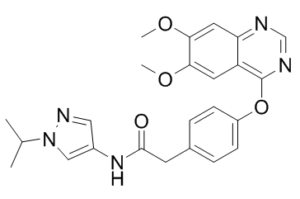
| SMILES | O=C(CC1C=CC(OC2C3C(=CC(=C(C=3)OC)OC)N=CN=2)=CC=1)NC1=CN(C(C)C)N=C1 |
|
| InChi Key | TWYCZJMOEMMCGC-UHFFFAOYSA-N | |
| InChi Code | InChI=1S/C24H25N5O4/c1-15(2)29-13-17(12-27-29)28-23(30)9-16-5-7-18(8-6-16)33-24-19-10-21(31-3)22(32-4)11-20(19)25-14-26-24/h5-8,10-15H,9H2,1-4H3,(H,28,30) | |
| Chemical Name | 2-[4-(6,7-dimethoxyquinazolin-4-yl)oxyphenyl]-N-(1-propan-2-ylpyrazol-4-yl)acetamide | |
| Synonyms |
|
|
| HS Tariff Code | 2934.99.9001 | |
| Storage |
Powder-20°C 3 years 4°C 2 years In solvent -80°C 6 months -20°C 1 month |
|
| Shipping Condition | Room temperature (This product is stable at ambient temperature for a few days during ordinary shipping and time spent in Customs) |
Biological Activity
| Targets |
VEGFR2 (IC50 = 8 nM); PDGFRβ (IC50 = 4 nM); FLT3 (IC50 = 7 nM); c-Kit (IC50 = 9 nM) AZD2932 is a quinazoline ether-derived tyrosine kinase inhibitor with high affinity for VEGFR-2 and PDGFR family members, with the following IC50 values: VEGFR-2 (KDR): 1.2 nM, PDGFRα: 3.5 nM, PDGFRβ: 2.8 nM. It shows no significant inhibition (IC50 > 1 μM) against EGFR, HER2, Src, or FLT3 kinases [1] |
| ln Vitro |
AZD2932 is effective and well-balanced against c-Kit, VEGFR-2, Flt-3, and PDGFβ. With the worst IC50 against 2C9 (8.0 μM), it does not inhibit any of the different cytochrome P450 isoforms. There is no activity of AZD2932 against hERG (IC50=137 μM)[1]. 1. Enzyme activity inhibition: AZD2932 (0.1-10 nM) dose-dependently inhibits recombinant human VEGFR-2 and PDGFR kinase activity. At 1 nM, it inhibits VEGFR-2 activity by ~90% and PDGFRα/β activity by ~85% compared to the untreated enzyme control [1] 2. HUVEC (human umbilical vein endothelial cell) experiments: AZD2932 (0.5-10 μM) suppresses VEGF-induced cell proliferation and tube formation. At 2 μM, HUVEC proliferation is reduced by ~75% (72-hour treatment), and tube length is decreased by ~80% (6-hour treatment) compared to the VEGF-stimulated group [1] 3. Tumor cell proliferation: AZD2932 inhibits the growth of VEGFR/PDGFR-expressing human tumor cell lines, with IC50 values of 2.1 μM for A549 (lung cancer), 1.8 μM for HT-29 (colon cancer), and 2.5 μM for SK-OV-3 (ovarian cancer) after 72-hour treatment [1] 4. Western blot analysis: In A549 cells, AZD2932 (2 μM) reduces phosphorylation of VEGFR-2 (Tyr1175) by ~85% and PDGFRβ (Tyr751) by ~80%, as well as downstream p-AKT (Ser473) and p-ERK1/2 by ~75% and ~70% respectively, compared to the control [1] |
| ln Vivo |
When AZD2932 is administered orally twice a day, spaced 10 hours apart, on the day the control animals are put to sleep, there is a notable 64% tumor growth inhibition for both the 50 and 12.5 mg/kg doses. The growth of Calu-6 tumors is inhibited by 81% and 72% at 50 and 12.5 mg/kg b.i.d., and that of LoVo tumors by 67% at 50 mg/kg b.i.d. Xenografts containing tumor cells that do not express PDGFβ are also sensitive to AZD2932 treatment. This can be attributed to the potent activity of AZD2932 against VEGFR2, as well as its possible impact on pericytes and tumor-associated fibroblasts through inhibition of PDGFR a and b. Both p-VEGFR2 and p-PDGFβ are 60–80% inhibited by AZD2932 at 3–50 mg/kg b.i.d. spaced 10 hours apart[1]. 1. Nude mouse xenograft model (A549 lung cancer): Oral administration of AZD2932 (15 mg/kg, once daily for 21 days) results in a tumor growth inhibition (TGI) rate of ~70%. Tumor volume in the treated group is ~30% of the vehicle control (0.5% methylcellulose + 0.1% Tween 80), and tumor weight is reduced by ~65% [1] 2. Nude mouse xenograft model (HT-29 colon cancer): AZD2932 (20 mg/kg, oral gavage, once daily for 28 days) achieves a TGI of ~65%. Intratumoral microvessel density (CD31-positive vessels) is decreased by ~60% compared to the vehicle group [1] |
| Enzyme Assay |
AZD2932 is a highly effective and versatile kinase inhibitor that targets VEGFR2, PDGFβ, Flt-3, and c-Kit. In a cell assay, its IC50 values are 8, 4, 7, and 9 nM, respectively. 1. Recombinant VEGFR-2 kinase activity assay: The assay is performed in a reaction buffer containing 50 mM Tris-HCl (pH 7.5), 10 mM MgCl2, 1 mM dithiothreitol (DTT), 25 μM ATP, and 1 μg/well Poly(Glu,Tyr)4:1 (substrate). Different concentrations of AZD2932 (0.05 nM-10 nM) are pre-incubated with recombinant human VEGFR-2 kinase (5 ng/well) for 15 minutes at 30°C. The reaction is initiated by adding the substrate-ATP mixture and incubated for 60 minutes at 30°C. Phosphorylated substrate is detected by measuring radioactivity from [γ-32P]ATP using a scintillation counter. IC50 is calculated via nonlinear regression of inhibition curves [1] 2. Recombinant PDGFRα/β kinase activity assay: The protocol is identical to the VEGFR-2 assay, with recombinant human PDGFRα (8 ng/well) or PDGFRβ (6 ng/well) used instead of VEGFR-2. AZD2932 concentrations range from 0.1 nM-10 nM, and kinase activity inhibition is measured by the same radioactive detection method to determine IC50 values [1] |
| Cell Assay |
1. HUVEC proliferation assay (MTT method): HUVECs are seeded in 96-well plates at a density of 3×10³ cells/well and cultured overnight in EGM-2 medium. AZD2932 (0.5-10 μM) and VEGF (50 ng/mL) are added, and cells are incubated for 72 hours at 37°C. MTT reagent (5 mg/mL, 10 μL/well) is added, followed by 4 hours of incubation. Formazan crystals are dissolved in DMSO (100 μL/well), and absorbance is measured at 570 nm. Cell viability is expressed as a percentage of the VEGF-stimulated control, and IC50 is derived from dose-response curves [1] 2. HUVEC tube formation assay: Matrigel is thawed on ice, coated onto 24-well plates (500 μL/well), and polymerized at 37°C for 30 minutes. HUVECs (2×10⁴ cells/well) are suspended in medium containing AZD2932 (0.5-10 μM) and VEGF (50 ng/mL), then seeded onto Matrigel. After 6 hours of incubation, tube-like structures are photographed under a microscope. Total tube length per well is quantified using image analysis software, and inhibition rate is calculated relative to the VEGF control [1] 3. A549 cell Western blot analysis: A549 cells are seeded in 6-well plates at 5×10⁵ cells/well and cultured overnight. AZD2932 (2 μM) is added, and cells are incubated for 2 hours. Cells are lysed in RIPA buffer containing protease and phosphatase inhibitors. Protein concentration is measured by BCA assay. Equal amounts of protein (40 μg) are separated by 10% SDS-PAGE, transferred to PVDF membranes, and probed with antibodies against p-VEGFR-2 (Tyr1175), VEGFR-2, p-PDGFRβ (Tyr751), PDGFRβ, p-AKT (Ser473), AKT, p-ERK1/2, and ERK1/2. HRP-conjugated secondary antibodies and ECL reagent are used for detection, and band intensity is quantified with ImageJ [1] |
| Animal Protocol |
Mice: In order to verify that AZD2932 exhibits comparable effectiveness against PDGFβ and VEGFR-2 phosphorylation, the female nude mice with C6 tumors receive intravenous doses of VEGF-A and PDGFBB five minutes before culling and six hours after the final AZD2932 dose, with the lungs promptly removed. Western blot analysis of lung lysates is performed to detect PDGFβ and total and phosphorylated VEGFR-2 [1]. 1. Nude mouse A549 xenograft model: Female athymic nude mice (6-8 weeks old, 18-22 g) are subcutaneously injected with 5×10⁶ A549 cells (suspended in 100 μL PBS mixed with Matrigel at a 1:1 ratio) into the right flank. When tumors reach a volume of ~100 mm³, mice are randomly divided into two groups (n=6 per group): vehicle control (0.5% methylcellulose + 0.1% Tween 80) and AZD2932-treated (15 mg/kg). The drug is administered by oral gavage once daily for 21 days. Tumor volume is measured every 3 days using a vernier caliper (volume = length × width² / 2), and body weight is monitored to assess potential toxicity [1] 2. Nude mouse HT-29 xenograft model: Female nude mice (6-8 weeks old) are subcutaneously injected with 5×10⁶ HT-29 cells (suspended in 100 μL PBS/Matrigel 1:1). When tumors reach ~100 mm³, mice are randomized into two groups (n=6 per group): vehicle and AZD2932 (20 mg/kg, oral gavage once daily for 28 days). At the end of treatment, mice are euthanized, tumors are excised and weighed, and tumor tissues are fixed for CD31 immunohistochemical staining to measure microvessel density [1] |
| ADME/Pharmacokinetics |
1. In mice: After oral administration of AZD2932 (15 mg/kg), the oral bioavailability (F) is 58%, peak plasma concentration (Cmax) is 1.6 μg/mL, time to reach Cmax (Tmax) is 1.8 hours, and terminal half-life (t1/2) is 7.2 hours [1] 2. In rats: Intravenous administration of AZD2932 (5 mg/kg) results in a t1/2 of 6.5 hours and a clearance rate of 1.4 mL/min/kg. Oral administration (10 mg/kg) shows F=51%, Cmax=1.1 μg/mL, and Tmax=2.0 hours [1] 3. Plasma protein binding rate: In human plasma, AZD2932 exhibits >92% protein binding, measured using an ultrafiltration method [1] |
| Toxicity/Toxicokinetics |
1. Acute toxicity in mice: Single oral administration of AZD2932 (up to 200 mg/kg) does not cause mortality within 7 days. Mice in the 150-200 mg/kg group show transient weight loss (4-6% at 48 hours) and reduced locomotor activity, which fully recover within 7 days [1] 2. Subchronic toxicity in rats (28-day oral administration): At doses of 10 mg/kg and 25 mg/kg, AZD2932 does not cause significant changes in body weight, organ weight (liver, kidney, spleen), or serum biochemical parameters (ALT, AST, creatinine, urea nitrogen). No histopathological abnormalities are observed in major organs [1] 3. No drug-drug interaction data or median lethal dose (LD50) values for AZD2932 were described in the literature [1] |
| References |
[1]. Discovery of AZD2932, a new Quinazoline Ether Inhibitor with high affinity for VEGFR-2 and PDGFR tyrosine kinases. Bioorg Med Chem Lett. 2012 Jan 1;22(1):262-6. |
| Additional Infomation |
1. AZD2932 belongs to the quinazoline ether class of small-molecule inhibitors, with a chemical structure optimized to enhance binding affinity for VEGFR-2 and PDGFR while minimizing off-target activity against other kinases [1] 2. It exerts antitumor effects through a dual mechanism: inhibiting VEGFR-2 to suppress tumor angiogenesis and blocking PDGFRα/β to inhibit tumor stromal support and cancer cell proliferation [1] 3. In preclinical studies, AZD2932 shows better in vivo efficacy than the earlier VEGFR inhibitor sunitinib in A549 xenograft models (TGI: 70% vs. 55% at equivalent doses), likely due to its higher affinity for VEGFR-2 [1] |
Solubility Data
| Solubility (In Vitro) |
|
|||
| Solubility (In Vivo) |
Solubility in Formulation 1: ≥ 1 mg/mL (2.23 mM) (saturation unknown) in 10% DMSO + 40% PEG300 + 5% Tween80 + 45% Saline (add these co-solvents sequentially from left to right, and one by one), clear solution. For example, if 1 mL of working solution is to be prepared, you can add 100 μL of 10.0 mg/mL clear DMSO stock solution to 400 μL of PEG300 and mix evenly; then add 50 μL of Tween-80 to the above solution and mix evenly; then add 450 μL of normal saline to adjust the volume to 1 mL. Preparation of saline: Dissolve 0.9 g of sodium chloride in 100 mL ddH₂ O to obtain a clear solution. Solubility in Formulation 2: 1 mg/mL (2.23 mM) in 10% DMSO + 90% (20% SBE-β-CD in Saline) (add these co-solvents sequentially from left to right, and one by one), suspension solution; with ultrasonication. For example, if 1 mL of working solution is to be prepared, you can add 100 μL of 10.0 mg/mL clear DMSO stock solution to 900 μL of 20% SBE-β-CD physiological saline solution and mix evenly. Preparation of 20% SBE-β-CD in Saline (4°C,1 week): Dissolve 2 g SBE-β-CD in 10 mL saline to obtain a clear solution. Solubility in Formulation 3: ≥ 1 mg/mL (2.23 mM) (saturation unknown) in 10% DMSO + 90% Corn Oil (add these co-solvents sequentially from left to right, and one by one), clear solution. For example, if 1 mL of working solution is to be prepared, you can add 100 μL of 10.0 mg/mL clear DMSO stock solution to 900 μL of corn oil and mix evenly. (Please use freshly prepared in vivo formulations for optimal results.) |
| Preparing Stock Solutions | 1 mg | 5 mg | 10 mg | |
| 1 mM | 2.2347 mL | 11.1734 mL | 22.3469 mL | |
| 5 mM | 0.4469 mL | 2.2347 mL | 4.4694 mL | |
| 10 mM | 0.2235 mL | 1.1173 mL | 2.2347 mL |