Decription: AMBMP (also known as BML-284; BML284) is a novel, potent and cell-permeable Wnt signaling activator with anti-inflammatory activity. AMBMP induces TCF-dependent transcriptional activity(EC50 =700 nM).
Physicochemical Properties
| Molecular Formula | C19H19CLN4O3 |
| Molecular Weight | 350.37 |
| Exact Mass | 350.137 |
| Elemental Analysis | C, 65.13; H, 5.18; N, 15.99; O, 13.70 |
| CAS # | 853220-52-7 |
| Related CAS # | BML-284 hydrochloride;2095432-75-8 |
| PubChem CID | 11210285 |
| Appearance | White to yellow solid powder |
| Density | 1.4±0.1 g/cm3 |
| Boiling Point | 623.4±65.0 °C at 760 mmHg |
| Flash Point | 330.8±34.3 °C |
| Vapour Pressure | 0.0±1.8 mmHg at 25°C |
| Index of Refraction | 1.684 |
| LogP | 3.72 |
| Hydrogen Bond Donor Count | 2 |
| Hydrogen Bond Acceptor Count | 7 |
| Rotatable Bond Count | 5 |
| Heavy Atom Count | 26 |
| Complexity | 455 |
| Defined Atom Stereocenter Count | 0 |
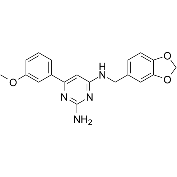
| SMILES | N1C(NCC2C=C3C(OCO3)=CC=2)=CC(C2C=C(OC)C=CC=2)=NC=1N |
| InChi Key | FABQUVYDAXWUQP-UHFFFAOYSA-N |
| InChi Code | InChI=1S/C19H18N4O3/c1-24-14-4-2-3-13(8-14)15-9-18(23-19(20)22-15)21-10-12-5-6-16-17(7-12)26-11-25-16/h2-9H,10-11H2,1H3,(H3,20,21,22,23) |
| Chemical Name | C19H18N4O3 |
| Synonyms | AMBMP; BML284; BML 284; BML-284; Wnt Agonist |
| HS Tariff Code | 2934.99.9001 |
| Storage |
Powder-20°C 3 years 4°C 2 years In solvent -80°C 6 months -20°C 1 month |
| Shipping Condition | Room temperature (This product is stable at ambient temperature for a few days during ordinary shipping and time spent in Customs) |
Biological Activity
| Targets |
TCF-dependent transcriptional activity (EC50 = 700 nM)
AMBMP acts as a small-molecule agonist of the Wnt signaling pathway, targeting the glycogen synthase kinase 3β (GSK-3β) as a direct molecular target (IC50 = 6.8 μM for inhibiting recombinant human GSK-3β enzymatic activity) [1] AMBMP enhances the transcriptional activity of the β-catenin/TCF complex (a key downstream effector of Wnt signaling) with an EC50 = 4.2 μM in Wnt reporter gene assays [1] |
| ln Vitro |
The migration and invasion capacities of MNK45 and AGS cells are markedly enhanced by BML-284 (10 μM; 24 hours), while the migration and invasion capacities of cells blocked by pizotifen are partially restored [1]. When compared to the NC group, BML-284 (10 μM; 24 h) dramatically increased the expression of β-catenin. Additionally, as compared to the pizotifen treatment group, it partially counteracted the effects of pizotifen on the expression of N- and E-cadherin in MNK45 and AGS cells [1]. 1. In HEK293 cells transfected with a TCF/LEF luciferase Wnt reporter plasmid, AMBMP (1–20 μM) dose-dependently activates Wnt signaling, with a maximal 3.8-fold increase in luciferase activity at 10 μM and an EC50 of 4.2 μM; this activation is abrogated by siRNA-mediated knockdown of β-catenin, confirming Wnt/β-catenin pathway specificity [1] 2. In recombinant human GSK-3β enzymatic assays, AMBMP (2–20 μM) inhibits GSK-3β activity in a dose-dependent manner, with an IC50 of 6.8 μM; 10 μM AMBMP reduces GSK-3β activity by 75% (measured by phosphorylation of the GSK-3β substrate glycogen synthase) [1] 3. In mouse L cells (which lack endogenous Wnt secretion), AMBMP (5–15 μM) induces cytoplasmic and nuclear accumulation of β-catenin (2.5-fold increase at 10 μM, detected by Western blot) and upregulates Wnt target genes (e.g., c-Myc, cyclin D1) by 2.1-fold and 1.9-fold respectively (RT-PCR analysis) [1] 4. AMBMP (≤20 μM) shows no significant cytotoxicity in HEK293 cells or mouse L cells (cell viability >90% by MTT assay) [1] |
| ln Vivo |
In a study where Tg (myl7:EGFP) transgenic embryos at 5.5 hpf were placed into cells with 20 embryos on a plate, BML-284 (10 ng) and pyrimethanil (4 mg/L) together partially prevented the teratogenic phenotype and heart abnormalities produced by pyrimethanil[1]. AMBMP Targets CaMKIIβ In Vivo, https://pmc.ncbi.nlm.nih.gov/articles/PMC7659555/ Researchers next asked whether AMBMP acts by enhancing CaMKIIβ activity in vivo. C3KO and C57BL/6 WT mice were treated with AMBMP and then their muscles were evaluated for CaMKIIβ and other signaling pathways. The activation of signaling was carried out by western blotting with antibodies specific for the active forms of these signaling pathways. Treatment with AMBMP (daily i.p. injection 7.5 mg/kg) led to CaMKIIβ activation in both WT and C3KO mice (Figures 4C and 4D). The drug appears to engage CaMKIIβ specifically as it does not activate AKT nor AMPK (nor other pathways that control muscle remodeling and oxidative metabolism) (Figures 4E and 4F). Furthermore, the effect of AMBMP on CaMKIIβ was likely post-transcriptional, and there was no significant change in the expression level of the Camk2b gene (Figure 4G). Thus, these studies establish proof of concept for the ability of AMBMP to activate CaMKII and subsequently to promote oxidative metabolism and benefit the LGMDR1 phenotype. 1. In Xenopus laevis embryo development assays, microinjection of AMBMP (50 μM in 2 nL injection volume) into ventral blastomeres induces axis duplication in 65% of embryos (a classic Wnt signaling-dependent phenotype), similar to the effect of Wnt3a protein injection; this phenotype is blocked by co-injection of a dominant-negative TCF plasmid [1] 2. In zebrafish embryos, treatment with AMBMP (10 μM) from the 1-cell stage to 24 hours post-fertilization (hpf) enhances Wnt signaling in the developing neural tube, as shown by increased expression of the Wnt target gene engrailed-2 (detected by whole-mount in situ hybridization) [1] |
| Enzyme Assay |
1. Recombinant human GSK-3β activity assay: Purified recombinant human GSK-3β (0.5 μg) was incubated with serial concentrations of AMBMP (1–20 μM) in GSK-3β assay buffer (20 mM Tris-HCl, 10 mM MgCl₂, 1 mM DTT, pH 7.5) for 15 minutes at 30°C. The glycogen synthase peptide substrate (200 μM) and [γ-³²P]ATP (0.5 μCi) were added to initiate the phosphorylation reaction, which proceeded for 30 minutes at 30°C. The reaction was terminated by adding phosphoric acid (1 M), and phosphorylated substrate was spotted onto P81 phosphocellulose paper. Unincorporated radioactivity was washed away with phosphoric acid, and bound radioactivity was measured by liquid scintillation counting to calculate GSK-3β inhibition and IC50 values [1] 2. Wnt/β-catenin TCF/LEF reporter gene assay: HEK293 cells were seeded in 96-well plates (1×10⁴ cells/well) and co-transfected with a TCF/LEF firefly luciferase reporter plasmid and a Renilla luciferase reference plasmid (for normalization). After 24 hours of transfection, cells were treated with AMBMP (1–20 μM) for 18 hours at 37°C. Luciferase activity was measured using a dual-luciferase assay kit, and firefly luciferase activity was normalized to Renilla luciferase activity to calculate fold activation of Wnt signaling and EC50 values [1] |
| Cell Assay |
Western Blot Analysis[2] Cell Types: Human gastric cancer cell lines MNK45 and AGS[1] Tested Concentrations: 10 µM Incubation Duration: 24 hrs (hours) Experimental Results: β-catenin expression was induced in MNK45 and AGS and E-cadherin and N-cadherin expressing cells were retained . 1. HEK293 Wnt reporter cell assay: HEK293 cells were cultured in DMEM supplemented with 10% fetal bovine serum under 5% CO₂ at 37°C. Cells were seeded in 96-well plates and transfected with the TCF/LEF luciferase reporter construct using a lipid-based transfection reagent. After transfection, cells were treated with serial concentrations of AMBMP (1–20 μM) and incubated for 18 hours. Luciferase activity was measured, and dose-response curves were fitted to determine the EC50 for Wnt pathway activation. For siRNA knockdown experiments, cells were transfected with β-catenin siRNA or control siRNA 24 hours before AMBMP treatment, and luciferase activity was measured to confirm pathway specificity [1] 2. Mouse L cell β-catenin accumulation and gene expression assay: Mouse L cells were seeded in 6-well plates (5×10⁵ cells/well) and treated with AMBMP (5–15 μM) for 24 hours at 37°C. Cells were lysed, and cytoplasmic/nuclear fractions were prepared for Western blot analysis using a β-catenin-specific antibody (GAPDH and lamin B were used as cytoplasmic and nuclear loading controls, respectively). For RT-PCR analysis, total RNA was extracted from treated cells, and cDNA was synthesized; qPCR was performed to quantify the expression of Wnt target genes (c-Myc, cyclin D1) with GAPDH as a reference gene [1] 3. Cell viability MTT assay: HEK293 cells and mouse L cells were seeded in 96-well plates (5×10³ cells/well) and treated with AMBMP (1–20 μM) for 48 hours at 37°C. MTT reagent (0.5 mg/mL) was added for 4 hours, formazan crystals were dissolved in DMSO, and absorbance at 570 nm was measured using a microplate reader to calculate cell viability as a percentage of vehicle-treated controls [1] |
| Animal Protocol |
Compound pharmacokinetics assay For pharmacokinetics, AMBMP was administered by different routes of delivery (subcutaneous, intraperitoneal, and oral, in food or by gavage) at two different dosages (10 mg/kg and 30 mg/kg). The blood was collected at 0.5 h, 1 h, 2 h, 4 h and 6 h post treatment by heart puncture. The concentrations of compounds in plasma were analyzed by Integrated Analytical Solutions, Inc.https://pmc.ncbi.nlm.nih.gov/articles/PMC7659555/ Seahorse analysis of Extracts from Frozen Muscle For Seahorse analysis, frozen soleus muscles from DMSO or AMBMP-treated mice (daily IP injections at 7.5 mg/kg) were homogenized by hand in a Dounce homogenizer in 200 mL of mitochondrial buffer (70 mM sucrose, 220 mM mannitol, 5 mM KH2PO4, 5 mM MgCl2, 1 mM EGTA, 2 mM HEPES, adjusted to pH 7.4 with KOH) on ice. Muscle homogenates were centrifuged at 900xg for 5 min at 4°C. Supernatants were transferred to new tubes; protein concentrations were measured using BCA protein Assay Kit. The samples (4 μg/well) were analyzed in the UCLA Mitochondrial and Metabolism Core using a Seahorse XF96 Analyzer. Data were normalized to total protein. Seahorse analysis was carried out according to Acin-Perez et al.https://pmc.ncbi.nlm.nih.gov/articles/PMC7659555/ 1. Xenopus laevis embryo axis duplication assay: Xenopus laevis eggs were fertilized in vitro and cultured in 0.1× Marc’s modified Ringer’s (MMR) solution at 22°C. At the 4-cell stage, ventral blastomeres were microinjected with AMBMP (50 μM in 2 nL of 0.1× MMR) or vehicle (0.1× MMR + 0.1% DMSO). For rescue experiments, a dominant-negative TCF plasmid (50 pg) was co-injected with AMBMP. Embryos were cultured to the tadpole stage (stage 35/36), and the percentage of embryos with secondary axis formation was scored under a dissecting microscope (n=50 embryos per group) [1] 2. Zebrafish embryo Wnt signaling assay: Zebrafish embryos were obtained by natural spawning and maintained in E3 medium at 28.5°C. Embryos were treated with AMBMP (10 μM) or vehicle (E3 medium + 0.1% DMSO) from the 1-cell stage to 24 hpf. Whole-mount in situ hybridization was performed using a digoxigenin-labeled engrailed-2 riboprobe to detect Wnt target gene expression; stained embryos were imaged, and the intensity of engrailed-2 expression in the neural tube was quantified by image analysis software [1] |
| Toxicity/Toxicokinetics |
1. In vitro cytotoxicity: AMBMP (≤20 μM) shows no significant cytotoxicity in HEK293 cells, mouse L cells, or Xenopus laevis embryo blastomeres (cell/embryo viability >90%) [1] 2. Embryonic toxicity: AMBMP at concentrations >20 μM in Xenopus embryos causes developmental arrest (15% of embryos at 30 μM) and mild malformations (e.g., tail curvature) in 10% of embryos at 25 μM, but no lethal toxicity at concentrations up to 50 μM [1] |
| References |
[1]. A small-molecule agonist of the Wnt signaling pathway. Angew Chem Int Ed Engl. 2005 Mar 18;44(13):1987-90. [2]. Pizotifen inhibits the proliferation and invasion of gastric cancer cells. Exp Ther Med. 2020 Feb;19(2):817-824. [3]. Exposure to pyrimethanil induces developmental toxicity and cardiotoxicity in zebrafish. Chemosphere. 2020 Sep;255:126889. |
| Additional Infomation |
N4-(1,3-benzodioxol-5-ylmethyl)-6-(3-methoxyphenyl)pyrimidine-2,4-diamine is a member of pyrimidines. 1. AMBMP (2-amino-4-methyl-5-bromo-6-phenylpyrimidine) is the first synthetic small-molecule agonist of the Wnt signaling pathway, identified in a high-throughput screen for compounds that activate Wnt/β-catenin signaling [1] 2. AMBMP exerts its Wnt-activating effect by directly inhibiting GSK-3β, a key negative regulator of the Wnt pathway; inhibition of GSK-3β prevents β-catenin phosphorylation and degradation, leading to β-catenin accumulation and activation of TCF/LEF-mediated transcription of Wnt target genes [1] 3. AMBMP induces Wnt-dependent developmental phenotypes (axis duplication) in Xenopus laevis embryos, confirming its in vivo activity as a Wnt agonist; this effect is dependent on functional β-catenin/TCF signaling [1] 4. AMBMP is a valuable preclinical research tool for studying Wnt signaling in development and disease (e.g., cancer, neurodegeneration), as Wnt pathway dysregulation is linked to numerous human disorders [1] |
Solubility Data
| Solubility (In Vitro) | DMSO : ≥ 100 mg/mL (~285.41 mM) |
| Solubility (In Vivo) |
Solubility in Formulation 1: ≥ 2.08 mg/mL (5.94 mM) (saturation unknown) in 10% DMSO + 40% PEG300 + 5% Tween80 + 45% Saline (add these co-solvents sequentially from left to right, and one by one), clear solution. For example, if 1 mL of working solution is to be prepared, you can add 100 μL of 20.8 mg/mL clear DMSO stock solution to 400 μL PEG300 and mix evenly; then add 50 μL Tween-80 to the above solution and mix evenly; then add 450 μL normal saline to adjust the volume to 1 mL. Preparation of saline: Dissolve 0.9 g of sodium chloride in 100 mL ddH₂ O to obtain a clear solution. Solubility in Formulation 2: ≥ 2.08 mg/mL (5.94 mM) (saturation unknown) in 10% DMSO + 90% (20% SBE-β-CD in Saline) (add these co-solvents sequentially from left to right, and one by one), clear solution. For example, if 1 mL of working solution is to be prepared, you can add 100 μL of 20.8 mg/mL clear DMSO stock solution to 900 μL of 20% SBE-β-CD physiological saline solution and mix evenly. Preparation of 20% SBE-β-CD in Saline (4°C,1 week): Dissolve 2 g SBE-β-CD in 10 mL saline to obtain a clear solution. Solubility in Formulation 3: ≥ 2.08 mg/mL (5.94 mM) (saturation unknown) in 10% DMSO + 90% Corn Oil (add these co-solvents sequentially from left to right, and one by one), clear solution. For example, if 1 mL of working solution is to be prepared, you can add 100 μL of 20.8 mg/mL clear DMSO stock solution to 900 μL of corn oil and mix evenly. (Please use freshly prepared in vivo formulations for optimal results.) |
| Preparing Stock Solutions | 1 mg | 5 mg | 10 mg | |
| 1 mM | 2.8541 mL | 14.2706 mL | 28.5413 mL | |
| 5 mM | 0.5708 mL | 2.8541 mL | 5.7083 mL | |
| 10 mM | 0.2854 mL | 1.4271 mL | 2.8541 mL |