ACY-957 is a novel, potent and selective inhibitor of HDAC1 and HDAC2 isoforms with IC50s of 7 nM, 18 nM, and 1300 nM against HDAC1/2/3, respectively. It exhibits no inhibition on other isoforms such as HDAC4/5/6/7/8/9.
Physicochemical Properties
| Molecular Formula | C24H23N5OS |
| Molecular Weight | 429.537323236465 |
| Exact Mass | 429.162 |
| CAS # | 1609389-52-7 |
| PubChem CID | 72374405 |
| Appearance | Light yellow to yellow solid powder |
| Density | 1.3±0.1 g/cm3 |
| Boiling Point | 602.7±55.0 °C at 760 mmHg |
| Flash Point | 318.3±31.5 °C |
| Vapour Pressure | 0.0±1.7 mmHg at 25°C |
| Index of Refraction | 1.726 |
| LogP | 2.73 |
| Hydrogen Bond Donor Count | 3 |
| Hydrogen Bond Acceptor Count | 6 |
| Rotatable Bond Count | 4 |
| Heavy Atom Count | 31 |
| Complexity | 615 |
| Defined Atom Stereocenter Count | 0 |
| InChi Key | VURDNNVAYZDGDK-UHFFFAOYSA-N |
| InChi Code | InChI=1S/C24H23N5OS/c25-19-6-3-17(22-2-1-13-31-22)15-21(19)28-24(30)18-4-7-20-16(14-18)5-8-23(27-20)29-11-9-26-10-12-29/h1-8,13-15,26H,9-12,25H2,(H,28,30) |
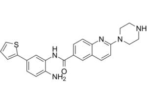
| Chemical Name | N-(2-amino-5-thiophen-2-ylphenyl)-2-piperazin-1-ylquinoline-6-carboxamide |
| Synonyms | ACY-957; ACY957; ACY 957 |
| HS Tariff Code | 2934.99.9001 |
| Storage |
Powder-20°C 3 years 4°C 2 years In solvent -80°C 6 months -20°C 1 month |
| Shipping Condition | Room temperature (This product is stable at ambient temperature for a few days during ordinary shipping and time spent in Customs) |
Biological Activity
| Targets |
Histone deacetylase 1 (HDAC1) (IC50 = 7 nM) [1] Histone deacetylase 2 (HDAC2) (IC50 = 18 nM) [1] Histone deacetylase 3 (HDAC3) (IC50 = 1300 nM) [1] |
| ln Vitro |
ACY-957 has no inhibitory impact on HDAC4/5/6/7/8/9, but it is a selective inhibitor of HDAC1 and HDAC2, with IC50s of 7 nM, 18 nM, and 1300 nM for HDAC1/2/3, respectively. For HDAC2 in primary hematopoietic progenitor cells, ACY-957 has an IC50 of 304 nM [1]. Treatment of primary human erythroblasts with 1 μM or 5 μM ACY-957 led to a dose-dependent accumulation of acetylation on histone H3 lysine 9 and 14 (H3K9/14ac), H3 lysine 56 (H3K56ac), H3 lysine 79 (H3K79ac), and H2B lysine 5 (H2BK5ac), while histone H3 lysine 9 tri-methylation (H3K9me3) was unaffected. [1] In two distinct erythroid progenitor culture systems (CS1 and CS2), treatment with 1 μM ACY-957 induced a significant time-dependent increase in the percentage of γ-globin (HBG) mRNA relative to total β-like globin mRNA, reaching approximately 35% in CS1 and 24% in CS2 at day 5, representing an approximately 3.5-fold increase over vehicle-treated controls. This induction exceeded that observed with 30 μM hydroxyurea. [1] Treatment with 1 μM ACY-957 increased the percentage of HbF-positive cells by 2 to 3-fold, as measured by flow cytometry, and also increased the mean fluorescent intensity (MFI) of HbF per cell by up to 2-fold. [1] ACY-957 treatment (1 μM) significantly increased levels of embryonic ε-globin (HBE) and fetal γ-globin (HBG) mRNA while decreasing levels of adult δ-globin (HBD) and β-globin (HBB) mRNA, consistent with a delay or reversal of globin switching. [1] In burst forming unit-erythroid (BFU-E) colonies cultured for 14 days, ACY-957 increased the percentage of HBG mRNA in a dose-dependent manner: 15% in vehicle, 48% at 1 μM, and 82% at 2 μM. [1] In peripheral blood mononuclear cells (PBMCs) from four sickle cell disease (SCD) donors, treatment with 1 μM ACY-957 significantly elevated HBG mRNA levels (e.g., from 12% to 58% in one donor). It also induced a dose-dependent increase in the number of HbF-positive cells and HbF protein abundance per cell. [1] Gene expression profiling showed that ACY-957 treatment (1 μM) induced 1294 genes and suppressed 681 genes. The expression changes significantly overlapped with those resulting from genetic knockdown of HDAC1 or HDAC2. Key changes included downregulation of the HBG repressors BCL11A (1.3-fold) and SOX6 (2.5-fold), and upregulation of GATA2 (2.8-fold). [1] Quantitative PCR confirmed that ACY-957 treatment prevented the normal suppression of GATA2 during erythroid differentiation, resulting in a 3.3-fold increase relative to controls at day 4. GATA1 and KLF1 expression were unaffected. [1] Lentiviral overexpression of GATA2 in primary erythroid progenitors significantly increased the percentage of HBG mRNA and HBG transcript levels while decreasing HBB mRNA levels, mimicking the effect of ACY-957. [1] Knockdown of GATA2 using shRNA attenuated the induction of HBG mRNA by ACY-957 by approximately 25%. [1] Chromatin immunoprecipitation sequencing (ChIP-Seq) in vehicle-treated primary erythroid progenitors showed high occupancy of both HDAC1 and HDAC2 across a 15 kb region of the GATA2 locus, encompassing known autoregulatory enhancer regions (+9.5 kb, -1.8 kb, -2.8 kb, -3.9 kb). [1] Treatment with ACY-957 led to significant increases in histone acetylation (H2BK5ac, H3K9ac, H3K27ac) at the GATA2 enhancer regions (up to 8-fold increase at the -1.8 kb region) and increased GATA2 protein binding at these same regions (up to 3-fold increase at the -1.8 kb region). [1] GATA2 ChIP-Seq also revealed that ACY-957 treatment increased GATA2 binding (1.8-fold) at a region near the HBD gene promoter within the β-globin locus. [1] Treatment with 1 μM ACY-957 led to an accumulation of TFRCposGYPAmid proerythroblasts and inhibited their differentiation into TFRCposGYPApos basophilic erythroblasts over an 8-day culture period. [1] |
| Enzyme Assay |
An in vitro biochemical assay was performed to assess inhibition of purified HDAC enzymes by ACY-957. The compound was dissolved and diluted in assay buffer. HDAC enzymes were diluted in assay buffer and pre-incubated with ACY-957 for 24 hours before adding the substrate (acetyl-lysine or trifluoroacetyl-lysine tripeptide, depending on the HDAC isoform). The substrate was used at a concentration equal to its Michaelis constant (Km). The enzymatic reaction was monitored over 30 minutes for the release of 7-amino-4-methoxy-coumarin after deacetylation, and the linear reaction rate was calculated to determine IC50 values. [1] A cellular HDAC2 inhibition assay was performed using a bioluminescent HDAC2-specific substrate. Primary human bone marrow-derived hematopoietic progenitors were expanded in culture for 6 days, then treated with ACY-957 for an additional 48 hours. Luminescence was detected in a lytic format according to the assay protocol to determine cellular IC50. [1] |
| Cell Assay |
Primary human CD34+ cells from bone marrow (healthy donors) or peripheral blood (SCD patients) were cultured using two distinct two-phase erythroid differentiation systems (Culture System 1, CS1, and Culture System 2, CS2). Cells were expanded in specific cytokine-supplemented media, then shifted to differentiation media containing erythropoietin and other factors. ACY-957 or vehicle was added at the start of differentiation. [1] Cells were harvested at various time points. Erythroid maturation stage was assessed by flow cytometry using antibodies against transferrin receptor (TFRC) and glycophorin A (GYPA). [1] Total RNA was isolated, followed by DNase digestion. cDNA was synthesized. Globin mRNA species (HBB, HBD, HBG, HBE) and other genes of interest (e.g., GATA2, BCL11A) were quantified by quantitative real-time PCR (QPCR) using specific TaqMan probes or SYBR green. The percentage of HBG mRNA was calculated relative to the sum of all β-like globin transcripts. [1] HbF protein was detected by flow cytometry. Cells were stained with a FITC-conjugated anti-human HbF antibody, and the percentage of positive cells and mean fluorescence intensity were measured. [1] For histone acetylation analysis by western blot, histones were extracted. Lysates were separated by gel electrophoresis, transferred to a membrane, and probed with antibodies specific for various acetylated histone marks (H3K9/14ac, H3K56ac, H3K79ac, H2BK5ac) and a total histone H4 antibody for normalization. [1] For GATA2 protein detection by western blot, cells were lysed in denaturing buffer, and proteins were separated by capillary electrophoresis or standard western blotting, followed by detection with an anti-GATA2 antibody. [1] For chromatin immunoprecipitation (ChIP), cells were fixed with formaldehyde. Chromatin was sheared by sonication and immunoprecipitated with antibodies against specific histone modifications (H3K9ac, H2BK5ac, H3K27ac) or GATA2. Precipitated DNA was analyzed either by next-generation sequencing (ChIP-Seq) or by QPCR (ChIP-QPCR) with primer sets specific for genomic regions of interest (e.g., GATA2 enhancers, HBD promoter). [1] For gene expression profiling, total RNA from vehicle- or ACY-957-treated cells was analyzed using Affymetrix GeneChip arrays. Data was processed and analyzed for differential expression and gene set enrichment. [1] For functional studies, lentiviral vectors encoding GATA2-specific shRNAs or a GATA2 overexpression construct were used to transduce primary erythroid progenitors. Transduced cells were selected with puromycin and then treated with ACY-957 or vehicle in expansion or differentiation media, followed by analysis of mRNA and protein. [1] Burst forming unit-erythroid (BFU-E) assays were performed by culturing normal human bone marrow mononuclear cells in semi-solid methylcellulose media containing cytokines (IL3, CSF2, KITLG, EPO) and ACY-957 or vehicle. After 14-16 days, BFU-E colonies were picked, and RNA was isolated for QPCR analysis. [1] |
| Toxicity/Toxicokinetics |
The literature states that non-selective HDAC inhibitors have been associated with significant toxicity and adverse events in the clinical setting. The development of ACY-957 as an HDAC1/2-selective inhibitor aims to minimize such potential toxicity. [1] The study notes that HDAC1/2 inhibition by ACY-957 at 1 μM blocked the differentiation of proerythroblasts to basophilic erythroblasts in vitro, suggesting a potential on-target effect on erythropoiesis that may require intermittent dosing in vivo to allow maturation of HbF-containing cells. [1] |
| References |
[1]. Chemical Inhibition of Histone Deacetylases 1 and 2 Induces Fetal Hemoglobin through Activation of GATA2. PLoS One. 2016 Apr 13;11(4):e0153767. |
| Additional Infomation |
ACY-957 is a novel, orally bioavailable, biaryl aminobenzamide-class small molecule inhibitor designed to be selective for HDAC1 and HDAC2 over other HDAC isoforms. It inhibits VEGF protein synthesis at the post-transcriptional level. [1] The compound was developed with the goal of inducing fetal hemoglobin (HbF) for the treatment of sickle cell disease (SCD) and β-thalassemia, while potentially minimizing toxicity associated with non-selective HDAC inhibitors. [1] The proposed mechanism is that ACY-957 inhibits HDAC1/2, leading to elevated histone acetylation at GATA2 enhancer regions. This promotes GATA2 binding and sustained GATA2 gene expression during erythroid maturation via a positive autoregulatory loop. Elevated GATA2 contributes to HBG induction, possibly through increased GATA2 binding at the HBD promoter region within the β-globin locus. [1] The study suggests that due to HDAC1's more dominant role in erythroid development, future drug development might focus on achieving HDAC2 selectivity to minimize potential erythroblast toxicity. [1] The induction of GATA2 by ACY-957 also suggests potential therapeutic utility in myelodysplastic syndrome (MDS) and acute myeloid leukemia (AML), where GATA2 haploinsufficiency or epigenetic silencing is implicated. [1] |
Solubility Data
| Solubility (In Vitro) | DMSO : ~83.33 mg/mL (~194.00 mM) |
| Solubility (In Vivo) |
Solubility in Formulation 1: ≥ 2.08 mg/mL (4.84 mM) (saturation unknown) in 10% DMSO + 40% PEG300 + 5% Tween80 + 45% Saline (add these co-solvents sequentially from left to right, and one by one), clear solution. For example, if 1 mL of working solution is to be prepared, you can add 100 μL of 20.8 mg/mL clear DMSO stock solution to 400 μL PEG300 and mix evenly; then add 50 μL Tween-80 to the above solution and mix evenly; then add 450 μL normal saline to adjust the volume to 1 mL. Preparation of saline: Dissolve 0.9 g of sodium chloride in 100 mL ddH₂ O to obtain a clear solution. Solubility in Formulation 2: ≥ 2.08 mg/mL (4.84 mM) (saturation unknown) in 10% DMSO + 90% (20% SBE-β-CD in Saline) (add these co-solvents sequentially from left to right, and one by one), clear solution. For example, if 1 mL of working solution is to be prepared, you can add 100 μL of 20.8 mg/mL clear DMSO stock solution to 900 μL of 20% SBE-β-CD physiological saline solution and mix evenly. Preparation of 20% SBE-β-CD in Saline (4°C,1 week): Dissolve 2 g SBE-β-CD in 10 mL saline to obtain a clear solution. Solubility in Formulation 3: ≥ 2.08 mg/mL (4.84 mM) (saturation unknown) in 10% DMSO + 90% Corn Oil (add these co-solvents sequentially from left to right, and one by one), clear solution. For example, if 1 mL of working solution is to be prepared, you can add 100 μL of 20.8 mg/mL clear DMSO stock solution to 900 μL of corn oil and mix evenly. (Please use freshly prepared in vivo formulations for optimal results.) |
| Preparing Stock Solutions | 1 mg | 5 mg | 10 mg | |
| 1 mM | 2.3281 mL | 11.6404 mL | 23.2807 mL | |
| 5 mM | 0.4656 mL | 2.3281 mL | 4.6561 mL | |
| 10 mM | 0.2328 mL | 1.1640 mL | 2.3281 mL |