Physicochemical Properties
| Molecular Formula | C11H12O2 |
| Molecular Weight | 176.2118 |
| Exact Mass | 176.083 |
| CAS # | 6836-19-7 |
| PubChem CID | 81276 |
| Appearance | Off-white to yellow solid powder |
| Density | 1.1±0.1 g/cm3 |
| Boiling Point | 312.3±31.0 °C at 760 mmHg |
| Melting Point | 59-63 °C(lit.) |
| Flash Point | 145.8±18.4 °C |
| Vapour Pressure | 0.0±0.7 mmHg at 25°C |
| Index of Refraction | 1.549 |
| LogP | 2.79 |
| Hydrogen Bond Donor Count | 0 |
| Hydrogen Bond Acceptor Count | 2 |
| Rotatable Bond Count | 1 |
| Heavy Atom Count | 13 |
| Complexity | 200 |
| Defined Atom Stereocenter Count | 0 |
| SMILES | O=C1C2C([H])=C(C([H])=C([H])C=2C([H])([H])C([H])([H])C1([H])[H])OC([H])([H])[H] |
| InChi Key | GABLTKRIYDNDIN-UHFFFAOYSA-N |
| InChi Code | InChI=1S/C11H12O2/c1-13-9-6-5-8-3-2-4-11(12)10(8)7-9/h5-7H,2-4H2,1H3 |
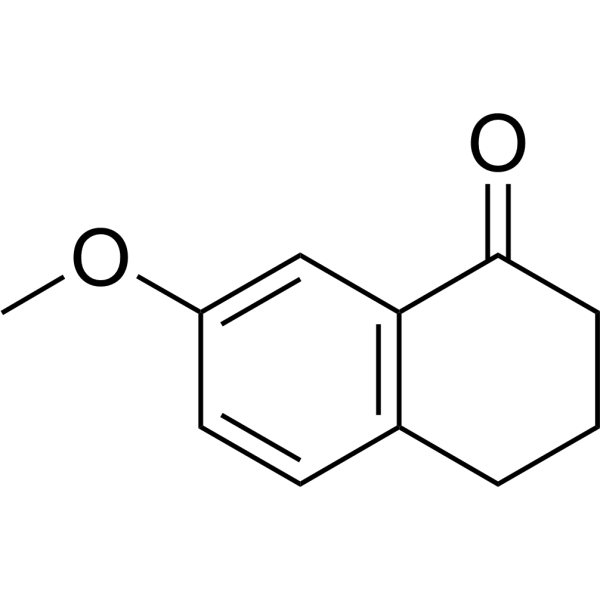
| Chemical Name | 7-methoxy-3,4-dihydro-2H-naphthalen-1-one |
| Synonyms | 7-Methoxy-1-tetralone; 6836-19-7; 7-METHOXY-3,4-DIHYDRONAPHTHALEN-1(2H)-ONE; 7-methoxy-3,4-dihydro-2H-naphthalen-1-one; 7-Methoxy-1,2,3,4-tetrahydronaphthalen-1-one; 1(2H)-Naphthalenone, 3,4-dihydro-7-methoxy-; MFCD00001696; H4FZ2W25VZ; |
| HS Tariff Code | 2934.99.9001 |
| Storage |
Powder-20°C 3 years 4°C 2 years In solvent -80°C 6 months -20°C 1 month |
| Shipping Condition | Room temperature (This product is stable at ambient temperature for a few days during ordinary shipping and time spent in Customs) |
Biological Activity
| Targets | Anticancer; NF-κB;matrix metallopeptidase 2 (MMP2)/MMP9; p-AKT. |
| ln Vitro | 7-Methoxy-1-tetralone (31.25-1000 μM; 48 h) suppresses LO2 and HepG2 cell growth [1]. HepG2 cells exposed to 40 μM, 100 μM, or 250 μM of 7-Methoxy-1-tetralone for 48 hours experience apoptosis, and the expression levels of c-Met, p-AKT, AKT, NF-κB, MMP2, and MMP9 proteins are regulated in the cells. [1]. |
| ln Vivo | In HepG2 cell subcutaneously implanted tumor models, 7-Methoxy-1-tetralone (80, 120, or 160 mg/kg/d; intraperitoneal injection; 19 doses total) suppresses tumor growth [1]. |
| Cell Assay |
Western Blot Analysis[1] Cell Types: HepG2 cells Tested Concentrations: 40 μM, 100 μM, and 250 μM Incubation Duration: 48 h Experimental Results: diminished the protein expression levels of c-Met, p-AKT, NF-κB, MMP2, and MMP9. Cell Proliferation Assay[1] Cell Types: LO2 and HepG2 cells Tested Concentrations: 31.25 μM, 62.5 μM, 125 μM, 250 μM, 500 μM, and 1000 μM Incubation Duration: 24 h, 48 h, and 72 h Experimental Results: demonstrated anti-proliferative activity of MT on LO2 and HepG2 cells. |
| Animal Protocol |
Animal/Disease Models: BALB/c nude mice (5weeks old) with subcutaneously (sc) implanted HepG2 cells[1] Doses: 80, 120, or 160 mg/ kg/d Route of Administration: intraperitoneal (ip)injection; sacrificed after 19 days Experimental Results: Resulted the tumor inhibition rates of 40.57% (80 mg/kg), 51.43% (120 mg/kg), 79.43% (160 mg/kg), respectively. |
| References | [1]. Wen Y, et al. 7-Methoxy-1-Tetralone Induces Apoptosis, Suppresses Cell Proliferation and Migration in Hepatocellular Carcinoma via Regulating c-Met, p-AKT, NF-κB, MMP2, and MMP9 Expression. Front Oncol. 2020 Feb 7;10:58. |
| Additional Infomation |
7-Methoxy-3,4-dihydro-1(2H)-naphthalenone is a member of tetralins. This study aimed to determine the anti-proliferative and anti-migratory effects of 7-methoxy-1-tetralone (MT) in hepatocellular carcinoma (HCC) cells. MTT assay assessed HCC cell viability; cell apoptosis of HCC cells was determined by flow cytometry; wound healing assay evaluated HCC cell migratory ability; protein expression levels were assessed using western blot assay; the in vivo antitumor effects of MT were tested in BALB/c nude mice and the pathological changes within the tumor tissues were evaluated by immunohistochemistry. MT treatment significantly suppressed the cell proliferative and migratory potentials of HepG2 cells, and induced HepG2 cell apoptosis. The western blot assay showed that MT treatment caused a suppression on c-Met, phosphorylated AKT (p-AKT), NF-κB, matrix metallopeptidase 2 (MMP2)/MMP9 protein levels in HepG2 cells. Further in vivo animal studies deciphered that MT treatment suppressed tumor growth of HepG2 cells in the nude mice, but had no effect on the body weight and the organ index of liver and spleen. Further immunohistochemistry analysis of the dissected tumor tissues showed that MT treatment significantly suppressed the protein expression levels of NF-κB, MMP9, MMP2, and p-AKT. In summary, the present study demonstrated the anti-tumor effects of MT on the HCC, and MT suppressed HCC progression possibly via regulating proliferation- and migration-related mediators including c-Met, p-AKT, NF-κB, MMP2, and MMP9 in HepG2 cells.[1] |
Solubility Data
| Solubility (In Vitro) | DMSO : 100 mg/mL (567.50 mM) |
| Solubility (In Vivo) |
Solubility in Formulation 1: ≥ 2.5 mg/mL (14.19 mM) (saturation unknown) in 10% DMSO + 90% Corn Oil (add these co-solvents sequentially from left to right, and one by one), clear solution. For example, if 1 mL of working solution is to be prepared, you can add 100 μL of 25.0 mg/mL clear DMSO stock solution to 900 μL of corn oil and mix evenly. (Please use freshly prepared in vivo formulations for optimal results.) |
| Preparing Stock Solutions | 1 mg | 5 mg | 10 mg | |
| 1 mM | 5.6750 mL | 28.3752 mL | 56.7505 mL | |
| 5 mM | 1.1350 mL | 5.6750 mL | 11.3501 mL | |
| 10 mM | 0.5675 mL | 2.8375 mL | 5.6750 mL |