iCRT 14 is a novel and potent inhibitor of β-catenin-responsive transcription (CRT) with IC50 of 40.3 nM against Wnt responsive STF16 luciferase in a homogeneous fluorescence polarization (FP) assay. Immunofluorescence staining of β-catenin in TNBC cell lines showed both nuclear and cytoplasmic localization, indicating activation of Wnt pathway in TNBC cells. iCRT-14 was an effective compound for inhibiting proliferation and antagonizing Wnt signaling in TNBC cells. In addition, treatment with iCRT-14 resulted in increased apoptosis in vitro. Knockdown of the Wnt pathway transcription factor, SOX4 in triple negative BT-549 cells resulted in decreased cell proliferation and migration, and combination treatment of iCRT-3 with SOX4 knockdown had a synergistic effect on inhibition of cell proliferation and induction of apoptosis.
Physicochemical Properties
| Molecular Formula | C21H17N3O2S |
| Molecular Weight | 375.44358 |
| Exact Mass | 375.104 |
| Elemental Analysis | C, 67.18; H, 4.56; N, 11.19; O, 8.52; S, 8.54 |
| CAS # | 677331-12-3 |
| Related CAS # | 901751-47-1(iCRT3); 18623-44-4 (iCRT5) |
| PubChem CID | 5967294 |
| Appearance | Light yellow to yellow solid powder |
| LogP | 4.795 |
| Hydrogen Bond Donor Count | 0 |
| Hydrogen Bond Acceptor Count | 4 |
| Rotatable Bond Count | 3 |
| Heavy Atom Count | 27 |
| Complexity | 617 |
| Defined Atom Stereocenter Count | 0 |
| SMILES | CC1=CC(=C(N1C2=CN=CC=C2)C)/C=C\3/C(=O)N(C(=O)S3)C4=CC=CC=C4 |
| InChi Key | NCSHZXNGQYSKLR-XDHOZWIPSA-N |
| InChi Code | InChI=1S/C21H17N3O2S/c1-14-11-16(15(2)23(14)18-9-6-10-22-13-18)12-19-20(25)24(21(26)27-19)17-7-4-3-5-8-17/h3-13H,1-2H3/b19-12+ |
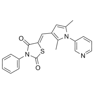
| Chemical Name | 5-[[2,5-dimethyl-1-(3-pyridinyl)-1H-pyrrol-3-yl]methylene]-3-phenyl-2,4-thiazolidinedione |
| Synonyms | iCRT14; iCRT-14; 677331-12-3; iCRT-14; iCRT14; (5Z)-5-[(2,5-dimethyl-1-pyridin-3-ylpyrrol-3-yl)methylidene]-3-phenyl-1,3-thiazolidine-2,4-dione; CHEMBL3589010; (5Z)-5-{[2,5-Dimethyl-1-(3-pyridinyl)-1H-pyrrol-3-yl]methylene}-3-phenyl-1,3-thiazolidine-2,4-dione; (5Z)-5-{[2,5-dimethyl-1-(pyridin-3-yl)-1H-pyrrol-3-yl]methylidene}-3-phenyl-1,3-thiazolidine-2,4-dione; iCRT 14 |
| HS Tariff Code | 2934.99.9001 |
| Storage |
Powder-20°C 3 years 4°C 2 years In solvent -80°C 6 months -20°C 1 month |
| Shipping Condition | Room temperature (This product is stable at ambient temperature for a few days during ordinary shipping and time spent in Customs) |
Biological Activity
| Targets |
β-catenin-responsive transcription (CRT; IC50 = 40.3 nM) β-catenin/TCF4 transcription complex iCRT14 is an inhibitor of catenin responsive transcription (CRT). It disrupts the interaction between β-catenin and TCF4, and may also interfere with TCF binding to DNA.[1] |
| ln Vitro |
Apart from its impact on the TCF-β-cat response, iCRT 14 has the ability to obstruct TCF's DNA binding [1]. Although it was still less than iCRT 3, iCRT 14 (10, 25, 50 μM) efficiently and time- and dose-dependently reduced the growth of BT-549 cells [2]. iCRT14 inhibits the Wnt-responsive luciferase reporter dTF12 in Drosophila Cl8 cells when Wnt signaling is activated by dsRNA-mediated knockdown of Axin. It inhibits the Wnt-responsive STF16 luciferase reporter in HEK293 cells with an IC50 in the nanomolar to micromolar range (specific value not provided). iCRT14 reduces the interaction between purified recombinant His-tagged β-catenin and GST-tagged TCF4 N-terminal domain in vitro. In HEK293 cells transfected with a degradation-resistant β-catenin (S37Aβ-cat), iCRT14 treatment significantly inhibited the β-catenin-TCF interaction in co-immunoprecipitation assays. Thermal melt analysis showed that iCRT14 caused a change in the melting temperature of the β-catenin-TCF4 complex by as much as 2°C, suggesting direct binding to the complex. iCRT14 (50 µM) treatment of HCT116 colon cancer cells (which harbor a β-catenin S45 deletion) resulted in significant reduction of Axin2 and Cyclin D1 mRNA levels, similar to the effect of β-catenin siRNA. Western blot analysis showed a corresponding reduction in Cyclin D1 protein levels in iCRT14-treated HCT116 cells. Flow cytometry analysis revealed that treatment of HCT116 cells with 50 µM iCRT14 caused a marked cell cycle arrest in the G0/G1 phase. Similar G0/G1 arrest was observed in HT29 colon cancer cells (which have an APC mutation) treated with 50 µM iCRT14. This cell cycle arrest correlated with a drastic reduction in the number of phospho-histone H3 positive cells and BrdU incorporation in HCT116 cells. iCRT14 reduced the invasive capacity of MCF7 breast cancer cells stably expressing S37Aβ-cat-HA in Matrigel-coated Boyden chamber assays. iCRT14 inhibited the down-regulation of membranous E-cadherin in MCF7-S37Aβ-cat-HA cells. In C57MG mouse mammary epithelial cells, iCRT14 inhibited Wnt3a-induced morphological transformation and reduced mRNA levels of the β-catenin target gene WISP1.[1] |
| ln Vivo |
In HCT116 xenografts, iCRT 14 (50 mg/kg, i.p.) dramatically decreases CycD1, tumor swelling [1]. In HCT116 and HT29 xenograft models established in athymic nude mice, intraperitoneal administration of iCRT14 (50 mg/kg, three times a week for 3 weeks) resulted in a marked decrease in Cyclin D1 staining and reduced numbers of phospho-histone H3 positive cells in tumor sections compared to DMSO-treated controls. This coincided with a ~50% reduction in the initial tumor growth rate within the first 3 weeks (~day 19) of treatment. After day 19, the growth rate became comparable to controls. Administration of a lower concentration (20 mg/kg) via minipump produced identical effects in HCT116 xenografts. No signs of systemic toxicity or weight loss were observed in mice throughout the study.[1] |
| Enzyme Assay |
Thermal Stability Analysis.[1] Purified β-cat-His and TCF4-N-GST were mixed in a 1:2 molar ratio in 1× PBS at room temperature in the presence of various concentrations of iCRTs (such as iCRT-14) and 0.5× SYPRO Orange. Samples were heated in 96-well plates using a LightCycler 480 System from 20 °C to 95 °C at a ramp rate of 0.06 °C per s. Fluorescence readings were acquired at 0.1-s intervals during the heating phase. Melting temperatures were calculated by plotting negative derivates of fluorescence intensity reading against temperature. Inflection point on the negative derivative curve is considered the melting temperature (Tm). |
| Cell Assay |
For cell proliferation assay in each cell line, cells were treated with DMSO as the vehicle or different concentrations of each Wnt inhibitor: iCRT-3 (25, 50, 75 μM), iCRT-5 (50, 100, 200 μM), iCRT-14 (10, 25, 50 μM), IWP-4 (1, 2.5, 5 μM), and XAV-939 (5, 10 μM). For cell proliferation, migration and invasion assays in BT549 cells with SOX4 knockdown, cells were treated with DMSO or 25 μM iCRT-3. The upper chamber of CIM-plate 16 was coated with Matrigel (1:40 dilution) for cell invasion assay. In addition, cell proliferation was measured in BT-549 cells with SOX4 knockdown that were treated with 50 μM genistein for six days, and 25 μM iCRT-3 at the time of the experiment. Each sample was assayed in triplicate, and three independent experiments were performed. Cell proliferation assays were run for 48 hours, and cell migration and invasion experiments for 24 hours. Cell index value, which is used to measure the relative change in electrical impedance to represent cell morphology, adhesion or viability, was calculated for each sample by the RTCA Software Package 1.2.[2] Cells were seeded at 20,000 cells/well into 96-well plates. After overnight incubation, cells were treated with DMSO or each Wnt inhibitor (iCRT-3, 75 μM; iCRT-5, 200 μM; iCRT-14, 50 μM; IWP-4, 5 μM and XAV-939, 10 μM) for 48 hours. Cell viability was determined using the Cell Titer-Glo luminescent cell viability assay kit according to the manufacturer’s instructions. Luminescence was measured using FLUOstar microplate reader. All treatments were performed in triplicate, and each experiment was repeated three times[2]. Primary Screen (Wnt Reporter Assay): Drosophila Cl8 cells were transfected with the Wnt-responsive dTF12-luciferase reporter and dsRNA targeting Axin to activate signaling. After 4 days, compounds from a small-molecule library were added at a final concentration of 9 ng/mL. Luciferase activity was measured after ~16 hours using a dual-luciferase system. The Z-factor for the assay was 0.77. Secondary Epistasis Assay: Cl8 cells were transfected with dsRNA targeting Slimb/βTrCP (downstream of the Axin/APC/GSK-3β complex) and the dTF12 reporter. The effect of compounds on reporter activity was assessed. β-catenin-TCF Interaction Assay (Co-IP): HEK293 cells were transfected with S37Aβ-cat and treated overnight with iCRT14. Whole-cell lysates were immunoprecipitated with anti-β-catenin antibody, and the precipitates were blotted for endogenous TCF4. In Vitro Protein Binding Assay: Purified recombinant His-tagged β-catenin was pre-incubated with iCRT14 at different concentrations, and its ability to bind purified GST-tagged TCF4 N-terminal domain was assessed. Thermal Melt Analysis: Purified β-catenin-His and TCF4-N-GST were mixed in a 1:2 molar ratio in PBS with iCRT14 and SYPRO Orange dye. Samples were heated from 20°C to 95°C at a rate of 0.06°C per second in a thermal cycler. Fluorescence was monitored, and melting temperatures were calculated from the negative derivative of the fluorescence curve. Quantitative RT-PCR (qRT-PCR): Cells were treated with iCRT14 for specified durations (1 day for MCF7/HCT116, 5 days for C57MG), lysed, and cDNA was synthesized. Gene expression (e.g., Axin2, Cyclin D1, WISP1, c-Myc, GAPDH) was analyzed using SYBR Green chemistry and specific primers. Cell Cycle Analysis (Flow Cytometry): Treated cells were fixed, stained with propidium iodide, and analyzed by flow cytometry to determine DNA content and cell cycle distribution. Invasion Assay (Boyden Chamber): MCF7-S37Aβ-cat-HA cells were seeded in the upper chamber of Matrigel-coated transwell inserts. The lower chamber contained medium with or without iCRT14. After incubation, cells that invaded through the Matrigel to the lower side of the membrane were counted. Immunofluorescence/Immunohistochemistry: Cells or tumor sections were fixed, permeabilized, blocked, and stained with primary antibodies (e.g., against E-cadherin, phospho-histone H3, Cyclin D1), followed by fluorescent or enzyme-conjugated secondary antibodies and imaging.[1] |
| Animal Protocol |
Xenograft Model: HCT116 or HT29 colon cancer cells were implanted subcutaneously in athymic nude mice. When tumors reached 80-120 mm³, mice were treated with iCRT14 dissolved in DMSO. Dosing: iCRT14 was administered via intraperitoneal injection at a dose of 50 mg/kg, three times a week for 3 weeks. In a separate experiment, a lower dose of 20 mg/kg was administered via minipump. Control: Control animals received DMSO vehicle. Monitoring: Tumor volume was measured periodically. Mice were monitored for signs of systemic toxicity and body weight changes.[1] |
| Toxicity/Toxicokinetics |
In the mouse xenograft study, administration of iCRT14 at 50 mg/kg (i.p., 3 times/week for 3 weeks) did not cause any signs of systemic toxicity or weight loss in the animals.[1] |
| References |
[1]. An RNAi-based chemical genetic screen identifies three small-molecule inhibitors of the Wnt/wingless signaling pathway. Proc Natl Acad Sci USA. 2011 Apr 12;108(15):5954-63. [2]. Wnt signaling blockage inhibits cell proliferation and migration, and induces apoptosis in triple-negative breast cancer cells. J Transl Med. 2013 Nov 4;11:280. |
| Additional Infomation |
Misregulated β-catenin responsive transcription (CRT) has been implicated in the genesis of various malignancies, including colorectal carcinomas, and it is a key therapeutic target in combating various cancers. Despite significant effort, successful clinical implementation of CRT inhibitory therapeutics remains a challenging goal. This is, in part, because of the challenge of identifying inhibitory compounds that specifically modulate the nuclear transcriptional activity of β-catenin while not affecting its cytoskeletal function in stabilizing adherens junctions at the cell membrane. Here, we report an RNAi-based modifier screening strategy for the identification of CRT inhibitors. Our data provide support for the specificity of these inhibitory compounds in antagonizing the transcriptional function of nuclear β-catenin. We show that these inhibitors efficiently block Wnt/β-catenin-induced target genes and phenotypes in various mammalian and cancer cell lines. Importantly, these Wnt inhibitors are specifically cytotoxic to human colon tumor biopsy cultures as well as colon cancer cell lines that exhibit deregulated Wnt signaling. [1] Background: Triple-negative breast cancer (TNBC) is an aggressive clinical subtype of breast cancer that is characterized by the lack of estrogen receptor (ER) and progesterone receptor (PR) expression as well as human epidermal growth factor receptor 2 (HER2) overexpression. The TNBC subtype constitutes approximately 10%-20% of all breast cancers, but has no effective molecular targeted therapies. Previous meta-analysis of gene expression profiles of 587 TNBC cases from 21 studies demonstrated high expression of Wnt signaling pathway-associated genes in basal-like 2 and mesenchymal subtypes of TNBC. In this study, we investigated the potential of Wnt pathway inhibitors in effective treatment of TNBC. Methods: Activation of Wnt pathway was assessed in four TNBC cell lines (BT-549, MDA-MB-231, HCC-1143 and HCC-1937), and the ER+ cell line MCF-7 using confocal microscopy and Western blot analysis of pathway components. Effectiveness of five different Wnt pathway inhibitors (iCRT-3, iCRT-5, iCRT-14, IWP-4 and XAV-939) on cell proliferation and apoptosis were tested in vitro. The inhibitory effects of iCRT-3 on canonical Wnt signaling in TNBC was evaluated by quantitative real-time RT-PCR analysis of Axin2 and dual-luciferase reporter assays. The effects of shRNA knockdown of SOX4 in combination with iCRT-3 and/or genistein treatments on cell proliferation, migration and invasion on BT-549 cells were also evaluated. Results: Immunofluorescence staining of β-catenin in TNBC cell lines showed both nuclear and cytoplasmic localization, indicating activation of Wnt pathway in TNBC cells. iCRT-3 was the most effective compound for inhibiting proliferation and antagonizing Wnt signaling in TNBC cells. In addition, treatment with iCRT-3 resulted in increased apoptosis in vitro. Knockdown of the Wnt pathway transcription factor, SOX4 in triple negative BT-549 cells resulted in decreased cell proliferation and migration, and combination treatment of iCRT-3 with SOX4 knockdown had a synergistic effect on inhibition of cell proliferation and induction of apoptosis. Conclusions: These data suggest that targeting SOX4 and/or the Wnt pathway could have therapeutic benefit for TNBC patients.[2] iCRT14 is a thiazolidinedione-class small molecule identified from an RNAi-based chemical genetic screen as an inhibitor of catenin responsive transcription (CRT). It functions by disrupting the β-catenin/TCF4 transcription complex and may also interfere with TCF binding to DNA. It shows specific cytotoxicity towards cancer cell lines with constitutively active Wnt/β-catenin signaling (e.g., HCT116, HT29) but not towards normal cells or cell lines without such addiction. It demonstrates modest but consistent antitumor efficacy in xenograft models without significant toxicity, highlighting its potential as a lead compound for targeting Wnt-driven cancers.[1] |
Solubility Data
| Solubility (In Vitro) | DMSO : ≥ 29 mg/mL (~77.24 mM) |
| Solubility (In Vivo) |
Solubility in Formulation 1: ≥ 2.5 mg/mL (6.66 mM) (saturation unknown) in 10% DMSO + 40% PEG300 +5% Tween-80 + 45% Saline (add these co-solvents sequentially from left to right, and one by one), clear solution. For example, if 1 mL of working solution is to be prepared, you can add 100 μL of 25.0 mg/mL clear DMSO stock solution to 400 μL PEG300 and mix evenly; then add 50 μL Tween-80 + to the above solution and mix evenly; then add 450 μL normal saline to adjust the volume to 1 mL. Preparation of saline: Dissolve 0.9 g of sodium chloride in 100 mL ddH₂ O to obtain a clear solution. (Please use freshly prepared in vivo formulations for optimal results.) |
| Preparing Stock Solutions | 1 mg | 5 mg | 10 mg | |
| 1 mM | 2.6635 mL | 13.3177 mL | 26.6354 mL | |
| 5 mM | 0.5327 mL | 2.6635 mL | 5.3271 mL | |
| 10 mM | 0.2664 mL | 1.3318 mL | 2.6635 mL |