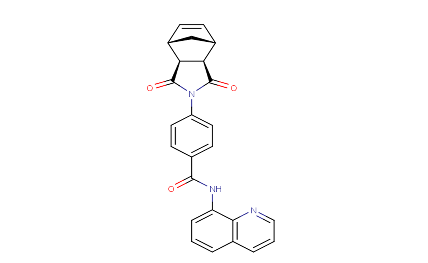
exo-IWR-1 is an inactive stereoisomer of Endo-IWR-1,and is used as a negative control of IWR-1 (V1351) which is a potent tankyrase inhibitor of the Wnt pathway with potential antitumor activity.
Physicochemical Properties
| Molecular Formula | C25H19N3O3 |
| Molecular Weight | 409.43700 |
| Exact Mass | 409.142 |
| CAS # | 1127442-87-8 |
| PubChem CID | 1163034 |
| Appearance | Light yellow to green yellow solid powder |
| Density | 1.4±0.1 g/cm3 |
| Boiling Point | 643.9±55.0 °C at 760 mmHg |
| Flash Point | 343.2±31.5 °C |
| Vapour Pressure | 0.0±1.9 mmHg at 25°C |
| Index of Refraction | 1.741 |
| LogP | 2.65 |
| Hydrogen Bond Donor Count | 1 |
| Hydrogen Bond Acceptor Count | 4 |
| Rotatable Bond Count | 3 |
| Heavy Atom Count | 31 |
| Complexity | 772 |
| Defined Atom Stereocenter Count | 4 |
| HS Tariff Code | 2934.99.9001 |
| Storage |
Powder-20°C 3 years 4°C 2 years In solvent -80°C 6 months -20°C 1 month |
| Shipping Condition | Room temperature (This product is stable at ambient temperature for a few days during ordinary shipping and time spent in Customs) |
Biological Activity
| Targets |
exo-IWR-1 targets the Wnt/β-catenin signaling pathway by stabilizing Axin2, thereby promoting β-catenin degradation, with an EC₅₀ value of 1.2 μM for inhibiting Wnt/β-catenin pathway activity (TOPFlash luciferase assay in Huh7 cells) [2] |
| ln Vitro |
The number of 293T cells infected with the bunyavirus remains unchanged in response to exo-IWR-1 (10 μM) [1]. When cells were pretreated or treated one hour post-infection, exo-IWR-1 had no effect on RVFV MP12-GFP infection [2]. Anti-RVFV activity: exo-IWR-1 (0.1–10 μM) dose-dependently inhibited RVFV replication in Huh7 and Vero cells, with EC₅₀ values of 1.5 μM (Huh7) and 1.8 μM (Vero) (qRT-PCR for viral RNA; viral plaque assay for titer) [2] - Wnt/β-catenin pathway inhibition: 1–5 μM reduced nuclear β-catenin accumulation by 45–78% (immunofluorescence/Western blot) and downregulated Wnt target genes (Axin2, Cyclin D1) by 2.3–3.1-fold (qRT-PCR) in RVFV-infected Huh7 cells [2] - Reduced viral protein expression: 3 μM decreased RVFV N protein levels by 65% (Western blot) and viral progeny release by 72% (plaque assay) [2] - Low cytotoxicity: CC₅₀ > 20 μM in Huh7 and Vero cells; cell viability >90% at concentrations up to 10 μM (MTT assay) [2] - No cross-resistance potential: Did not induce RVFV resistance after 10 serial passages in Huh7 cells treated with sub-EC₅₀ concentrations (0.5 μM) [1] |
| Enzyme Assay |
TOPFlash luciferase assay: Huh7 cells were co-transfected with TOPFlash (Wnt-responsive luciferase reporter plasmid) and Renilla luciferase (internal control plasmid). After 24 hours, cells were pretreated with exo-IWR-1 (0.1–10 μM) for 1 hour, then stimulated with Wnt3a (50 ng/mL) for 16 hours. Luciferase activity was measured, and pathway inhibition rate was calculated; EC₅₀ for pathway inhibition was 1.2 μM [2] |
| Cell Assay |
RVFV replication inhibition assay: Huh7/Vero cells were seeded in 24-well plates, pretreated with exo-IWR-1 (0.1–10 μM) for 1 hour, then infected with RVFV (MOI = 0.1) for 48 hours. Viral RNA was quantified by qRT-PCR; viral titer was determined by plaque assay [2] - Wnt pathway marker detection: RVFV-infected Huh7 cells were treated with exo-IWR-1 (1–5 μM) for 24 hours. Cells were lysed for Western blot (β-catenin, Axin2, RVFV N protein) or fixed for immunofluorescence (nuclear β-catenin staining with DAPI) [2] - Cytotoxicity assay: Huh7/Vero cells were seeded in 96-well plates, treated with exo-IWR-1 (0.1–50 μM) for 72 hours. MTT reagent was added, and absorbance at 570 nm was measured to calculate cell viability and CC₅₀ [2] - Resistance induction assay: Huh7 cells were infected with RVFV and cultured with exo-IWR-1 (0.5 μM, sub-EC₅₀) for 10 serial passages. Viral replication efficiency was measured by qRT-PCR to assess resistance development [1] |
| References |
[1]. Genome-wide RNA Interference Screen Identifies Novel Drug Targets for Rift Valley Fever Virus That Minimi the Potential for Drug Resistance. [2]. A Genome-Wide RNA Interference Screen Identifies a Role for Wnt/β-Catenin Signaling during Rift Valley Fever Virus Infection. J Virol. 2016 Jul 27;90(16):7084-7097. |
| Additional Infomation |
IWR-1-exo is a dicarboximide having an exo bridged phthalimide structure, substituted at nitrogen by a 4-(quinolin-8-ylcarbamoyl)benzoyl group. It is a weak axin stabilizer, an analogue of IWR-1-endo. It has a role as an axin stabilizer. It is a dicarboximide and a bridged compound. exo-IWR-1 is a small-molecule inhibitor of the Wnt/β-catenin signaling pathway, derived from IWR-1 with enhanced cell permeability [2] - Its anti-RVFV mechanism involves stabilizing the Axin2 scaffold protein, promoting β-catenin ubiquitination and degradation, thereby blocking the Wnt/β-catenin pathway required for RVFV replication [2] - It was identified as a potential anti-RVFV agent through genome-wide RNAi screening, which highlighted the dependency of RVFV on host Wnt/β-catenin signaling [1][2] - The compound shows low cytotoxicity and no obvious RVFV resistance induction, making it a promising lead for anti-RVFV drug development [1][2] |
Solubility Data
| Solubility (In Vitro) | DMSO : ~2 mg/mL (~4.88 mM) |
| Solubility (In Vivo) |
Note: Listed below are some common formulations that may be used to formulate products with low water solubility (e.g. < 1 mg/mL), you may test these formulations using a minute amount of products to avoid loss of samples. Injection Formulations (e.g. IP/IV/IM/SC) Injection Formulation 1: DMSO : Tween 80: Saline = 10 : 5 : 85 (i.e. 100 μL DMSO stock solution → 50 μL Tween 80 → 850 μL Saline) *Preparation of saline: Dissolve 0.9 g of sodium chloride in 100 mL ddH ₂ O to obtain a clear solution. Injection Formulation 2: DMSO : PEG300 :Tween 80 : Saline = 10 : 40 : 5 : 45 (i.e. 100 μL DMSO → 400 μLPEG300 → 50 μL Tween 80 → 450 μL Saline) Injection Formulation 3: DMSO : Corn oil = 10 : 90 (i.e. 100 μL DMSO → 900 μL Corn oil) Example: Take the Injection Formulation 3 (DMSO : Corn oil = 10 : 90) as an example, if 1 mL of 2.5 mg/mL working solution is to be prepared, you can take 100 μL 25 mg/mL DMSO stock solution and add to 900 μL corn oil, mix well to obtain a clear or suspension solution (2.5 mg/mL, ready for use in animals). Injection Formulation 4: DMSO : 20% SBE-β-CD in saline = 10 : 90 [i.e. 100 μL DMSO → 900 μL (20% SBE-β-CD in saline)] *Preparation of 20% SBE-β-CD in Saline (4°C,1 week): Dissolve 2 g SBE-β-CD in 10 mL saline to obtain a clear solution. Injection Formulation 5: 2-Hydroxypropyl-β-cyclodextrin : Saline = 50 : 50 (i.e. 500 μL 2-Hydroxypropyl-β-cyclodextrin → 500 μL Saline) Injection Formulation 6: DMSO : PEG300 : castor oil : Saline = 5 : 10 : 20 : 65 (i.e. 50 μL DMSO → 100 μLPEG300 → 200 μL castor oil → 650 μL Saline) Injection Formulation 7: Ethanol : Cremophor : Saline = 10: 10 : 80 (i.e. 100 μL Ethanol → 100 μL Cremophor → 800 μL Saline) Injection Formulation 8: Dissolve in Cremophor/Ethanol (50 : 50), then diluted by Saline Injection Formulation 9: EtOH : Corn oil = 10 : 90 (i.e. 100 μL EtOH → 900 μL Corn oil) Injection Formulation 10: EtOH : PEG300:Tween 80 : Saline = 10 : 40 : 5 : 45 (i.e. 100 μL EtOH → 400 μLPEG300 → 50 μL Tween 80 → 450 μL Saline) Oral Formulations Oral Formulation 1: Suspend in 0.5% CMC Na (carboxymethylcellulose sodium) Oral Formulation 2: Suspend in 0.5% Carboxymethyl cellulose Example: Take the Oral Formulation 1 (Suspend in 0.5% CMC Na) as an example, if 100 mL of 2.5 mg/mL working solution is to be prepared, you can first prepare 0.5% CMC Na solution by measuring 0.5 g CMC Na and dissolve it in 100 mL ddH2O to obtain a clear solution; then add 250 mg of the product to 100 mL 0.5% CMC Na solution, to make the suspension solution (2.5 mg/mL, ready for use in animals). Oral Formulation 3: Dissolved in PEG400 Oral Formulation 4: Suspend in 0.2% Carboxymethyl cellulose Oral Formulation 5: Dissolve in 0.25% Tween 80 and 0.5% Carboxymethyl cellulose Oral Formulation 6: Mixing with food powders Note: Please be aware that the above formulations are for reference only. InvivoChem strongly recommends customers to read literature methods/protocols carefully before determining which formulation you should use for in vivo studies, as different compounds have different solubility properties and have to be formulated differently. (Please use freshly prepared in vivo formulations for optimal results.) |
| Preparing Stock Solutions | 1 mg | 5 mg | 10 mg | |
| 1 mM | 2.4424 mL | 12.2118 mL | 24.4236 mL | |
| 5 mM | 0.4885 mL | 2.4424 mL | 4.8847 mL | |
| 10 mM | 0.2442 mL | 1.2212 mL | 2.4424 mL |