Physicochemical Properties
| Molecular Formula | C25H28O6 |
| Molecular Weight | 424.4862 |
| Exact Mass | 424.188 |
| CAS # | 20931-37-7 |
| Related CAS # | alpha-Mangostin;6147-11-1 |
| PubChem CID | 5495925 |
| Appearance | Off-white to yellow solid powder |
| Density | 1.2±0.1 g/cm3 |
| Boiling Point | 616.5±55.0 °C at 760 mmHg |
| Melting Point | 175-176ºC |
| Flash Point | 208.3±25.0 °C |
| Vapour Pressure | 0.0±1.8 mmHg at 25°C |
| Index of Refraction | 1.598 |
| LogP | 6.02 |
| Hydrogen Bond Donor Count | 2 |
| Hydrogen Bond Acceptor Count | 6 |
| Rotatable Bond Count | 6 |
| Heavy Atom Count | 31 |
| Complexity | 692 |
| Defined Atom Stereocenter Count | 0 |
| InChi Key | YRKKJHJIWCRNCW-UHFFFAOYSA-N |
| InChi Code | InChI=1S/C25H28O6/c1-13(2)7-9-15-18(29-5)12-20-22(23(15)27)24(28)21-16(10-8-14(3)4)25(30-6)17(26)11-19(21)31-20/h7-8,11-12,26-27H,9-10H2,1-6H3 |
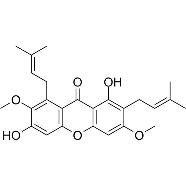
| Chemical Name | 1,6-dihydroxy-3,7-dimethoxy-2,8-bis(3-methylbut-2-enyl)xanthen-9-one |
| HS Tariff Code | 2934.99.9001 |
| Storage |
Powder-20°C 3 years 4°C 2 years In solvent -80°C 6 months -20°C 1 month |
| Shipping Condition | Room temperature (This product is stable at ambient temperature for a few days during ordinary shipping and time spent in Customs) |
Biological Activity
| Targets |
beta-Mangostin targets multiple biological molecules and pathogens, with key activity data as follows: - Matrix Metalloproteinase 2 (MMP-2): Concentration-dependent expression inhibition [1] - Matrix Metalloproteinase 9 (MMP-9): Concentration-dependent expression inhibition [1] - Heat Shock Protein 70 (HSP70): Concentration-dependent downregulation [2] - Plasmodium falciparum (3D7 strain): IC₅₀ = 3.5 μg/mL [3] - Mycobacterium tuberculosis H₃₇Rv: Minimum Inhibitory Concentration (MIC) = 32 μg/mL [4] |
| ln Vitro |
In HL60 cells linked to G0/G1 cell cycle arrest, beta-mangostin downregulates the HSP70 gene and activates the intrinsic apoptotic pathway via reactive oxygen species [2]. 1. Suppression of hepatocellular carcinoma cell invasion: - Human hepatocellular carcinoma HepG2 cells were treated with beta-Mangostin (5, 10, 20 μM) for 24 hours. Transwell invasion assay demonstrated dose-dependent inhibition of cell invasion, with inhibition rates of 31% (5 μM), 58% (10 μM), and 72% (20 μM) compared to vehicle control [1] - Western blot analysis showed reduced protein levels of MMP-2 and MMP-9 in a dose-dependent manner, while RT-PCR confirmed decreased mRNA expression of MMP-2 and MMP-9. Simultaneously, phosphorylation of ERK and JNK (p-ERK, p-JNK) was increased, indicating activation of ERK and JNK signaling pathways [1] - Gelatin zymography confirmed dose-dependent reduction in the enzymatic activity of secreted MMP-2 and MMP-9 from HepG2 cells [1] 2. Induction of apoptosis and cell cycle arrest in HL60 cells: - Human acute myeloid leukemia HL60 cells were treated with beta-Mangostin (2.5, 5, 10 μM) for 24–48 hours. Annexin V/PI double staining showed dose-dependent apoptosis, with apoptotic rates of 12.3% (2.5 μM), 28.7% (5 μM), and 45.2% (10 μM) after 48 hours (vs. 4.1% in vehicle control) [2] - Flow cytometry analysis revealed G₀/G₁ phase cell cycle arrest: 68.5% of cells were in G₀/G₁ phase at 10 μM (vs. 51.2% in vehicle control) [2] - Intracellular reactive oxygen species (ROS) levels were measured by fluorescent dye staining, showing a 2.8-fold increase at 10 μM compared to control [2] - Western blot and RT-PCR confirmed dose-dependent downregulation of HSP70 protein and mRNA levels. The intrinsic apoptosis pathway was activated, as evidenced by increased Bax/Bcl-2 ratio, cytochrome c release from mitochondria to cytoplasm, and cleavage of caspase-3 and caspase-9 [2] 3. Antimalarial activity: - Plasmodium falciparum (3D7 strain) was cultured in vitro with human red blood cells. beta-Mangostin was added at serial concentrations (0.1–10 μg/mL), and the culture was incubated at 37°C for 48 hours. Giemsa staining and microscopic counting of parasitemia showed growth inhibition of P. falciparum with an IC₅₀ of 3.5 μg/mL [3] 4. Antimycobacterial activity: - Mycobacterium tuberculosis H₃₇Rv was cultured in Middlebrook 7H9 broth supplemented with appropriate additives. beta-Mangostin was tested at concentrations ranging from 8 to 64 μg/mL, and the culture was incubated at 37°C for 7 days. Bacterial growth was monitored by measuring absorbance at 600 nm, and the MIC was determined as 32 μg/mL (inhibiting >90% bacterial growth) [4] |
| Enzyme Assay |
1. MMP-2/MMP-9 enzymatic activity assay (gelatin zymography): HepG2 cells were seeded in 6-well plates and treated with beta-Mangostin (5, 10, 20 μM) for 24 hours. Cell culture supernatants were collected and mixed with non-reducing sample buffer. Samples were separated by SDS-PAGE on a gel containing 10% gelatin (substrate for MMP-2 and MMP-9). After electrophoresis, the gel was washed twice with 2.5% Triton X-100 to remove SDS, then incubated in reaction buffer at 37°C for 24 hours to allow MMP-mediated gelatin degradation. The gel was stained with Coomassie Brilliant Blue R-250 and destained to visualize clear bands corresponding to MMP activity. Band intensity was quantified by imaging software, showing dose-dependent reduction in MMP-2 and MMP-9 activity [1] 2. Antimalarial activity assay: Plasmodium falciparum (3D7 strain) was maintained in RPMI 1640 medium supplemented with 10% human serum and 5% human red blood cells. beta-Mangostin was dissolved in an appropriate solvent and serially diluted to final concentrations of 0.1–10 μg/mL in the culture medium. The infected red blood cells were incubated with the compound at 37°C in a 5% CO₂, 5% O₂, and 90% N₂ atmosphere for 48 hours. After incubation, thin blood smears were prepared, stained with Giemsa, and examined under a microscope. Parasitemia was calculated by counting the number of infected red blood cells among 1000 total red blood cells. The IC₅₀ value was derived by fitting the dose-response curve of parasitemia inhibition [3] 3. Antimycobacterial activity assay: Mycobacterium tuberculosis H₃₇Rv was grown in Middlebrook 7H9 broth supplemented with 10% ADC enrichment and 0.05% Tween 80. beta-Mangostin was dissolved in a suitable solvent and added to the bacterial culture to final concentrations of 8, 16, 32, 64 μg/mL. The culture was incubated at 37°C with shaking for 7 days. Bacterial growth was assessed by measuring the optical density at 600 nm. The MIC was defined as the lowest concentration of beta-Mangostin that inhibited >90% of bacterial growth compared to the solvent control [4] |
| Cell Assay |
1. Hepatocellular carcinoma cell invasion assay: - HepG2 cells were trypsinized and resuspended in serum-free medium at a density of 5×10⁴ cells/mL. Matrigel was diluted with serum-free medium and coated onto the upper surface of transwell inserts (8 μm pore size), then incubated at 37°C for 4 hours to form a gel matrix [1] - beta-Mangostin was added to both the upper and lower chambers of the transwell plate at final concentrations of 5, 10, 20 μM. The upper chamber was seeded with 100 μL of cell suspension, and the lower chamber was filled with 600 μL of medium containing 10% fetal bovine serum (chemoattractant) [1] - The plate was incubated at 37°C with 5% CO₂ for 24 hours. Non-invasive cells on the upper surface of the insert were wiped off with a cotton swab. Invasive cells on the lower surface were fixed with 4% paraformaldehyde for 15 minutes, stained with 0.1% crystal violet for 20 minutes, and rinsed with PBS [1] - Invasive cells were counted under a light microscope (five random fields per insert), and the invasion inhibition rate was calculated relative to the vehicle control [1] 2. HL60 cell apoptosis, cell cycle, and ROS detection assays: - Apoptosis assay: HL60 cells were seeded in 6-well plates at 1×10⁶ cells/mL and treated with beta-Mangostin (2.5, 5, 10 μM) for 48 hours. Cells were harvested by centrifugation, washed twice with cold PBS, and resuspended in binding buffer. Annexin V-FITC and PI were added to the cell suspension, which was then incubated in the dark at room temperature for 15 minutes. Apoptotic cells were analyzed by flow cytometry [2] - Cell cycle assay: HL60 cells were treated with beta-Mangostin (10 μM) for 24 hours, harvested, and fixed with 70% cold ethanol at -20°C overnight. Fixed cells were washed with PBS, resuspended in PI staining solution (containing RNase A), and incubated in the dark at 37°C for 30 minutes. Cell cycle distribution was analyzed by flow cytometry [2] - ROS detection: HL60 cells were loaded with ROS-sensitive fluorescent dye at 37°C for 30 minutes, then treated with beta-Mangostin (2.5, 5, 10 μM) for 24 hours. Cells were washed with PBS, and fluorescent intensity (reflecting ROS levels) was measured by flow cytometry [2] 3. Western blot and RT-PCR assays for target expression: - Western blot: Cells (HepG2 or HL60) were treated with beta-Mangostin at specified concentrations for 24 hours, then lysed with RIPA buffer containing protease and phosphatase inhibitors. Equal amounts of protein (25–30 μg) were separated by SDS-PAGE, transferred to PVDF membranes, and blocked with 5% non-fat milk for 1 hour at room temperature. Membranes were incubated with primary antibodies against target proteins (MMP-2, MMP-9, p-ERK, ERK, p-JNK, JNK for HepG2; HSP70, Bax, Bcl-2, caspase-3, caspase-9, cytochrome c for HL60) and β-actin (loading control) overnight at 4°C. After washing, membranes were incubated with peroxidase-conjugated secondary antibodies for 1 hour at room temperature. Protein bands were visualized with chemiluminescent reagents, and band intensity was quantified by imaging software [1,2] - RT-PCR: Total RNA was extracted from treated cells using an RNA extraction method. Complementary DNA (cDNA) was synthesized from total RNA, and PCR amplification was performed with specific primers for target genes (MMP-2, MMP-9 for HepG2; HSP70 for HL60) and GAPDH (internal control). PCR products were separated by agarose gel electrophoresis, stained with ethidium bromide, and quantified by imaging software [1,2] |
| References |
[1]. β-mangostin suppresses human hepatocellular carcinoma cell invasion through inhibition of MMP-2 and MMP-9 expression and activating the ERK and JNK pathways. Environ Toxicol. 2017 Nov;32(11):2360-2370. [2]. Beta-mangostin from Cratoxylum arborescens activates the intrinsic apoptosis pathway through reactive oxygen species with downregulation of the HSP70 gene in the HL60 cells associated with a G 0/G 1 cell-cycle arrest. Tumour Biol. 2017 Nov;39(11):1010428317731451. [3]. Antimalarial xanthones from Garcinia cowa. Planta Med. 1998 Feb;64(1):70-2. [4]. Antimycobacterial activity of prenylated xanthones from the fruits of Garcinia mangostana. Chem Pharm Bull (Tokyo). 2003 Jul;51(7):857-9. |
| Additional Infomation |
Beta-Mangostin is a member of xanthones. beta-Mangostin has been reported in Garcinia cowa, Garcinia mangostana, and other organisms with data available. 1. Source and structural characteristics: beta-Mangostin is a natural prenylated xanthone compound. It is isolated from the fruits of Garcinia mangostana (mangosteen) and the plant Cratoxylum arborescens. Its chemical structure features a xanthone core scaffold with prenyl substituents, which is crucial for its biological activities [2,3,4] 2. Mechanisms of action: - Anti-hepatocellular carcinoma: Inhibits the expression and enzymatic activity of MMP-2 and MMP-9, which are key enzymes for cell invasion. Simultaneously activates ERK and JNK signaling pathways to suppress HepG2 cell invasion [1] - Anti-acute myeloid leukemia: Induces intracellular ROS accumulation, downregulates HSP70 expression, disrupts the balance between anti-apoptotic (Bcl-2) and pro-apoptotic (Bax) proteins, promotes cytochrome c release from mitochondria, activates caspase-3 and caspase-9, and triggers intrinsic apoptosis. It also induces G₀/G₁ phase cell cycle arrest to inhibit HL60 cell proliferation [2] - Antimalarial: Directly inhibits the growth and replication of Plasmodium falciparum (3D7 strain) in vitro [3] - Antimycobacterial: Directly inhibits the growth of Mycobacterium tuberculosis H₃₇Rv by suppressing bacterial proliferation [4] 3. Therapeutic potential: beta-Mangostin exhibits diverse biological activities, including anti-tumor (hepatocellular carcinoma, acute myeloid leukemia), antimalarial, and antimycobacterial effects. As a natural compound with multi-target actions, it holds promise as a lead compound for the development of novel therapeutic agents against these diseases [1,2,3,4] |
Solubility Data
| Solubility (In Vitro) | DMSO : ~25 mg/mL (~58.89 mM) |
| Solubility (In Vivo) |
Solubility in Formulation 1: ≥ 2.5 mg/mL (5.89 mM) (saturation unknown) in 10% DMSO + 40% PEG300 + 5% Tween80 + 45% Saline (add these co-solvents sequentially from left to right, and one by one), clear solution. For example, if 1 mL of working solution is to be prepared, you can add 100 μL of 25.0 mg/mL clear DMSO stock solution to 400 μL PEG300 and mix evenly; then add 50 μL Tween-80 to the above solution and mix evenly; then add 450 μL normal saline to adjust the volume to 1 mL. Preparation of saline: Dissolve 0.9 g of sodium chloride in 100 mL ddH₂ O to obtain a clear solution. Solubility in Formulation 2: ≥ 2.5 mg/mL (5.89 mM) (saturation unknown) in 10% DMSO + 90% (20% SBE-β-CD in Saline) (add these co-solvents sequentially from left to right, and one by one), clear solution. For example, if 1 mL of working solution is to be prepared, you can add 100 μL of 25.0 mg/mL clear DMSO stock solution to 900 μL of 20% SBE-β-CD physiological saline solution and mix evenly. Preparation of 20% SBE-β-CD in Saline (4°C,1 week): Dissolve 2 g SBE-β-CD in 10 mL saline to obtain a clear solution. Solubility in Formulation 3: ≥ 1.25 mg/mL (2.94 mM) (saturation unknown) in 10% DMSO + 90% Corn Oil (add these co-solvents sequentially from left to right, and one by one), clear solution. For example, if 1 mL of working solution is to be prepared, you can add 100 μL of 12.5 mg/mL clear DMSO stock solution to 900 μL of corn oil and mix evenly. (Please use freshly prepared in vivo formulations for optimal results.) |
| Preparing Stock Solutions | 1 mg | 5 mg | 10 mg | |
| 1 mM | 2.3558 mL | 11.7788 mL | 23.5577 mL | |
| 5 mM | 0.4712 mL | 2.3558 mL | 4.7115 mL | |
| 10 mM | 0.2356 mL | 1.1779 mL | 2.3558 mL |