YS-49 (YS49) is a novel and potent anti-inflammatory agent and PI3K/Akt signaling activator signaling that has the potential to treat vascular diseases like hypertension and atherosclerosis. Through the induction of HO-1, it prevents VSMC proliferation that is induced by Ang II. Through the induction of heme oxygenase-1, YS-49 controls angiotensin II-stimulated ROS production, JNK phosphorylation, and vascular smooth muscle cell proliferation.
Physicochemical Properties
| Molecular Formula | C20H22BRNO3 |
| Molecular Weight | 404.2976 |
| Exact Mass | 385.067 |
| Elemental Analysis | C, 59.42; H, 5.48; Br, 19.76; N, 3.46; O, 11.87 |
| CAS # | 132836-42-1 |
| Related CAS # | YS-49 monohydrate |
| PubChem CID | 10249534 |
| Appearance | White to off-white solid powder |
| LogP | 4.967 |
| Hydrogen Bond Donor Count | 4 |
| Hydrogen Bond Acceptor Count | 3 |
| Rotatable Bond Count | 2 |
| Heavy Atom Count | 24 |
| Complexity | 401 |
| Defined Atom Stereocenter Count | 0 |
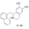
| SMILES | Br[H].O([H])C1=C(C([H])=C2C([H])([H])C([H])([H])N([H])C([H])(C([H])([H])C3=C([H])C([H])=C([H])C4=C([H])C([H])=C([H])C([H])=C34)C2=C1[H])O[H].O([H])[H] |
| InChi Key | QHCUNMLYODPTIU-UHFFFAOYSA-N |
| InChi Code | InChI=1S/C20H19NO2.BrH.H2O/c22-19-11-15-8-9-21-18(17(15)12-20(19)23)10-14-6-3-5-13-4-1-2-7-16(13)14;;/h1-7,11-12,18,21-23H,8-10H2;1H;1H2 |
| Chemical Name | 1,2,3,4-Tetrahydro-1-(1-naphthalenylmethyl)-6,7-Isoquinolinediol hydrobromide monohydrate |
| Synonyms | YS49 HBr; YS-49; YS 49; YS49; YS-49 hydrobromide |
| HS Tariff Code | 2934.99.9001 |
| Storage |
Powder-20°C 3 years 4°C 2 years In solvent -80°C 6 months -20°C 1 month Note: Please store this product in a sealed and protected environment (e.g. under nitrogen), avoid exposure to moisture. |
| Shipping Condition | Room temperature (This product is stable at ambient temperature for a few days during ordinary shipping and time spent in Customs) |
Biological Activity
| Targets |
PI3K; Akt; cardiac β-adrenoceptors
Heme Oxygenase-1 (HO-1) [1] c-Jun N-terminal Kinase (JNK) (inhibits Ang II-induced phosphorylation) [1] Reactive Oxygen Species (ROS) production pathway (inhibits Ang II-induced ROS generation) [1] Inducible Nitric Oxide Synthase (iNOS) (inhibits its expression, ) [2] Nuclear Factor-kappa B (NF-κB) (inhibits its activation, ) [2] Mitogen-Activated Protein Kinase (MAPK) (p38/ERK1/2, modulates phosphorylation,) [2] |
| ln Vitro |
YS-49 (1-100 μM; 18 hours; RAVSMC and RAW 264.7 cells) concentration-dependently inhibits the accumulation of nitrite in both RAVSMC and RAW 264.7 exposed to lipopolysaccharide (LPS) plus INF-γ, with IC50 values of 22 μM and 30 μM, respectively[2]. YS-49 (10-100 μM; 18 hours; RAVSMC and RAW 264.7 cells) inhibits the transcriptional expression of the iNOS gene in RAVSMC and RAW 264.7 cells when LPS and/or cytokines are present[2]. YS 49 (1-10 μM) dose-dependently induces the mRNA and protein expression of HO-1 in rat vascular smooth muscle cells (VSMC) (detected by RT-PCR and Western blotting); it inhibits angiotensin II (Ang II, 100 nM)-stimulated ROS production in VSMC by approximately 60% at 10 μM (measured by DCFH-DA fluorescence assay), suppresses Ang II-induced JNK phosphorylation by about 70% at 10 μM (Western blotting), and reduces Ang II-stimulated VSMC proliferation by 55% at 10 μM (BrdU incorporation assay); the HO-1 inhibitor ZnPP IX reverses all these effects of YS 49, indicating its actions are HO-1-dependent [1] YS 49 (1-30 μM) dose-dependently inhibits LPS (1 μg/mL) or IL-1β (10 ng/mL)-induced iNOS mRNA and protein expression in rat aortic VSMC (detected by RT-PCR and Western blotting), and reduces nitric oxide (NO) production by approximately 80% at 30 μM (Griess reagent assay); in RAW264.7 macrophages, YS 49 (10-30 μM) similarly inhibits LPS-induced iNOS expression and NO production; mechanistically, YS 49 suppresses NF-κB nuclear translocation (immunofluorescence and EMSA) and modulates the MAPK pathway: it decreases p38 phosphorylation and increases ERK1/2 phosphorylation, with no significant effect on JNK phosphorylation; additionally, YS 49 exhibits positive inotropic effects in cardiomyocytes [2] |
| ln Vivo |
YS-49 (5 mg/kg; intraperitoneal injection; 8 hours; male Sprague Dawley rats) treatment significantly reduces serum NOx levels in LPS-treated rats, the NOx levels reduce from 86 μM to 34 μM[2]. For HO-1 enzyme activity assay: Collect YS 49-treated VSMC, lyse the cells by ultrasonication and centrifuge to obtain the supernatant; adjust the protein concentration of the supernatant and incubate it with reaction buffer containing heme substrate and NADPH at 37°C for 30 minutes; terminate the reaction with HCl and measure the absorbance at 464 nm (characteristic absorption of biliverdin, the catalytic product of HO-1) using a spectrophotometer; calculate the HO-1 enzyme activity units based on the absorbance values [1] For iNOS enzyme activity assay: Harvest VSMC/RAW264.7 cells treated with YS 49 and LPS, lyse the cells and collect the supernatant; incubate the supernatant with reaction buffer containing L-arginine and NADPH at 37°C for 60 minutes; add Griess reagent A and B successively, stand at room temperature for 15 minutes, and measure the absorbance at 540 nm using a microplate reader; calculate the iNOS activity by comparing with the NO standard curve [2] |
| Enzyme Assay |
1. VSMC proliferation assay: Isolate primary rat aortic VSMC and culture to passages 3-5; seed the cells in 96-well plates at a density of 5×10³ cells/well, synchronize the cells and stimulate with Ang II (100 nM) while treating with different concentrations of YS 49 (1, 5, 10 μM) for 24 hours; add BrdU and incubate for 12 hours, fix the cells and add anti-BrdU antibody, measure the absorbance at 450 nm using a microplate reader to calculate the proliferation inhibition rate [1] 2. ROS detection assay: Seed VSMC in confocal dishes, synchronize and pretreat with YS 49 for 1 hour, then stimulate with Ang II for 30 minutes; load the cells with DCFH-DA fluorescent probe (10 μM final concentration) and incubate at 37°C for 20 minutes; observe the fluorescence intensity under a confocal microscope and quantitatively analyze the ROS level [1] 3. Western blot for HO-1 and p-JNK: After treating VSMC with YS 49 and Ang II, extract total cellular protein, separate the proteins by SDS-PAGE, transfer to membranes and incubate with antibodies against HO-1, p-JNK, total JNK and GAPDH; develop the bands by chemiluminescence and quantify the gray values of the bands [1] 4. RT-PCR for HO-1 mRNA: Extract total RNA from VSMC, reverse-transcribe into cDNA, perform PCR amplification with HO-1-specific primers (GAPDH as internal reference); separate the PCR products by agarose gel electrophoresis, quantify the bands and calculate the relative expression of HO-1 mRNA [1] |
| Cell Assay |
1. VSMC proliferation assay: Isolate primary rat aortic VSMC and culture to passages 3-5; seed the cells in 96-well plates at a density of 5×10³ cells/well, synchronize the cells and stimulate with Ang II (100 nM) while treating with different concentrations of YS 49 (1, 5, 10 μM) for 24 hours; add BrdU and incubate for 12 hours, fix the cells and add anti-BrdU antibody, measure the absorbance at 450 nm using a microplate reader to calculate the proliferation inhibition rate [1] 2. ROS detection assay: Seed VSMC in confocal dishes, synchronize and pretreat with YS 49 for 1 hour, then stimulate with Ang II for 30 minutes; load the cells with DCFH-DA fluorescent probe (10 μM final concentration) and incubate at 37°C for 20 minutes; observe the fluorescence intensity under a confocal microscope and quantitatively analyze the ROS level [1] 3. Western blot for HO-1 and p-JNK: After treating VSMC with YS 49 and Ang II, extract total cellular protein, separate the proteins by SDS-PAGE, transfer to membranes and incubate with antibodies against HO-1, p-JNK, total JNK and GAPDH; develop the bands by chemiluminescence and quantify the gray values of the bands [1] 4. RT-PCR for HO-1 mRNA: Extract total RNA from VSMC, reverse-transcribe into cDNA, perform PCR amplification with HO-1-specific primers (GAPDH as internal reference); separate the PCR products by agarose gel electrophoresis, quantify the bands and calculate the relative expression of HO-1 mRNA [1] 5. iNOS expression and NO detection assay: Seed rat VSMC/RAW264.7 cells in 96-well plates at 1×10⁴ cells/well, stimulate with LPS (1 μg/mL) or IL-1β (10 ng/mL) and treat with YS 49 (1, 10, 30 μM) for 24 hours; collect the supernatant and detect NO concentration with Griess reagent; extract total RNA/protein from the cells and detect iNOS mRNA and protein expression by RT-PCR and Western blotting, respectively [2] 6. NF-κB activity assay: Seed RAW264.7 cells in 6-well plates, treat with YS 49 and LPS, then extract nuclear protein and detect NF-κB DNA-binding activity by EMSA; simultaneously, detect the nuclear translocation of NF-κB p65 subunit by immunofluorescence: fix the cells, stain with anti-p65 antibody and DAPI (for nuclear staining), and observe the localization under a confocal microscope [2] 7. MAPK phosphorylation assay: After treating VSMC with YS 49 and LPS, extract total cellular protein, detect the expression of p-p38, total p38, p-ERK1/2, total ERK1/2, p-JNK and total JNK by Western blotting, and quantify the phosphorylation levels [2] |
| Animal Protocol |
Male Sprague Dawley rats (250-300 g)[2] 50 mg/kg Intraperitoneal injection; 8 hours |
| References |
[1]. YS49,1-(alpha-naphtylmethyl)-6,7-dihydroxy-1,2,3,4-tetrahydroisoquinoline,regulates angiotensin II-stimulated ROS production, JNK phosphorylation and vascular smooth muscle cell proliferation via the induction of heme oxygen [2]. Prevention of the expression of inducible nitric oxide synthase by a novel positive inotropic agent, YS 49, in rat vascular smooth muscle and RAW 264.7 macrophages. Br J Pharmacol. 1999 Sep;128(2):357-64. [3]. RhoA-mediated inhibition of vascular endothelial cell mobility: positive feedback through reduced cytosolic p21 and p27. J Cell Physiol. 2014 Oct;229(10):1455-65. |
| Additional Infomation |
YS 49 has a chemical name of 1-(alpha-naphtylmethyl)-6,7-dihydroxy-1,2,3,4-tetrahydroisoquinoline; it is a novel vasoactive compound that exerts antioxidant and antiproliferative effects by inducing HO-1 expression, with potential therapeutic applications in vascular remodeling-related diseases (e.g., hypertension, atherosclerosis); its mechanism of action relies on HO-1-mediated ROS scavenging and JNK pathway inhibition [1] YS 49 was initially identified as a novel positive inotropic agent with the ability to enhance myocardial contractility; it also exhibits anti-inflammatory activity by inhibiting NF-κB and modulating the MAPK pathway, thereby reducing excessive iNOS expression and NO production, and has potential for the treatment of inflammation-related vascular diseases and myocardial dysfunction; no cytotoxicity of YS 49 was observed in VSMC and RAW264.7 cells at experimental concentrations (≤30 μM) [2] |
Solubility Data
| Solubility (In Vitro) |
DMSO: 77~100 mg/mL (199.3~258.9 mM) Water: ~6 mg/mL (~15.5 mM) Ethanol: ~77 mg/mL (~199.3 mM) |
| Solubility (In Vivo) |
Solubility in Formulation 1: ≥ 2.5 mg/mL (6.47 mM) (saturation unknown) in 10% DMSO + 40% PEG300 + 5% Tween80 + 45% Saline (add these co-solvents sequentially from left to right, and one by one), clear solution. For example, if 1 mL of working solution is to be prepared, you can add 100 μL of 25.0 mg/mL clear DMSO stock solution to 400 μL PEG300 and mix evenly; then add 50 μL Tween-80 to the above solution and mix evenly; then add 450 μL normal saline to adjust the volume to 1 mL. Preparation of saline: Dissolve 0.9 g of sodium chloride in 100 mL ddH₂ O to obtain a clear solution. Solubility in Formulation 2: ≥ 2.5 mg/mL (6.47 mM) (saturation unknown) in 10% DMSO + 90% (20% SBE-β-CD in Saline) (add these co-solvents sequentially from left to right, and one by one), clear solution. For example, if 1 mL of working solution is to be prepared, you can add 100 μL of 25.0 mg/mL clear DMSO stock solution to 900 μL of 20% SBE-β-CD physiological saline solution and mix evenly. Preparation of 20% SBE-β-CD in Saline (4°C,1 week): Dissolve 2 g SBE-β-CD in 10 mL saline to obtain a clear solution. Solubility in Formulation 3: ≥ 2.5 mg/mL (6.47 mM) (saturation unknown) in 10% DMSO + 90% Corn Oil (add these co-solvents sequentially from left to right, and one by one), clear solution. For example, if 1 mL of working solution is to be prepared, you can add 100 μL of 25.0 mg/mL clear DMSO stock solution to 900 μL of corn oil and mix evenly. Solubility in Formulation 4: 6.67 mg/mL (17.27 mM) in PBS (add these co-solvents sequentially from left to right, and one by one), clear solution; with ultrasonication (<60°C). (Please use freshly prepared in vivo formulations for optimal results.) |
| Preparing Stock Solutions | 1 mg | 5 mg | 10 mg | |
| 1 mM | 2.4734 mL | 12.3671 mL | 24.7341 mL | |
| 5 mM | 0.4947 mL | 2.4734 mL | 4.9468 mL | |
| 10 mM | 0.2473 mL | 1.2367 mL | 2.4734 mL |