Y15 (also known as FAK inhibitor Y15 and FAK Inhibitor 14) is a novel, potent, specific and direct inhibitor of focal adhesion kinase (FAK) which blocks phosphorylation of Y397 with an IC50 value of about 1 μM. In thyroid cancer cell lines, Y15 reduces viability, clonogenicity, and cell attachment while working in concert with targeted therapies. Y15 has been demonstrated to slow the growth of cancer both in vivo and in vitro. Y15 prevents tumor growth, lowers the viability of cancer cells, and inhibits its autophosphorylation activity.
Physicochemical Properties
| Molecular Formula | C6H14CL4N4 |
| Molecular Weight | 284.0142 |
| Exact Mass | 281.997 |
| Elemental Analysis | C, 25.37; H, 4.97; Cl, 49.93; N, 19.73 |
| CAS # | 4506-66-5 |
| Related CAS # | 4506-66-5 (HCl) |
| PubChem CID | 78260 |
| Appearance | Light green to green solid powder |
| Boiling Point | 400.9ºC at 760mmHg |
| Melting Point | ≥300ºC(lit.) |
| Flash Point | 233.6ºC |
| Index of Refraction | 1.827 |
| LogP | 5.548 |
| Hydrogen Bond Donor Count | 8 |
| Hydrogen Bond Acceptor Count | 4 |
| Rotatable Bond Count | 0 |
| Heavy Atom Count | 14 |
| Complexity | 90.3 |
| Defined Atom Stereocenter Count | 0 |
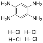
| SMILES | Cl[H].Cl[H].Cl[H].Cl[H].N([H])([H])C1C([H])=C(C(=C([H])C=1N([H])[H])N([H])[H])N([H])[H] |
| InChi Key | BZDGCIJWPWHAOF-UHFFFAOYSA-N |
| InChi Code | InChI=1S/C6H10N4.4ClH/c7-3-1-4(8)6(10)2-5(3)9;;;;/h1-2H,7-10H2;4*1H |
| Chemical Name | benzene-1,2,4,5-tetramine;tetrahydrochloride |
| Synonyms | FAK Inhibitor 14; FAK Inhibitor Y15; Y15 hydrochloride; Y15 tetrahydrochloride; 1,2,4,5-Benzenetetramine tetrahydrochloride; Benzene-1,2,4,5-tetraamine tetrahydrochloride; FAK Inhibitor 14; Y15; Benzene-1,2,4,5-tetramine 4HCl; 1,2,4,5-Tetraaminobenzene tetrahydrochloride; MFCD00012970; Y 15; Y-15. |
| HS Tariff Code | 292159 |
| Storage |
Powder-20°C 3 years 4°C 2 years In solvent -80°C 6 months -20°C 1 month Note: Please store this product in a sealed and protected environment, avoid exposure to moisture. |
| Shipping Condition | Room temperature (This product is stable at ambient temperature for a few days during ordinary shipping and time spent in Customs) |
Biological Activity
| Targets |
FAK
Y15 targets FAK (PTK2) with an IC50 of 1.5 μM (kinase activity inhibition)[1] Y15 targets FAK (PTK2) with an IC50 of 1.2 μM (kinase activity inhibition) [2] |
| ln Vitro |
Y15 treatment in vitro results in decreased cell viability, increased detachment, and increased apoptosis in colon cancer cells, breast cancer cells, and melanoma. Y15 dose-dependently inhibits the expression of total FAK and pY397 in TPC1, BCPAP, K1, and TT cell lines. All cell lines of thyroid cancer experience effective dose-dependent detachment. All medullary and papillary thyroid cancer cell lines exhibit an increase in necrosis, and all of them show a dose-dependent decrease in colony formation when exposed to Y15[1]. Y15 does not target homologous Pyk-2, c-Src, c-RAF, EGFR, IGFR, PDGFR, PI3K, VEGFR-3, and c-Met [3]. 1. Y15 exhibited antiproliferative effects on multiple tumor cell lines: the IC50 (48h, MTT assay) was 5 μM for MDA-MB-231 cells, 7 μM for MCF-7 cells, 6 μM for HepG2 cells, and 8 μM for A549 cells [1] 2. Y15 (5 μM) induced apoptosis in MDA-MB-231 cells, with the apoptotic rate increasing from 3.2% (control) to 28.5% after 48h treatment (Annexin V/PI staining, flow cytometry) [1] 3. Y15 reduced the protein expression of p-FAK (Tyr397) and p-STAT3 (Tyr705) in a concentration-dependent manner in MDA-MB-231 cells; at 5 μM, p-FAK expression decreased by approximately 70% and p-STAT3 by approximately 65% (Western blot) [1] 4. Y15 (2.5 μM, 5 μM) inhibited the migration of MDA-MB-231 cells by 40% and 75%, and invasion by 35% and 68% respectively after 24h treatment (Transwell assay) [1] 1. Y15 inhibited the proliferation of colon cancer cell lines: the IC50 (48h, CCK-8 assay) was 4 μM for HCT116 cells and 5 μM for SW480 cells [2] 2. Y15 (2 μM, 4 μM) reduced the colony formation of HCT116 cells from 120 colonies/well (control) to 45 and 15 colonies/well, with inhibition rates of 62.5% and 87.5% respectively [2] 3. Y15 (4 μM) arrested HCT116 cells in the G1 phase after 24h treatment: the proportion of G1-phase cells increased from 55% (control) to 72%, S-phase cells decreased from 30% to 18%, and G2/M-phase cells decreased from 15% to 10% (PI staining, flow cytometry) [2] 4. Y15 decreased the protein expression of p-FAK (Tyr397) and p-Src (Tyr416) in a concentration-dependent manner in HCT116 cells; at 4 μM, p-FAK expression decreased by approximately 60% and p-Src by approximately 55% (Western blot) [2] 1. Y15 showed cytotoxicity to human normal liver cell line L02: the IC50 was 25 μM (24h) and 18 μM (48h) (MTT assay); cell viability was over 85% when treated with Y15 at ≤10 μM for 48h [3] 2. Y15 showed cytotoxicity to human renal tubular epithelial cell line HK-2: the IC50 was 30 μM (24h) and 22 μM (48h) (MTT assay); cell viability was over 90% when treated with Y15 at ≤10 μM [3] 3. Y15 (15 μM, 30 μM) induced oxidative stress in L02 cells after 48h treatment: at 30 μM, ROS levels increased by approximately 40%, MDA content increased by approximately 35%, and SOD activity decreased by approximately 25% (kit detection) [3] |
| ln Vivo |
Y15 inhibits the growth of neuroblastoma, pancreatic, and breast tumors in vivo[2]. According to the pharmacokinetics study conducted on mice, Y15 is absorbed very quickly in these animals, with maximum plasma concentration being reached in 4.8 minutes after intraperitoneal administration at a dose of 30 mg/kg. With half-lives of 6.9 and 11.6 minutes, respectively, Y15 metabolizes quickly in mouse and human liver microsomes. Y15 has a maximum tolerated dose of 200 mg/kg for single-dose oral administration and a maximum tolerated dose of 100 mg/kg for multiple oral administration during a 7-day study. At 30 mg/kg by IP during the 28-day study and 100 mg/kg by PO during the 7-day study, Y15 does not result in any mortality or statistically significant changes in body weight. In various mouse organs, at 30 mg/kg by IP over 28 days and at 100 mg/kg dose by PO over 7 days, there are no clinical, chemical, hematological, or histopathological changes[3]. 1. In the MDA-MB-231 xenograft model in nude mice, Y15 (10 mg/kg, intraperitoneal injection, 5 times/week for 3 weeks) reduced tumor volume from 1200 mm³ (control) to 450 mm³, and tumor weight from 1.1 g (control) to 0.4 g, with a tumor inhibition rate of approximately 63.6% [1] 2. Immunohistochemistry showed that the positive expression rates of p-FAK and p-STAT3 in tumor tissues of the Y15-treated group decreased from 85% and 80% (control) to 25% and 20% respectively, and the Ki-67 positive rate decreased from 75% to 30% [1] 1. In the HCT116 xenograft model in nude mice, Y15 (8 mg/kg, oral administration, once daily for 21 days) reduced tumor volume from 1000 mm³ (control) to 380 mm³, and tumor weight from 0.95 g (control) to 0.32 g, with a tumor inhibition rate of approximately 66.3% [2] 2. qRT-PCR showed that the mRNA expression of Cyclin D1 and CDK4 in tumor tissues of the Y15-treated group decreased by approximately 50% and 45% respectively [2] 1. Acute toxicity in SD rats: single oral administration of Y15 at 50/100 mg/kg caused no death; 200 mg/kg caused mild diarrhea (recovered in 3 days); 400 mg/kg caused a mortality rate of 20% (2/10), with gastric mucosal congestion and mild hepatic steatosis in deceased rats [3] 2. Subchronic toxicity in SD rats: oral administration of Y15 at 10 mg/kg for 90 days caused no obvious abnormalities; 20 mg/kg caused mild elevation of liver enzymes (ALT increased by approximately 20%) and a small amount of inflammatory cell infiltration in the liver; 40 mg/kg caused a 50% increase in ALT/AST, a 30% increase in BUN, moderate hepatic steatosis, and mild edema of renal proximal tubular epithelial cells [3] |
| Enzyme Assay |
A kinase buffer containing 10 μCi of [γ-32P]-ATP A kinase buffer containing 10 μCi of [γ-32P]-ATP is incubated with 0.1 μg of purified FAK protein and 20 mM HEPES, pH 7.4, 5 mM MgCl2, 5 mM MnCl2, 0.1 mM Na3VO4. After the kinase reaction has been running for five minutes at room temperature, 2× Laemmli buffer is added to halt it. Proteins are separated using a Ready SDS-10% PAGE gel, and autoradiography is used to show the phosphorylated enolase. 1. FAK kinase activity assay: Recombinant human FAK protein was pre-incubated with different concentrations of Y15 (0.1-10 μM) in reaction buffer for 15 minutes, followed by the addition of ATP and substrate polypeptide (containing FAK phosphorylation site) and incubation at 30℃ for 30 minutes; the reaction was terminated, and the absorbance of phosphorylated substrate was detected by a microplate reader to calculate the kinase activity inhibition rate and IC50 via nonlinear regression analysis [1] 1. FAK-Src complex kinase activity assay: Recombinant FAK and Src proteins were incubated in reaction buffer to form a complex, then pre-incubated with different concentrations of Y15 (0.05-5 μM) for 20 minutes; ATP and specific substrate were added, and the mixture was incubated at 37℃ for 40 minutes; the radioactivity of phosphorylated substrate was detected using radioisotope-labeled ATP to calculate the kinase activity inhibition rate and determine the IC50 [2] |
| Cell Assay |
Ten thousand cells per well, in 100 μL of medium containing 10% FBS and 1% penicillin/streptomycin, are seeded onto 96-well dishes. After the inhibitor treatment for twenty-four hours, each well receives 20 μL of Cell Titer 96 Aqueous One Solution Cell Proliferation Assay. At 490 nm, the plate is read following a two-hour reagent incubation period. 1. Cell proliferation assay (MTT method): Logarithmic phase tumor cells (MDA-MB-231, MCF-7, etc.) were seeded in 96-well plates at 5×10³ cells/well, cultured for 24h, then treated with different concentrations of Y15 (0-20 μM) for 48h; MTT solution was added, and after 4h of incubation, the supernatant was discarded, organic solvent was added to dissolve formazan crystals, and the absorbance at 490 nm was detected by a microplate reader to calculate cell viability and IC50 [1] 2. Apoptosis assay (Annexin V/PI double staining): MDA-MB-231 cells were seeded in 6-well plates, cultured for 24h, then treated with 5 μM Y15 for 48h; cells were collected, washed twice with pre-cooled PBS, stained with Annexin V-FITC and PI for 15 minutes in the dark at room temperature, and the apoptotic rate was detected by flow cytometry [1] 3. Western blot assay: MDA-MB-231 cells treated with Y15 were collected, total protein was extracted, and protein concentration was determined; SDS-PAGE electrophoresis was performed, proteins were transferred to membranes, blocked, and incubated with primary antibodies (anti-p-FAK, anti-FAK, anti-p-STAT3, anti-STAT3, anti-β-actin) overnight, followed by secondary antibody incubation the next day; chemiluminescent reagents were used for development, and the gray values of protein bands were analyzed [1] 4. Cell migration and invasion assay (Transwell method): For migration assay, MDA-MB-231 cells suspended in serum-free medium with different concentrations of Y15 were seeded in the upper chamber of Transwell, and medium containing 10% fetal bovine serum was added to the lower chamber; after 24h of culture, cells were stained with crystal violet and the number of migrated cells was counted; for invasion assay, Matrigel was coated on the upper chamber, and the rest steps were the same as the migration assay [1] 1. Cell proliferation assay (CCK-8 method): HCT116 and SW480 cells were seeded in 96-well plates at 4×10³ cells/well, cultured for 24h, then treated with different concentrations of Y15 (0-15 μM) for 48h; CCK-8 solution was added, and after 2h of incubation, the absorbance at 450 nm was detected by a microplate reader to calculate cell viability and IC50 [2] 2. Colony formation assay: HCT116 cells were seeded in 6-well plates at 500 cells/well, cultured for 24h, then treated with different concentrations of Y15 for 14 days; cells were fixed with methanol, stained with crystal violet, and the number of colonies with more than 50 cells was counted to calculate the colony formation rate [2] 3. Cell cycle assay (PI staining): HCT116 cells were seeded in 6-well plates, treated with Y15 for 24h, collected, fixed with 70% cold ethanol overnight, washed with PBS, stained with PI and RNase A for 30 minutes in the dark at room temperature, and the cell cycle distribution was detected by flow cytometry [2] 4. qRT-PCR assay: Total RNA was extracted from HCT116 cells or tumor tissues treated with Y15, reverse-transcribed into cDNA, and PCR amplification was performed with specific primers using cDNA as the template; GAPDH was used as an internal reference to calculate the relative expression of target genes (Cyclin D1, CDK4) [2] 1. Cytotoxicity assay (MTT method) for liver and renal tubular epithelial cells: L02 and HK-2 cells were seeded in 96-well plates at 6×10³ cells/well, cultured for 24h, then treated with different concentrations of Y15 (0-50 μM) for 24h and 48h respectively; MTT solution was added for 4h of incubation, formazan crystals were dissolved, and the absorbance was detected to calculate cell viability and IC50 [3] 2. Oxidative stress assay: L02 cells were treated with Y15 (15 μM, 30 μM) for 48h, and intracellular ROS levels, MDA content, and SOD activity were detected using kits [3] |
| Animal Protocol |
Mice: Female, naked mice six weeks of age are employed. Athalic nude mice are given a subcutaneous injection of 5×10⁶ Panc si5-IGF-1R cells, which have been mixed with matrigel, in their flank on day zero. By day seven, the animals are split into two groups at random. 30 mg/kg of Y15 was administered to one group (n = 5), while PBS was given to the other group (n = 5). Using 5×10⁶ Panc si-ctrl cells combined with matrigel, subcutaneous injections are made into the flanks of naked mice to create Panc si-ctrl xenografts. On day 7, these animals are also split into two groups at random; five animals each group received TAE226 (30 mg/kg), while the other five animals received PBS as a control. Intraperitoneal injections of 0.1 mL in total volume are used to administer the medications and PBS. Beginning on day 10, tumor sizes are measured every three or four days in terms of length (mm) and width (mm). The formula to calculate the volume of a tumor is volume (cm3) = 1/2×length (cm)×width (cm)2. 1. MDA-MB-231 xenograft model in nude mice: 5×10⁶ MDA-MB-231 cells were inoculated into the right axilla of 4-6 week-old female nude mice; when the tumor volume reached approximately 100 mm³, the mice were randomly divided into control and treatment groups (n=6); Y15 was dissolved in DMSO and diluted with normal saline (final DMSO concentration <0.1%), administered intraperitoneally at 10 mg/kg, 5 times a week for 3 weeks; the control group was injected with an equal volume of solvent; tumor length and width were measured every 3 days to calculate tumor volume (volume = length × width²/2); mice were sacrificed at the end of the experiment, and tumor tissues were stripped and weighed [1] 1. HCT116 xenograft model in nude mice: 5×10⁶ HCT116 cells were inoculated into the right back of 4-6 week-old nude mice; when the tumor volume reached approximately 80 mm³, the mice were randomly divided into control and treatment groups (n=8); Y15 was suspended in 0.5% CMC-Na, administered by gavage at 8 mg/kg once daily for 21 days; the control group was given an equal volume of 0.5% CMC-Na; tumor volume was measured every 2 days; mice were sacrificed at the end of the experiment, and tumor tissues were stripped to extract RNA for PCR detection [2] 1. Acute toxicity assay in SD rats: Healthy SD rats (half male and half female, weight 180-220 g) were randomly divided into 5 groups (n=10), and given a single oral dose of Y15 at 0 (normal saline), 50, 100, 200, 400 mg/kg; Y15 was dissolved in a mixture of Tween-80 and normal saline (final Tween-80 concentration 5%); the death and clinical symptoms (activity, diet, feces) of rats were observed for 14 days, and deceased rats were dissected to observe organ pathological changes [3] 2. Subchronic toxicity assay in SD rats: Healthy SD rats (half male and half female, weight 180-220 g) were randomly divided into 4 groups (n=12), and given daily oral doses of Y15 at 0, 10, 20, 40 mg/kg for 90 days; Y15 was dissolved in the same way as the acute toxicity assay; rat body weight was weighed weekly; blood was collected at the end of the experiment to detect biochemical indicators, and organs (heart, liver, spleen, lung, kidney) were collected for HE staining to observe histopathological changes [3] |
| ADME/Pharmacokinetics |
1. Plasma protein binding rate: The plasma protein binding rate of Y15 in rat plasma was 85.2%±2.3% (detected by ultrafiltration) [3] 2. Metabolism: After oral administration of Y15 (20 mg/kg) to rats, two main metabolites (hydroxylated and demethylated products) were detected in the liver and identified by LC-MS [3] |
| Toxicity/Toxicokinetics |
1. Y15 (10 mg/kg, intraperitoneal injection) caused no significant weight loss or abnormal liver/kidney function (serum ALT, AST, BUN, Cr detection) in nude mice [1] 1. Y15 (8 mg/kg, oral administration) caused no abnormalities in diet, drinking water, activity, or body weight of nude mice, and no obvious pathological damage to major organs (heart, liver, spleen, lung, kidney) (HE staining) [2] 1. Acute toxicity: The oral LD50 of Y15 in SD rats was approximately 350 mg/kg (calculated by Bliss method) [3] 2. Subchronic toxicity: Y15 caused no obvious subchronic toxicity at 10 mg/kg; mild hepatotoxicity at 20 mg/kg; moderate hepatotoxicity and mild nephrotoxicity at 40 mg/kg [3] 3. Plasma protein binding rate: Y15 had a plasma protein binding rate of 85.2%±2.3% in rat plasma [3] 4. Oxidative stress toxicity: High-concentration Y15 (30 μM) induced oxidative stress in human liver L02 cells, characterized by increased ROS levels, elevated MDA content, and decreased SOD activity [3] |
| References |
[1]. Oncotarget . 2014 Sep 15;5(17):7945-59. [2]. Carcinogenesis, Volume 33, Issue 5, May 2012, Pages 1004–1013 [3]. Arch Toxicol . 2015 Jul;89(7):1095-101. |
| Additional Infomation |
Focal adhesion kinase (FAK) is up-regulated in thyroid cancer and small molecule FAK scaffolding inhibitor, Y15, was shown to decrease cancer growth in vitro and in vivo. We sought to test the effectiveness of Y15 in thyroid cancer cell lines, profile gene expression with Y15 compared with clinical trial FAK inhibitor PF-04554878, and use Y15 in novel drug combinations. Cell viability was decreased in a dose dependent manner in four thyroid cancer cell lines with Y15 and with higher doses in PF-04554878. Y397 FAK and total FAK were decreased with Y15 and decreased less with PF-04554878. Detachment and necrosis were increased in a dose-dependent manner in all cell lines with Y15. Clonogenicity was decreased in a dose-dependent manner for both Y15 and PF-04554878. We compared gene profiles between papillary thyroid cell lines, TPC1, BCPAP and K1, and 380, 109, and 74 genes were significantly >2-fold changed with Y15 treatment, respectively. Common up-regulated genes were involved in apoptosis, cell cycle, transcription and heat shock; down-regulated genes were involved in cell cycle, cell-to-cell interactions, and cancer stem cell markers. We also compared gene profiles of TT cells treated with Y15 versus PF-04554878. Y15 caused 144 genes to change over 4 fold and PF-04554878 caused 208 gene changes >4-fold (p<0.05). Among genes changed 4 fold, 11 were shared between the treatments, including those involved in metabolism, cell cycle, migration and transcription. Y15 demonstrated synergy with PF-04554878 in TT cells and also synergy with Cabozantinib, Sorafenib, Pazopanib, and strong synergy with Sunitinib in resistant K1 cells. This report revealed the biological effect of Y15 inhibitor, detected the unique and common gene signature profiles in response to Y15 in 4 different thyroid cancer cell lines, demonstrated differential response changes with Y15 and PF-04554878 treatment, and showed the synergy of Y15 with PF-04554878, Cabozantinib, Sorafenib, Pazopanib, and Sunitinib.[1] 1. Y15 is a small-molecule FAK inhibitor that blocks the FAK-STAT3 signaling pathway by inhibiting FAK phosphorylation (Tyr397), thereby inhibiting tumor cell proliferation, migration, and invasion, and inducing tumor cell apoptosis [1] 2. This study first reported the inhibitory effect of Y15 on triple-negative breast cancer cells, providing a new potential target and drug candidate for the treatment of triple-negative breast cancer [1] 1. Y15, as a FAK inhibitor, can regulate the expression of cell cycle-related genes (Cyclin D1, CDK4) by inhibiting the FAK-Src signaling pathway, arrest colon cancer cells in the G1 phase, and thus inhibit cell proliferation and colony formation [2] 2. This study confirmed the therapeutic potential of Y15 in colorectal cancer, providing experimental evidence for targeted therapy of colorectal cancer [2] 1. Y15 caused no obvious toxicity to normal cells and experimental animals within the therapeutic dose range (<10 mg/kg), but high doses (≥20 mg/kg) could induce liver and kidney toxicity, and its toxic mechanism may be related to the induction of oxidative stress [3] 2. Liver and kidney function should be monitored during clinical application, and high-dose use should be avoided [3] |
Solubility Data
| Solubility (In Vitro) |
DMSO: 25~56 mg/mL (197.2~88.0 mM) Water: ~56 mg/mL (~197.2 mM) |
| Solubility (In Vivo) |
Solubility in Formulation 1: 10 mg/mL (35.21 mM) in PBS (add these co-solvents sequentially from left to right, and one by one), clear solution; with sonication (<60°C). (Please use freshly prepared in vivo formulations for optimal results.) |
| Preparing Stock Solutions | 1 mg | 5 mg | 10 mg | |
| 1 mM | 3.5210 mL | 17.6050 mL | 35.2100 mL | |
| 5 mM | 0.7042 mL | 3.5210 mL | 7.0420 mL | |
| 10 mM | 0.3521 mL | 1.7605 mL | 3.5210 mL |