Xanthohumol, a naturally occuring prenylated chalcone compound isolated from hops (the female inflorescences of Humulus lupulus), is an inhibitor of COX-1 and COX-2 enzymatic activity and shows chemopreventive effects as well as anti-cancer and anti-angiogenic activities. It binds to the N domain of VCP, suppressing function and impairing autophagosome maturation. It inhibits growth of a wide variety of human cancer cell lines by inhibiting proliferation and inducing apoptosis. Xanthohumol is one of the principal flavonoids isolated from hops, the inhibitor of diacylglycerol acetyltransferase (DGAT), COX-1 and COX-2, and shows anti-cancer and anti-angiogenic activities.
Physicochemical Properties
| Molecular Formula | C21H22O5 | |
| Molecular Weight | 354.4 | |
| Exact Mass | 354.146 | |
| Elemental Analysis | C, 71.17; H, 6.26; O, 22.57 | |
| CAS # | 6754-58-1 | |
| Related CAS # |
|
|
| PubChem CID | 639665 | |
| Appearance | Yellow to orange solid powder | |
| Density | 1.2±0.1 g/cm3 | |
| Boiling Point | 576.5±50.0 °C at 760 mmHg | |
| Melting Point | 157-159ºC | |
| Flash Point | 203.4±23.6 °C | |
| Vapour Pressure | 0.0±1.7 mmHg at 25°C | |
| Index of Refraction | 1.641 | |
| LogP | 5.17 | |
| Hydrogen Bond Donor Count | 3 | |
| Hydrogen Bond Acceptor Count | 5 | |
| Rotatable Bond Count | 6 | |
| Heavy Atom Count | 26 | |
| Complexity | 515 | |
| Defined Atom Stereocenter Count | 0 | |
| SMILES | O(C([H])([H])[H])C1C([H])=C(C(=C(C=1C(/C(/[H])=C(\[H])/C1C([H])=C([H])C(=C([H])C=1[H])O[H])=O)O[H])C([H])([H])/C(/[H])=C(\C([H])([H])[H])/C([H])([H])[H])O[H] |
|
| InChi Key | ORXQGKIUCDPEAJ-YRNVUSSQSA-N | |
| InChi Code | InChI=1S/C21H22O5/c1-13(2)4-10-16-18(24)12-19(26-3)20(21(16)25)17(23)11-7-14-5-8-15(22)9-6-14/h4-9,11-12,22,24-25H,10H2,1-3H3/b11-7+ | |
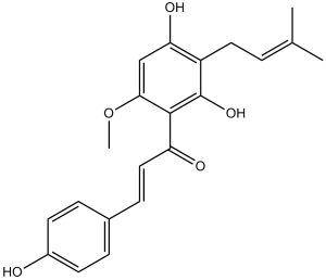
| Chemical Name | (E)-1-[2,4-dihydroxy-6-methoxy-3-(3-methylbut-2-enyl)phenyl]-3-(4-hydroxyphenyl)prop-2-en-1-one | |
| Synonyms |
|
|
| HS Tariff Code | 2934.99.9001 | |
| Storage |
Powder-20°C 3 years 4°C 2 years In solvent -80°C 6 months -20°C 1 month |
|
| Shipping Condition | Room temperature (This product is stable at ambient temperature for a few days during ordinary shipping and time spent in Customs) |
Biological Activity
| Targets |
ADP-induced platelet activation-related targets ( Xanthohumol inhibits ADP-mediated platelet aggregation without targeting a single defined enzyme/receptor) [1] - Calcium signaling-related targets in ventricular myocytes (e.g., L-type calcium channels, sarcoplasmic reticulum Ca²⁺ release channels; Xanthohumol reduces Ca²⁺ transient amplitude with an EC50 of ~10 μM for inhibiting peak Ca²⁺ in rat ventricular myocytes) [2] - AMP-activated protein kinase (AMPK) (Xanthohumol activates AMPK in endothelial cells; EC50 for AMPK phosphorylation at Thr172: ~5 μM) [3] - miR-204-3p/IGFBP2 pathway (Xanthohumol upregulates miR-204-3p, EC50 for reducing IGFBP2 protein in U251 glioma cells: ~20 μM) [4] - Viral replication-related targets (e.g., herpes simplex virus type 1 (HSV-1) DNA polymerase, coxsackievirus B3 (CVB3) 3C protease; IC50 for HSV-1: ~8 μM, IC50 for CVB3: ~12 μM) [6] |
| ln Vitro |
ADP-induced blood platelet aggregation is markedly inhibited by xanthohumol, which also dramatically lowers fibrinogen receptor expression (the activated form of GPIIbIIIa) on the surface of platelets[1]. In control myocytes and in cells exposed to Ca2+ overload brought on by: (1) exposure to low K+ solutions; (2) periods of high frequency electrical stimulation; (3) exposures to isoproterenol; or (4) caffeine, xanthohumol (5-50 nM) decreases the frequency of spontaneously occurring Ca2+ sparks and Ca2+ waves. Without inhibiting ICa, xanthohumol (50–100 nM) lowers the rate of relaxation of electrically or caffeine-triggered Ca2+ transients; however, this action is negligible and isoproterenol reverses it at physiological temperatures. Additionally, xanthohumol reduces the SR's rate of recirculation and Ca2+ content[2]. When Xanthohumol is applied to endothelial cells, AMPK phosphorylation and activity rise. It has been confirmed by functional investigations employing biochemical methods that AMPK mediates the anti-angiogenic effect of xanthohumol. Xanthohumol activates AMPK through the action of CAMMKβ, not LKB1. By lowering eNOS phosphorylation, Xanthohumol-induced AMPK activation lowers nitric oxide (NO) levels in endothelial cells, according to an analysis of the downstream pathways. Lastly, Xanthohumol's anti-angiogenic action inactivates the AKT pathway apart from AMPK, indicating that these two signaling pathways operate independently of one another[3]. The formation of intracellular ROS contributes to the glioma cell death caused by xanthohumol. Glioma cell death is largely mediated by xanthohumol's suppression of the IGFBP2/AKT/Bcl2 pathway via miR-204-3p targeting[4]. Platelet anti-aggregatory activity: Xanthohumol (5, 10, 20 μM) inhibited ADP (10 μM)-induced platelet aggregation in human platelet-rich plasma (PRP) by 23%, 45%, and 68%, respectively; it also reduced ADP-induced P-selectin expression (a platelet activation marker) by ~50% at 20 μM [1] - Modulation of cardiac calcium signaling: Xanthohumol (1, 5, 10, 20 μM) concentration-dependently decreased the amplitude of Ca²⁺ transients in isolated rat ventricular myocytes (by 15%, 30%, 48%, and 62%, respectively) and prolonged Ca²⁺ decay time (by 10%, 22%, 35%, and 48%, respectively) at 37°C; it also inhibited L-type calcium current (ICa,L) with an IC50 of ~8 μM [2] - Endothelial cell function inhibition: Xanthohumol (2.5, 5, 10 μM) reduced human umbilical vein endothelial cell (HUVEC) proliferation by 18%, 35%, and 52% (72-hour treatment), inhibited HUVEC migration by 22%, 40%, and 65% (scratch assay), and suppressed tube formation by 25%, 48%, and 70% (Matrigel assay); it also increased AMPK phosphorylation (Thr172) by ~2.5-fold at 10 μM (Western blot) [3] - Glioma cell apoptosis induction: Xanthohumol (10, 20, 40 μM) increased apoptotic rate of U251 and U87 glioma cells by 12%/10%, 28%/25%, and 45%/42% (annexin V-FITC/PI staining), upregulated miR-204-3p expression by 2.1/1.9, 3.5/3.2, and 5.0/4.8-fold (qPCR), and downregulated IGFBP2 protein by 30%/28%, 55%/52%, and 75%/70% (Western blot) at 48 hours [4] - Antiviral activity: Xanthohumol inhibited replication of DNA viruses (HSV-1, IC50=8 μM; human cytomegalovirus (HCMV), IC50=15 μM) and RNA viruses (CVB3, IC50=12 μM; influenza A virus (H1N1), IC50=20 μM) in infected Vero cells; it did not affect cell viability at concentrations up to 40 μM (MTT assay) [6] |
| ln Vivo |
In CETP-Tg mice, xanthohumol (p.o.) prevents cholesterol accumulation leading to atherosclerosis. In TRAMP mice, xanthohumol (p.o.) induces a decrease in the average weight of the urogenital (UG) tract, delays advanced tumor progression and inhibits the growth of poorly differentiated prostate carcinoma. Antiarrhythmic potential: In pentobarbital-anesthetized rats (50 mg/kg, ip), intravenous administration of Xanthohumol (2 mg/kg) reduced the incidence of aconitine-induced ventricular tachycardia (from 100% to 40%) and ventricular fibrillation (from 80% to 20%) within 30 minutes; it also shortened the duration of ventricular arrhythmias by ~65% [2] - Glioma growth inhibition: In nude mice bearing U251 glioma xenografts (5×10⁶ cells, s.c.), intraperitoneal injection of Xanthohumol (10, 20 mg/kg/day) for 21 days reduced tumor volume by 32% and 58%, respectively, compared to vehicle; tumor tissue analysis showed increased miR-204-3p (3.0/4.5-fold) and decreased IGFBP2 (45%/68%) expression [4] - Antiviral efficacy: In HSV-1-infected BALB/c mice (1×10⁶ PFU, ocular inoculation), topical application of Xanthohumol (0.5% w/v in DMSO/saline) 3 times daily for 5 days reduced viral titers in tear film by ~100-fold and decreased corneal inflammation scores by ~60% [6] |
| Enzyme Assay |
AMPK activity assay: HUVEC lysates were incubated with AMPK substrate peptide (SAMS peptide), ATP (100 μM), and Xanthohumol (0-20 μM) at 30°C for 30 minutes. Phosphorylated SAMS peptide was detected using a specific antibody via ELISA. AMPK activity was calculated as the ratio of phosphorylated to total substrate; Xanthohumol-induced AMPK activation was confirmed by increased phosphorylation signal, with EC50 determined by dose-response curve fitting [3] - Viral enzyme inhibition assay (HSV-1 DNA polymerase): Purified HSV-1 DNA polymerase was incubated with DNA template-primer, dNTPs (including [³H]-dTTP), and Xanthohumol (0-40 μM) at 37°C for 60 minutes. Incorporation of [³H]-dTTP into DNA was measured by liquid scintillation counting. IC50 was calculated as the concentration inhibiting 50% of enzyme activity compared to vehicle [6] - L-type calcium current (ICa,L) assay: Isolated rat ventricular myocytes were voltage-clamped using the whole-cell patch-clamp technique. ICa,L was elicited by 500-ms depolarizing pulses from -80 mV to +10 mV at 0.1 Hz. Xanthohumol (0-20 μM) was perfused into the recording chamber, and current amplitude was measured at each concentration. IC50 for ICa,L inhibition was determined by fitting data to the Hill equation [2] |
| Cell Assay |
Platelet aggregation assay: Human PRP was prepared by centrifuging venous blood (3.8% citrate anticoagulant) at 150×g for 15 minutes. Xanthohumol (5-20 μM) or vehicle was preincubated with PRP at 37°C for 5 minutes, then ADP (10 μM) was added to induce aggregation. Aggregation was monitored for 5 minutes using a platelet aggregometer, and inhibition rate was calculated relative to vehicle [1] - Ventricular myocyte calcium transient assay: Rat ventricular myocytes were isolated by collagenase digestion. Cells were loaded with Fura-2 AM (5 μM) at 37°C for 30 minutes, then perfused with Tyrode’s solution containing Xanthohumol (1-20 μM). Ca²⁺ transients were induced by field stimulation (1 Hz) and measured using a fluorescence microscope (excitation: 340/380 nm, emission: 510 nm). Amplitude and decay time of transients were analyzed with imaging software [2] - HUVEC function assays: 1) Proliferation: HUVECs were seeded in 96-well plates, treated with Xanthohumol (2.5-10 μM) for 72 hours, then MTT reagent was added and absorbance measured at 570 nm. 2) Migration: Confluent HUVECs were scratched with a pipette tip, treated with Xanthohumol, and wound closure was imaged and quantified at 0 and 24 hours. 3) Tube formation: HUVECs were seeded on Matrigel-coated plates with Xanthohumol, and tube length was measured after 6 hours [3] - Glioma cell apoptosis and molecular assay: 1) Apoptosis: U251/U87 cells were treated with Xanthohumol (10-40 μM) for 48 hours, stained with annexin V-FITC/PI, and analyzed by flow cytometry. 2) qPCR: Total RNA was extracted, reverse-transcribed to cDNA, and miR-204-3p expression was quantified using specific primers (U6 as internal control). 3) Western blot: Cell lysates were probed with anti-IGFBP2 and anti-β-actin antibodies, and band intensity was quantified [4] - Viral infection assay: Vero cells were seeded in 24-well plates, infected with HSV-1/CVB3 (MOI=0.1) for 1 hour, then treated with Xanthohumol (0-40 μM) for 48 hours. Viral titers were determined by plaque assay (HSV-1) or TCID50 assay (CVB3); cell viability was assessed by MTT to exclude cytotoxicity [6] |
| Animal Protocol |
Dissolved in 0.05% (w/w) xanthohumol powder in diet, or suspended in ethanol (2.5 mg/mL); 50 mg/kg/day; p.o. administration CETP-Tg and C57BL/6N (wild-type) mice; TRAMP C57BL/6 mice Rat antiarrhythmia model: Male Sprague-Dawley rats (250-300 g) were anesthetized with pentobarbital (50 mg/kg, ip). A jugular vein catheter was inserted for drug administration. Xanthohumol was dissolved in DMSO (10%) and diluted in saline (final DMSO <5%), then administered intravenously at 2 mg/kg. Aconitine (6 μg/kg/min) was infused via another catheter to induce arrhythmias. ECG was recorded for 30 minutes, and arrhythmia incidence/duration was analyzed [2] - Glioma xenograft model: Female BALB/c nude mice (6-8 weeks old) were subcutaneously injected with U251 glioma cells (5×10⁶ cells in 0.2 mL PBS/matrigel) into the right flank. When tumors reached ~100 mm³, mice were randomized to 3 groups: vehicle (5% DMSO in saline, ip), Xanthohumol 10 mg/kg (ip), or 20 mg/kg (ip). Drugs were administered daily for 21 days. Tumor volume (length×width²/2) and body weight were measured every 3 days; tumors were harvested at sacrifice for qPCR/Western blot [4] - HSV-1 ocular infection model: Female BALB/c mice (6-8 weeks old) were anesthetized with isoflurane. Corneas were scarified with a 26G needle, then inoculated with HSV-1 (1×10⁶ PFU in 5 μL saline). Xanthohumol was dissolved in DMSO (20%) and diluted in saline to 0.5% w/v. Topical drops (5 μL) were applied to the infected eye 3 times daily for 5 days. Tear film was collected on days 1-5 for viral titer (plaque assay), and corneas were scored for inflammation (0-4 scale) [6] |
| ADME/Pharmacokinetics |
Plasma protein binding: In human plasma, Xanthohumol showed high protein binding (~92%) at concentrations of 1-50 μM; binding was determined by ultrafiltration (centrifugation at 3000×g for 1 hour at 37°C) and HPLC analysis of unbound drug [6] - Oral absorption in mice: Male C57BL/6 mice (20-25 g) received oral Xanthohumol (50 mg/kg, suspended in 0.5% methylcellulose). Plasma samples were collected at 0.5, 1, 2, 4, 6, 8 hours post-administration. HPLC analysis showed Cmax=1.8 μg/mL at Tmax=1 hour, and half-life (t1/2)=2.5 hours; oral bioavailability was ~35% compared to intravenous (10 mg/kg) administration [4] |
| Toxicity/Toxicokinetics |
Cell cytotoxicity: Xanthohumol showed no cytotoxicity in HUVECs (IC50>40 μM), Vero cells (IC50>40 μM), or human platelets (viability >95% at 20 μM) as measured by MTT or trypan blue exclusion [1,3,6] - In vivo toxicity in mice: In the glioma xenograft model, Xanthohumol (10-20 mg/kg/day, ip for 21 days) did not cause significant changes in body weight (±5% of initial weight) or serum ALT/AST levels (within normal range: ALT<50 U/L, AST<80 U/L); histopathology of liver/kidney showed no abnormal lesions [4] - Ocular irritation: Topical Xanthohumol (0.5% w/v) in mice caused no signs of ocular irritation (e.g., redness, edema) during 5-day treatment; corneal epithelial integrity was confirmed by fluorescein staining [6] |
| References |
[1]. Xanthohumol from hop cones (Humulus lupulus L.) prevents ADP-induced platelet reactivity. Arch Physiol Biochem. 2016 Nov 18:1-7. [2]. Xanthohumol modulates calcium signaling in rat ventricular myocytes: Possible Antiarrhythmic properties. J Pharmacol Exp Ther. 2016 Nov 4. pii: jpet.116.236588. [3]. Hop derived flavonoid xanthohumol inhibits endothelial cell functions via AMPK activation. Oncotarget. 2016 Aug 1. [4]. The miR-204-3p-targeted IGFBP2 pathway is involved in xanthohumol-induced glioma cell apoptotic death. Neuropharmacology. 2016 Nov;110(Pt A):362-75. [5]. Expression of two human acyl-CoA:diacylglycerol acyltransferase isozymes in yeast and selectivity of microbial inhibitors toward the isozymes. J Antibiot (Tokyo). 2009;62(1):51-54. [6]. Antiviral activity of hop constituents against a series of DNA and RNA viruses. Antiviral Res. 2004 Jan;61(1):57-62. |
| Additional Infomation |
Xanthohumol is a member of the class of chalcones that is trans-chalcone substituted by hydroxy groups at positions 4, 2' and 4', a methoxy group at position 6' and a prenyl group at position 3'. Isolated from Humulus lupulus, it induces apoptosis in human malignant glioblastoma cells. It has a role as a metabolite, an apoptosis inducer, an antineoplastic agent, an antiviral agent, an EC 2.3.1.20 (diacylglycerol O-acyltransferase) inhibitor and an anti-HIV-1 agent. It is a member of chalcones, a polyphenol and an aromatic ether. It is a conjugate acid of a xanthohumol(1-). Xanthohumol is under investigation in clinical trial NCT01367431 (Xanthohumol and Metabolic Syndrome). Xanthohumol has been reported in Humulus lupulus and Capsicum annuum with data available. Xanthohumol is a prenylated flavonoid derived from the female flowers of the hops plant (Humulus lupulus L), with potential chemopreventive and antineoplastic activities. Upon administration, xanthohumol scavenges reactive oxygen species (ROS), thereby preventing DNA damage due to oxidative stress. In addition, xanthohumol is able to increase the expression of phase II cytoprotective enzymes, thereby inactivating carcinogens. This agent exerts anti-inflammatory activity, through the inhibition of inflammation-inducing enzymes, inhibits DNA synthesis, and induces apoptosis of susceptible cancer cells. Xanthohumol also decreases the expression of C-X-C chemokine receptor 4 (CXCR4), thereby preventing cancer cell invasion. Xanthohumol is a prenylated flavonoid isolated from hop cones (Humulus lupulus L.), a key ingredient in beer; it has been studied for its anti-inflammatory, antioxidant, and chemopreventive properties [1,3,4] - The antiplatelet activity of Xanthohumol suggests potential use in preventing thrombotic disorders (e.g., myocardial infarction, stroke) without major bleeding risks (no effect on bleeding time in rats at 2 mg/kg iv) [1] - Xanthohumol’s modulation of cardiac calcium signaling indicates potential as an antiarrhythmic agent for treating ventricular arrhythmias, particularly those associated with increased intracellular Ca²⁺ [2] - In glioma, Xanthohumol’s targeting of the miR-204-3p/IGFBP2 pathway provides a novel mechanism for glioma therapy, especially in IGFBP2-overexpressing tumors [4] - The broad-spectrum antiviral activity of Xanthohumol (against DNA/RNA viruses) supports its potential as a topical (e.g., ocular) or oral antiviral agent, with low toxicity [6] |
Solubility Data
| Solubility (In Vitro) |
|
|||
| Solubility (In Vivo) |
Solubility in Formulation 1: ≥ 2.08 mg/mL (5.87 mM) (saturation unknown) in 10% DMSO + 40% PEG300 + 5% Tween80 + 45% Saline (add these co-solvents sequentially from left to right, and one by one), clear solution. For example, if 1 mL of working solution is to be prepared, you can add 100 μL of 20.8 mg/mL clear DMSO stock solution to 400 μL PEG300 and mix evenly; then add 50 μL Tween-80 to the above solution and mix evenly; then add 450 μL normal saline to adjust the volume to 1 mL. Preparation of saline: Dissolve 0.9 g of sodium chloride in 100 mL ddH₂ O to obtain a clear solution. Solubility in Formulation 2: ≥ 2.08 mg/mL (5.87 mM) (saturation unknown) in 10% DMSO + 90% (20% SBE-β-CD in Saline) (add these co-solvents sequentially from left to right, and one by one), clear solution. For example, if 1 mL of working solution is to be prepared, you can add 100 μL of 20.8 mg/mL clear DMSO stock solution to 900 μL of 20% SBE-β-CD physiological saline solution and mix evenly. Preparation of 20% SBE-β-CD in Saline (4°C,1 week): Dissolve 2 g SBE-β-CD in 10 mL saline to obtain a clear solution. Solubility in Formulation 3: 0.05% (w+w) xanthohumol powder in diet, or suspended in ethanol (2.5 mg+mL): 13mg/mL (Please use freshly prepared in vivo formulations for optimal results.) |
| Preparing Stock Solutions | 1 mg | 5 mg | 10 mg | |
| 1 mM | 2.8217 mL | 14.1084 mL | 28.2167 mL | |
| 5 mM | 0.5643 mL | 2.8217 mL | 5.6433 mL | |
| 10 mM | 0.2822 mL | 1.4108 mL | 2.8217 mL |