WHI-P180 (also known as Janex 3) is a multi-kinase inhibitor which inhibits RET, KDR and EGFR with IC50s of 5 nM, 66 nM and 4 μM, respectively. WHI-P180 is a potent in vitro and in vivo inhibitor of IgE-mediated mast cell responses. Additional preclinical research on WHI-P180 could enhance its in vivo efficacy and serve as a foundation for the development of efficient mast cell-mediated allergy reaction treatment and prevention initiatives. After being administered intravenously, intraperitoneally, or topically, WHI-P180 had an elimination half-life of less than 10 minutes in CD-1 (BALB/c) mice. WHI-P180 had a systemic clearance of 8188 mL/h/kg in BALB/c mice and 6742 mL/h/kg in CD-I mice. Interestingly, WHI-P180 inhibited IgE/antigen-induced vascular hyperpermeability in a well-characterized murine model of passive cutaneous anaphylaxis when given in two consecutive nontoxic intraperitoneal bolus doses of 25 mg/kg.
Physicochemical Properties
| Molecular Formula | C16H15N3O3 | |
| Molecular Weight | 297.31 | |
| Exact Mass | 297.111 | |
| Elemental Analysis | C, 64.64; H, 5.09; N, 14.13; O, 16.14 | |
| CAS # | 211555-08-7 | |
| Related CAS # | WHI-P180 hydrochloride;153437-55-9 | |
| PubChem CID | 5687 | |
| Appearance | White to off-white solid powder | |
| Density | 1.3±0.1 g/cm3 | |
| Boiling Point | 476.5±40.0 °C at 760 mmHg | |
| Flash Point | 242.0±27.3 °C | |
| Vapour Pressure | 0.0±1.2 mmHg at 25°C | |
| Index of Refraction | 1.689 | |
| LogP | 2.44 | |
| Hydrogen Bond Donor Count | 2 | |
| Hydrogen Bond Acceptor Count | 6 | |
| Rotatable Bond Count | 4 | |
| Heavy Atom Count | 22 | |
| Complexity | 358 | |
| Defined Atom Stereocenter Count | 0 | |
| SMILES | O(C([H])([H])[H])C1=C(C([H])=C2C(=C1[H])C(=NC([H])=N2)N([H])C1C([H])=C([H])C([H])=C(C=1[H])O[H])OC([H])([H])[H] |
|
| InChi Key | BNDYIYYKEIXHNK-UHFFFAOYSA-N | |
| InChi Code | InChI=1S/C16H15N3O3/c1-21-14-7-12-13(8-15(14)22-2)17-9-18-16(12)19-10-4-3-5-11(20)6-10/h3-9,20H,1-2H3,(H,17,18,19) | |
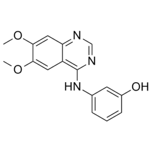
| Chemical Name | 3-[(6,7-dimethoxyquinazolin-4-yl)amino]phenol | |
| Synonyms |
|
|
| HS Tariff Code | 2934.99.9001 | |
| Storage |
Powder-20°C 3 years 4°C 2 years In solvent -80°C 6 months -20°C 1 month |
|
| Shipping Condition | Room temperature (This product is stable at ambient temperature for a few days during ordinary shipping and time spent in Customs) |
Biological Activity
| Targets |
EGFR (IC50 = 4 μM); KDR (IC50 = 66 nM); RET (IC50 = 5 nM) WHI-P180 is also a potent inhibitor of mast cell responses mediated by IgE. WHI-P180 has an elimination half-life of less than ten minutes in CD-1 (BALB/c) mice after intravenous, intraperitoneal, or po administration. In CD-I mice, the systemic clearance of WHI-P180 is 6742 mL/h/kg, whereas in BALB/c mice, it is 8188 mL/h/kg. Interestingly, in a well-established murine model of passive cutaneous anaphylaxis, WHI-P180 inhibits IgE/antigen-induced vascular hyperpermeability when given in two consecutive nontoxic intraperitoneal bolus doses of 25 mg/kg[3]. |
| ln Vitro |
WHI-P180 is also a potent inhibitor of mast cell responses mediated by IgE. WHI-P180 has an elimination half-life of less than ten minutes in CD-1 (BALB/c) mice after intravenous, intraperitoneal, or po administration. In CD-I mice, the systemic clearance of WHI-P180 is 6742 mL/h/kg, whereas in BALB/c mice, it is 8188 mL/h/kg. Interestingly, in a well-established murine model of passive cutaneous anaphylaxis, WHI-P180 inhibits IgE/antigen-induced vascular hyperpermeability when given in two consecutive nontoxic intraperitoneal bolus doses of 25 mg/kg[3]. In cellular assays using BaF3 cells engineered to be dependent on KIF5B-RET, compound 36 inhibited RET-driven cell proliferation with an IC₅₀ of 2100 nM. In parallel KDR-dependent BaF3 cells, it showed an IC₅₀ of >10000 nM, resulting in a cellular selectivity ratio of ≥5 for RET over KDR. [1] Compound 36 was devoid of non-specific cytotoxicity in wild-type BaF3 cells (IC₅₀ >10000 nM), suggesting meaningful kinase selectivity in a cellular context. [1] |
| ln Vivo |
WHI-P180 is also a potent inhibitor of mast cell responses mediated by IgE. In CD-1 mice (BALB/c mice), the elimination half-life of WHI-P180 is less than 10 minutes following intravenous, intraperitoneal, or po injection. For CD-I mice, the systemic clearance rate of WHI-P180 was 6742 mL/h/kg, but for BALB/c mice, it was 8188 mL/h/kg. Notably, WHI-P180, when given as two consecutive intraperitoneal nontoxic bolus doses of 25 mg/kg, decreased IgE/antigen-induced vascular permeability in a well-characterized mouse model of passive cutaneous allergy. excessive sexuality [3]. In diet-induced obese (DIO) mice with hepatic steatosis, treatment with SR9238 (30 mg/kg/day, i.p., for 30 days) substantially repressed hepatic expression of lipogenic genes (Fasn, ~60%; Srebp1c, ~80%; Scd1, ~90%). [2] Treatment reduced hepatic lipid accumulation (visualized and quantified by Bodipy 493/503 staining). [2] It suppressed hepatic expression of pro-inflammatory cytokines (Tnfa, ~80%; Il1b, >95%) and reduced infiltration of Kupffer cells (liver macrophages, assessed by F4/80 immunohistochemistry). [2] Treatment reduced plasma markers of hepatocellular injury: alkaline phosphatase (ALP), alanine transaminase (ALT), and aspartate aminotransferase (AST). [2] Treatment significantly reduced total plasma cholesterol, HDL, and LDL levels. [2] It suppressed hepatic expression of Cyp7a1 and Hmgcr (~80%), the rate-limiting enzyme in cholesterol synthesis. [2] No significant changes were observed in plasma glucose, insulin, triglycerides, body weight, or body fat composition in DIO mice. [2] In brown adipose tissue of DIO mice, no significant changes in the expression of LXR target genes Fasn and Abca1 were observed after treatment, indicating liver-specific effects. [2] In chow-fed mice treated for 7 days, SR9238 increased hepatic Il1b expression while decreasing Fasn and Srebp1c, indicating a potential initial pro-inflammatory effect that is countered in the long-term within an inflammatory disease model. [2] |
| Enzyme Assay |
For fifteen minutes, inhibitors (WHI-P180) are pre-incubated in the plate containing five microliters of kinase and assay buffer at concentrations of thirteen pM RET and one hundred pM KDR. 5 μL of ATP and substrate added at 2×final reaction concentrations start the reaction. This is 18 μM and 2 μM for RET and 16 μM and 1 μM for KDR, respectively. At ATP Km, reactions are carried out for every target. After 20 minutes of assaying at room temperature, 10 μL of HTRF detection buffer supplemented with TK-antibody labelled with Eu3+-Cryptate (1:100 dilution) and streptavidin-XL665 (128 nM) is added to end the experiment. The FRET signal is measured after a one-hour incubation period at room temperature[1]. Biochemical kinase inhibition assays for RET and KDR were performed using a time-resolved fluorescence resonance energy transfer (FRET) method. The assay was conducted in a 10 µL reaction volume in 384-well plates. For the RET assay, the buffer contained magnesium chloride, DTT, and other components. The reaction mixture was pre-incubated with the kinase (at a specified concentration) and the test compound for 15 minutes. The reaction was then initiated by adding ATP and a biotinylated substrate peptide at concentrations near the Km for each kinase (RET: 18 µM ATP, 2 µM substrate; KDR: 16 µM ATP, 1 µM substrate). After a 20-minute incubation at room temperature, the reaction was stopped by adding a detection buffer containing EDTA, a europium cryptate-labeled anti-phospho-tyrosine antibody, and streptavidin-XL665. Following a 1-hour incubation, the FRET signal was measured. IC₅₀ values were determined from dose-response curves. [1] |
| Cell Assay |
A recombinant kinase that is activated is expressed by BaF3 cells that are dependent on IL3. Recombinant kinase activity is necessary for the survival and proliferation of the altered cells after IL3 is removed. In RPMI-1640 medium supplemented with 10% FBS and suitable antibiotics, BaF3 cell lines that express KIF5B-RET and KDR are raised. Recombinant mouse IL-3 (10 ng/mL) is added to 10% FBS-containing RPMI-1640 medium to sustain unmodified BaF3 cells (WT). An acoustic liquid handling platform is used to dispense compounds after cells are plated into 384-well plates at a density of 1500 or 3000 per well in 30 μL culture medium in order to determine the compound IC50. The viability of the cells is assessed by adding 10 μL of CellTiter-Glo reagent and measuring luminescence after they have been incubated for 48 hours at 37 °C in a humidified 5% CO2 atmosphere[1]. Cellular potency was assessed using engineered BaF3 cell lines. These IL3-dependent cells were modified to express and depend on either an activated RET fusion (KIF5B-RET) or KDR for survival and proliferation. For the assay, cells were plated in 384-well plates. Test compounds were dispensed using an acoustic liquid handler. After incubating the cells with compounds for 48 hours at 37°C in a humidified 5% CO₂ atmosphere, cell viability was determined by adding a luminescent cell viability reagent and measuring the signal. IC₅₀ values were calculated from the dose-response curves. Non-specific cytotoxicity was evaluated similarly using the parental wild-type BaF3 cell line maintained with IL3. [1] |
| Animal Protocol |
Mice: The measurement of mouse plasma WHI-P180 levels is done using a quantitative detection method based on high performance liquid chromatography (HPLC). The WinNonlin program is used to compute the pharmacokinetic parameters in order to fit the plasma concentration-time data to a single compartment pharmacokinetic model. The pharmacodynamic effects of WHI-P180 on vascular hyperpermeability associated with anaphylaxis are investigated using a cutaneous anaphylaxis model[3]. Pharmacokinetic studies for compound 36 were conducted in male CD-1 mice following a single intravenous or oral administration. Blood samples were collected as dried blood spots at various time points. The samples were processed through solvent extraction and a phospholipid removal plate, followed by LC-MS/MS analysis to determine drug concentration. Pharmacokinetic parameters were calculated from the concentration-time data using non-compartmental analysis. [1] |
| ADME/Pharmacokinetics |
The intrinsic clearance (CLint) of compound 36 was higher in human hepatocytes than in human liver microsomes (6.2 µL/min/mg in microsomes), indicating the involvement of Phase II metabolism. Metabolism was more rapid in mouse systems (microsomes: 28.2 µL/min/mg; hepatocytes: 38.1 µL/min/10⁶ cells). [1] Compound 36 showed good aqueous solubility (>100 µM). Its permeability in Caco-2 cells was moderate (Papp = 8.2 × 10⁻⁶ cm/s) with an efflux ratio of 4.9. [1] In mice, after intravenous administration, the total blood clearance was low (<10% of liver blood flow). Following oral administration, the bioavailability was approximately 35%, and the oral half-life was about 2 hours. [1] |
| Toxicity/Toxicokinetics |
In a cellular cytotoxicity assay using wild-type BaF3 cells (the parental line for the engineered cells), compound 36 showed no non-specific toxicity at concentrations up to 10000 nM (IC₅₀ >10000 nM). [1] |
| References |
[1]. The discovery of 2-substituted phenol quinazolines as potent RET kinase inhibitors with improved KDR selectivity. Eur J Med Chem. 2016 Apr 13;112:20-32. [2]. 4-[3-Bromo-4-hydroxyphenyl)amino]-6,7-dimethoxyquinazolin-1-ium chloride methanol solvateand 4-[(3-hydroxyphenyl)amino]-6,7-dimethoxy-1-quinazolinium chloride. Acta Crystallogr C. 2001 Jan;57(Pt 1):76-8. [3]. Pharmacokinetics and biologic activity of the novel mast cell inhibitor, 4-(3-hydroxyphenyl)-amino-6,7-dimethoxyquinazoline in mice. Pharm Res. 1999 Jan;16(1):117-22. |
| Additional Infomation |
The study was driven by the clinical need for selective RET kinase inhibitors, as existing multi-target drugs like vandetanib cause dose-limiting toxicities due to off-target inhibition of kinases like KDR. [1] Compound 36 was discovered through structure-based design starting from a vandetanib-like scaffold. The incorporation of a phenol at the R² position and a methyl group at the R¹ position of the aniline ring was key. The phenol forms hydrogen bonds with Glu775 and Asp892 in the RET kinase active site, boosting affinity. The R¹ methyl group, while slightly disfavored in RET due to proximity to Ser891, causes a larger drop in affinity for KDR (where Ser891 is replaced by a bulkier cysteine, Cys1045), thereby improving selectivity. [1] The initial phenolic lead compound suffered from high metabolic clearance. The introduction of the flanking methyl substituent at R¹ was found to improve metabolic stability in hepatocytes. [1] |
Solubility Data
| Solubility (In Vitro) |
|
|||
| Solubility (In Vivo) |
Solubility in Formulation 1: ≥ 2.5 mg/mL (8.41 mM) (saturation unknown) in 10% DMSO + 40% PEG300 + 5% Tween80 + 45% Saline (add these co-solvents sequentially from left to right, and one by one), clear solution. For example, if 1 mL of working solution is to be prepared, you can add 100 μL of 25.0 mg/mL clear DMSO stock solution to 400 μL PEG300 and mix evenly; then add 50 μL Tween-80 to the above solution and mix evenly; then add 450 μL normal saline to adjust the volume to 1 mL. Preparation of saline: Dissolve 0.9 g of sodium chloride in 100 mL ddH₂ O to obtain a clear solution. Solubility in Formulation 2: ≥ 2.5 mg/mL (8.41 mM) (saturation unknown) in 10% DMSO + 90% Corn Oil (add these co-solvents sequentially from left to right, and one by one), clear solution. For example, if 1 mL of working solution is to be prepared, you can add 100 μL of 25.0 mg/mL clear DMSO stock solution to 900 μL of corn oil and mix evenly. Solubility in Formulation 3: 10 mg/mL (33.63 mM) in 50% PEG300 50% Saline (add these co-solvents sequentially from left to right, and one by one), suspension solution; with ultrasonication. Preparation of saline: Dissolve 0.9 g of sodium chloride in 100 mL ddH₂ O to obtain a clear solution. (Please use freshly prepared in vivo formulations for optimal results.) |
| Preparing Stock Solutions | 1 mg | 5 mg | 10 mg | |
| 1 mM | 3.3635 mL | 16.8175 mL | 33.6349 mL | |
| 5 mM | 0.6727 mL | 3.3635 mL | 6.7270 mL | |
| 10 mM | 0.3363 mL | 1.6817 mL | 3.3635 mL |