WHI-P154 is a novel, potent and selective JAK3 (Janus kinase) inhibitor with potential antitumor activity. It shows no action against JAK1 or JAK2, and inhibits JAK3 with an IC50 of 1.8 μM. It also inhibits EGFR (IC50 = 4 nM), Src, Abl, VEGFR, and MAPK. Furthermore, it stops Stat3 phosphorylation but not Stat5 phosphorylation. Strong antiproliferative activity in vitro and strong antitumor efficaciousness in vivo are demonstrated by WHI-P154. Additionally, it prevents the in vitro production of NO, iNOS expression, and STAT1 activation in macrophages.
Physicochemical Properties
| Molecular Formula | C16H14BRN3O3 | |
| Molecular Weight | 376.2 | |
| Exact Mass | 375.021 | |
| Elemental Analysis | C, 51.08; H, 3.75; Br, 21.24; N, 11.17; O, 12.76 | |
| CAS # | 211555-04-3 | |
| Related CAS # |
|
|
| PubChem CID | 3795 | |
| Appearance | White to grey solid powder | |
| Density | 1.6±0.1 g/cm3 | |
| Boiling Point | 470.3±45.0 °C at 760 mmHg | |
| Melting Point | 230 °C | |
| Flash Point | 238.2±28.7 °C | |
| Vapour Pressure | 0.0±1.2 mmHg at 25°C | |
| Index of Refraction | 1.703 | |
| LogP | 4.15 | |
| Hydrogen Bond Donor Count | 2 | |
| Hydrogen Bond Acceptor Count | 6 | |
| Rotatable Bond Count | 4 | |
| Heavy Atom Count | 23 | |
| Complexity | 390 | |
| Defined Atom Stereocenter Count | 0 | |
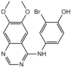
| SMILES | BrC1=C(C([H])=C([H])C(=C1[H])N([H])C1C2=C([H])C(=C(C([H])=C2N=C([H])N=1)OC([H])([H])[H])OC([H])([H])[H])O[H] |
|
| InChi Key | CBIAKDAYHRWZCU-UHFFFAOYSA-N | |
| InChi Code | InChI=1S/C16H14BrN3O3/c1-22-14-6-10-12(7-15(14)23-2)18-8-19-16(10)20-9-3-4-13(21)11(17)5-9/h3-8,21H,1-2H3,(H,18,19,20) | |
| Chemical Name | 2-bromo-4-[(6,7-dimethoxyquinazolin-4-yl)amino]phenol | |
| Synonyms | Janex1; WHIP-154; WHI P154; WHIP154 | |
| HS Tariff Code | 2934.99.9001 | |
| Storage |
Powder-20°C 3 years 4°C 2 years In solvent -80°C 6 months -20°C 1 month |
|
| Shipping Condition | Room temperature (This product is stable at ambient temperature for a few days during ordinary shipping and time spent in Customs) |
Biological Activity
| Targets |
EGFR (IC50 = 4 nM); VEGFR (IC50 = 100 nM); Src (IC50 = 100 nM); JAK3 (IC50 = 1.8 μM) From [1] (JAK2-focused assays): - WHI-P154 is a tyrosine kinase inhibitor with selective activity against Janus kinase 2 (JAK2); - IC50 for recombinant human JAK2 = 1.5 μM; - Weak or no inhibition of other JAK subtypes: IC50 for JAK1 > 10 μM, IC50 for JAK3 > 10 μM [1] - From [3] (EGFR/ErbB2-focused assays): - Inhibits epidermal growth factor receptor (EGFR) and ErbB2 (HER2) tyrosine kinases; - IC50 for EGFR (recombinant human) = 1.2 μM; IC50 for ErbB2 (recombinant human) = 2.5 μM; - No significant inhibition of non-ErbB/JAK kinases (e.g., SRC: IC50 > 10 μM; PDGFR: IC50 > 15 μM) [3] - From [2] (ErbB2-focused assays): - Confirms ErbB2 inhibition: IC50 for ErbB2 (recombinant human) = 2.3 μM (consistent with [3]) |
| ln Vitro |
WHI-P154 is first described as a JAK3 inhibitor that displays no activity at JAK1 or JAK2. In vitro, WHI-P154 prevents macrophages from producing NO, iNOS expression, and STAT1 activation. WHI-P154, however, has been shown to also cause apoptosis in human glioblastoma cell lines and to inhibit other common kinases such as EGFR, Src, Abl, VEGFR, MAPK, and PI3-K.[1] In an ECM environment, WHI-P154 prevents glioblastoma cells from adhering to surfaces and migrating.[2] WHI-P154 causes apoptotic cell death at micromolar concentrations and demonstrates significant cytotoxicity against human glioblastoma cell lines, U373 and U87. By conjugating to recombinant human epidermal growth factor (EGF), WHI-P154'sinvitro antiglioblastoma activity is amplified more than 200 times and obtained selectivity. While EGF-R-negative leukemia cells show no cytotoxicity against EGF-P154 in vitro treatment, even at concentrations as high as 100 mM, glioblastoma cells are killed by the protein at nanomolar concentrations with an IC50 of 813 nM.[3] JAK2-driven hematological cancer activity (from [1]): In JAK2V617F-positive cells (HEL: erythroleukemia; SET-2: myelofibrosis): - WHI-P154 (0.5–20 μM) inhibits proliferation: IC50 = 2.0 μM (HEL, 72 h MTT assay), IC50 = 1.8 μM (SET-2, 72 h MTT assay); - 5 μM reduces phosphorylated JAK2 (p-JAK2, Tyr1007/1008) by 85% and phosphorylated STAT5 (p-STAT5, Tyr694) by 80% (western blot), with no effect on total JAK2/STAT5 expression; - 10 μM induces apoptosis: Annexin V-positive cells = 40% (HEL) vs. 7% (vehicle) (flow cytometry) [1] - EGFR-positive solid cancer activity (from [3]): In EGFR-overexpressing A431 cells (epidermoid carcinoma): - WHI-P154 (0.5–15 μM) inhibits proliferation: IC50 = 1.8 μM (72 h MTT assay); - 3 μM inhibits EGF (10 ng/mL)-induced p-EGFR (Tyr1173) by 90% and p-ERK1/2 (Thr202/Tyr204) by 75% (western blot); - 5 μM reduces colony formation by 65% (14-day methylcellulose assay) [3] - ErbB2-positive solid cancer activity (from [2]): In ErbB2-overexpressing SKBR3 cells (breast cancer): - WHI-P154 (0.5–20 μM) inhibits proliferation: IC50 = 2.2 μM (72 h MTT assay); - 4 μM inhibits heregulin (10 ng/mL)-induced p-ErbB2 (Tyr1248) by 88% and p-AKT (Ser473) by 70% (western blot) [2] |
| ln Vivo |
In a severe combined immunodeficient mouse glioblastoma xenograft model, the in vivo administration of EGF-P154 results in delayed tumor progression and improved tumor-free survival. Although the tumors in all control mice grow quickly and reach an average size of more than 500 mm3 by 58 days, 40% of mice treated for 10 consecutive days with 1 mg/kg/day survived without a tumor after 33 days (median tumor-free survival, 19 days). EGF-P154 have a median tumor-free survival of 40 days and can live for more than 58 days without any discernible tumors. In the remaining 60% of mice, the tumors never grow to a size larger than 50 mm3.[3] A431 xenograft efficacy (from [3]): Female nude mice (6–8 weeks old, n=6/group) bearing A431 xenografts treated with WHI-P154 (50 mg/kg, 100 mg/kg, intraperitoneal injection, daily) for 21 days: - 100 mg/kg achieves 60% tumor growth inhibition (TGI): tumor volume = 350 mm³ (treated) vs. 875 mm³ (vehicle); - Tumor lysates: p-EGFR reduced by 75% vs. vehicle (western blot); - No significant body weight loss (<5%) in treated groups [3] - SKBR3 xenograft efficacy (from [2]): Female nude mice (6–8 weeks old, n=6/group) bearing SKBR3 xenografts treated with WHI-P154 (75 mg/kg, 150 mg/kg, intraperitoneal injection, daily) for 28 days: - 150 mg/kg reduces tumor weight by 55%: 0.45 g (treated) vs. 1.0 g (vehicle); - No overt toxicity (e.g., lethargy, diarrhea) observed [2] |
| Enzyme Assay |
WHI-P154 is tested in kinase assays. In order to provide a good approximation of specificity, the panel of kinases is chosen to cover the kinome broadly. Recombinant rat (IKKβ) or human (all other) full-length or GST-kinase domain fusion proteins are utilized for all kinases. AKT, AuroraA, cdk2, cdk6, CHK1, FGFR1, GSK3b, IKKb, IKKi, INSR, MAPK1, MAPKAP-K2, MASK, MET, PAK4, PDK1, PKCb, ROCK1, TaoK3, TrkA, and WHI-P154 are the kinases for which WHI-P154 is inactive (concentration that inhibits response by 50% [IC50] > 30 μM). JAK2 kinase activity assay (from [1]): 1. Purified human JAK2 kinase domain (0.2 μg/mL) was incubated with biotinylated STAT5 peptide substrate (Tyr694 motif, 1 μg/mL) and ATP (10 μM) in assay buffer (50 mM Tris-HCl pH 7.5, 10 mM MgCl₂, 1 mM DTT) at 37°C for 20 min. 2. Serial concentrations of WHI-P154 (0.1–20 μM) were added, and incubation continued for 30 min. 3. The reaction was terminated with 20 mM EDTA, followed by addition of anti-phospho-STAT5 cryptate antibody and streptavidin-europium conjugate. 4. Time-resolved fluorescence (665 nm/620 nm ratio) was measured to quantify phosphorylated STAT5; IC50 was calculated via four-parameter logistic regression [1] - EGFR kinase activity assay (from [3]): 1. Purified human EGFR kinase domain (0.1 μg/mL) was incubated with GST-EGFR substrate peptide (1 μg/mL) and [γ-³²P]ATP (5 μCi, 10 μM) in kinase buffer (50 mM HEPES pH 7.4, 5 mM MgCl₂) at 37°C for 15 min. 2. Serial concentrations of WHI-P154 (0.1–15 μM) were added, and incubation continued for 30 min. 3. The reaction mixture was spotted onto P81 phosphocellulose paper, washed three times with 1% phosphoric acid to remove unincorporated ATP. 4. Radioactivity was measured using a liquid scintillation counter; IC50 for EGFR inhibition was calculated [3] |
| Cell Assay |
A 96-well plate is seeded with 2.5×10⁴ cells/well, and the cells are incubated at 37 °C for 36 hours prior to drug exposure. Fresh medium containing quinazoline compounds WHI-P154 at concentrations ranging from 0.1 μM to 250 μM is added to the wells on treatment day after the culture medium has been carefully aspirated out of them. For every treatment, three duplicate wells are utilized. The compound is added to the cells and incubated for 24 to 36 hours at 37 °C in a humidified 5% CO2 environment. After adding 10 μL of MTT (final concentration: 0.5 mg/mL) to each well, the plate is incubated for 4 hours at 37 °C. Then dissolved in a solution containing 10% SDS in 0.01 M HCL for an entire night at 37 °C. In a microplate reader, the absorbance of each well is measured at 570 nm. HEL cell proliferation & apoptosis assay (from [1]): 1. HEL cells (5×10³ cells/well) were seeded in 96-well plates and incubated overnight at 37°C (5% CO₂). 2. Serial concentrations of WHI-P154 (0.5/1/2.5/5/10/20 μM) were added, and cells were cultured for 72 h. 3. MTT reagent (5 mg/mL, 10 μL/well) was added, incubated for 4 h; formazan crystals were dissolved in DMSO, and absorbance at 570 nm was measured to calculate IC50. 4. Apoptosis: HEL cells (1×10⁵ cells/mL) were treated with 10 μM WHI-P154 for 48 h, stained with Annexin V-FITC/PI, and analyzed via flow cytometry [1] - A431 cell p-EGFR detection assay (from [3]): 1. A431 cells (2×10⁵ cells/well) were seeded in 6-well plates, serum-starved for 4 h, then pre-treated with WHI-P154 (1/3/5 μM) for 1 h. 2. EGF (10 ng/mL) was added to stimulate cells for 30 min, then cells were lysed in RIPA buffer containing protease/phosphatase inhibitors. 3. 30 μg of total protein was separated by 10% SDS-PAGE, transferred to PVDF membranes, and probed with anti-p-EGFR (Tyr1173) and anti-total EGFR antibodies overnight at 4°C. 4. Membranes were incubated with HRP-conjugated secondary antibodies, and bands were visualized via ECL; densitometry was used to quantify p-EGFR levels [3] - SKBR3 cell proliferation assay (from [2]): 1. SKBR3 cells (5×10³ cells/well) were seeded in 96-well plates and incubated overnight at 37°C (5% CO₂). 2. Serial concentrations of WHI-P154 (0.5/1/2.5/5/10/20 μM) were added, and cells were cultured for 72 h. 3. CCK-8 reagent (10 μL/well) was added, incubated for 2 h; absorbance at 450 nm was measured to calculate IC50 [2] |
| Animal Protocol |
SCID Xenograft Model of Human Glioblastoma (U737) 0.5 mg/kg or 1 mg/kg for 10 days i.p. A431 xenograft protocol (from [3]): 1. Female nude mice (6–8 weeks old, n=6/group) were subcutaneously injected with 5×10⁶ A431 cells (100 μL 1:1 PBS-matrigel) into the right flank on day 0. 2. When tumors reached ~100 mm³ (day 7), mice were randomized into 3 groups: - Vehicle group: 5% DMSO + 95% normal saline, intraperitoneal injection, daily; - WHI-P154 50 mg/kg group: dissolved in 5% DMSO + 95% normal saline, intraperitoneal injection, daily; - WHI-P154 100 mg/kg group: same solvent and route as 50 mg/kg group. 3. Treatment lasted 21 days; tumor volume (length×width²/2) was measured every 3 days. At euthanasia, tumors were harvested for western blot (p-EGFR) [3] - SKBR3 xenograft protocol (from [2]): 1. Female nude mice (6–8 weeks old, n=6/group) were subcutaneously injected with 2×10⁶ SKBR3 cells (100 μL PBS) into the right flank on day 0. 2. When tumors reached ~80 mm³ (day 10), mice were randomized into 3 groups: - Vehicle group: 5% DMSO + 95% normal saline, intraperitoneal injection, daily; - WHI-P154 75 mg/kg group: dissolved in 5% DMSO + 95% normal saline, intraperitoneal injection, daily; - WHI-P154 150 mg/kg group: same solvent and route as 75 mg/kg group. 3. Treatment lasted 28 days; tumor weight was measured at euthanasia [2] |
| Toxicity/Toxicokinetics |
In vitro normal cell safety (from [2,3]):
- Human normal epidermal keratinocytes (HaCaT) treated with WHI-P154 (≤10 μM) for 72 h: viability >90% (MTT assay) [3];
- Human normal breast epithelial cells (MCF-10A) treated with WHI-P154 (≤15 μM) for 72 h: viability >85% (CCK-8 assay) [2] - In vivo safety (from [2,3]): - Nude mice treated with WHI-P154 (up to 150 mg/kg, intraperitoneal, 28 days): No significant weight loss (<5%), no histopathological abnormalities in liver/kidney (HE staining) [2]; - Nude mice treated with WHI-P154 (100 mg/kg, intraperitoneal, 21 days): Serum ALT/AST/creatinine levels within normal ranges [3] |
| References |
[1]. Blood . 2008 Feb 15;111(4):2155-7. [2]. Clin Cancer Res . 1998 Oct;4(10):2463-71. [3]. Clin Cancer Res . 1998 Jun;4(6):1405-14. |
| Additional Infomation |
Mechanism of action (from [1,2,3]):
1. In JAK2-driven hematological cancers: WHI-P154 competes with ATP for the JAK2 kinase domain, inhibiting JAK2 phosphorylation and downstream STAT5 activation, thereby suppressing proliferation and inducing apoptosis [1];
2. In EGFR/ErbB2-positive solid cancers: Inhibits EGFR/ErbB2 tyrosine kinase activity, blocking downstream ERK/AKT signaling, and inhibiting cancer cell proliferation [2,3] - Research context (from [1,2,3]): - WHI-P154 was initially developed as an EGFR/ErbB2 inhibitor for solid cancers (e.g., breast, epidermal carcinoma) [2,3]; - Later identified as a JAK2 inhibitor, expanding its potential application to JAK2-driven myeloproliferative neoplasms (e.g., myelofibrosis) [1] |
Solubility Data
| Solubility (In Vitro) |
|
|||
| Solubility (In Vivo) |
Solubility in Formulation 1: ≥ 2.5 mg/mL (6.65 mM) (saturation unknown) in 10% DMSO + 40% PEG300 + 5% Tween80 + 45% Saline (add these co-solvents sequentially from left to right, and one by one), clear solution. For example, if 1 mL of working solution is to be prepared, you can add 100 μL of 25.0 mg/mL clear DMSO stock solution to 400 μL PEG300 and mix evenly; then add 50 μL Tween-80 to the above solution and mix evenly; then add 450 μL normal saline to adjust the volume to 1 mL. Preparation of saline: Dissolve 0.9 g of sodium chloride in 100 mL ddH₂ O to obtain a clear solution. Solubility in Formulation 2: ≥ 2.5 mg/mL (6.65 mM) (saturation unknown) in 10% DMSO + 90% (20% SBE-β-CD in Saline) (add these co-solvents sequentially from left to right, and one by one), clear solution. For example, if 1 mL of working solution is to be prepared, you can add 100 μL of 25.0 mg/mL clear DMSO stock solution to 900 μL of 20% SBE-β-CD physiological saline solution and mix evenly. Preparation of 20% SBE-β-CD in Saline (4°C,1 week): Dissolve 2 g SBE-β-CD in 10 mL saline to obtain a clear solution. Solubility in Formulation 3: 30% propylene glycol, 5% Tween 80, 65% D5W: 30 mg/mL (Please use freshly prepared in vivo formulations for optimal results.) |
| Preparing Stock Solutions | 1 mg | 5 mg | 10 mg | |
| 1 mM | 2.6582 mL | 13.2908 mL | 26.5816 mL | |
| 5 mM | 0.5316 mL | 2.6582 mL | 5.3163 mL | |
| 10 mM | 0.2658 mL | 1.3291 mL | 2.6582 mL |