VLX1570 is a competitive inhibitor of the 19S proteasome-specific DUB (deubiquitylating enzymes) activity with IC50 ranging from 4.2 uM to 8.6 uM. VLX1570 preferentially inhibits proteasomal DUB activity while not inhibiting the activities of a panel of non-proteasomal DUBs. VLX1570 binds to and inhibits the activity of ubiquitin-specific protease-14 (USP14) in vitro ipon administration, with comparatively weaker inhibitory activity towards UCHL5 (ubiquitin-C-terminal hydrolase-5). This blocks the ubiquitin proteasome degradation pathway, prevents the degradation of defective proteins, and leads to an accumulation of poly-ubiquitylated proteins, which induces the unfolded protein response (UPR) and results in both the induction of tumor cell apoptosis and the inhibition of tumor cell growth. Therefore, VLX1570 has apoptosis-inducing ability and anticancer activities.
Physicochemical Properties
| Molecular Formula | C23H17F2N3O6 | |
| Molecular Weight | 469.39 | |
| Exact Mass | 469.108 | |
| Elemental Analysis | C, 58.85; H, 3.65; F, 8.09; N, 8.95; O, 20.45 | |
| CAS # | 1431280-51-1 | |
| Related CAS # |
|
|
| PubChem CID | 71523377 | |
| Appearance | Light yellow to yellow solid powder | |
| Density | 1.5±0.1 g/cm3 | |
| Boiling Point | 722.3±60.0 °C at 760 mmHg | |
| Flash Point | 390.6±32.9 °C | |
| Vapour Pressure | 0.0±2.3 mmHg at 25°C | |
| Index of Refraction | 1.662 | |
| LogP | 4.19 | |
| Hydrogen Bond Donor Count | 0 | |
| Hydrogen Bond Acceptor Count | 8 | |
| Rotatable Bond Count | 3 | |
| Heavy Atom Count | 34 | |
| Complexity | 909 | |
| Defined Atom Stereocenter Count | 0 | |
| SMILES | FC1C=CC(=CC=1[N+](=O)[O-])/C=C1\C(/C(=C\C2C=CC(=C(C=2)[N+](=O)[O-])F)/CN(C(C=C)=O)CC\1)=O |
|
| InChi Key | SCKXBVLYWLLALY-CQRYCMKKSA-N | |
| InChi Code | InChI=1S/C23H17F2N3O6/c1-2-22(29)26-8-7-16(9-14-3-5-18(24)20(11-14)27(31)32)23(30)17(13-26)10-15-4-6-19(25)21(12-15)28(33)34/h2-6,9-12H,1,7-8,13H2/b16-9-,17-10- | |
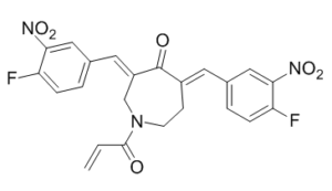
| Chemical Name | 1-acryloyl-3,5-bis((Z)-4-fluoro-3-nitrobenzylidene)azepan-4-one | |
| Synonyms | VLX-1570;VLX1570;VLX 1570 | |
| HS Tariff Code | 2934.99.9001 | |
| Storage |
Powder-20°C 3 years 4°C 2 years In solvent -80°C 6 months -20°C 1 month |
|
| Shipping Condition | Room temperature (This product is stable at ambient temperature for a few days during ordinary shipping and time spent in Customs) |
Biological Activity
| Targets |
Deubiquitinase(IC50= appr 10 μM)
The targets of VLX1570 are ubiquitin-specific protease 14 (USP14) and ubiquitin C-terminal hydrolase L5 (UCHL5)[2][3] - USP14: IC50 = 3.2 μM (fluorogenic substrate assay)[2]; Ki = 0.9 μM (competitive binding assay)[2] - UCHL5: IC50 = 5.8 μM (fluorogenic substrate assay)[2] |
| ln Vitro |
USP14 and UCHL5 activity of 19S regulatory particles are inhibited by VLX1570, with USP14 being more strongly inhibited. In KMS-11 myeloma cells, VLX1570 (1 μM) exhibits inhibitory activity against USP14. VLX1570 on HCT116 cells shows an IC50 of 0.58 μM[1].Using two distinct sources of recombinant protein, VLX1570 binds to recombinant USP14 with a Kd of 1.5–18 μM; in contrast, recombinant UCHL5 has a higher Kd (14–18 μM) than recombinant USP14. With IC50s of 43 ± 2 nM, 74 ± 2 nM, 126 ± 3 nM, and 191 ± 1 nM for KMS-11, RPMI8226, OPM-2, and OPM-2-BZR cells, respectively, VLX1570 exhibits strong antiproliferative activities on multiple myeloma cells[2]. BCWM.1 cell viability is suppressed by VLX1570, with an EC50 of 20.22 nM. In all Waldenstrom macroglobulinemia (WM) cell lines tested, including BCWM.1/IR (IR) and BCWM.1/BR (BR) subclones, VLX1570 (100, 250, and 500 nM) dose-dependently induces significant apoptosis by 12 hours. In WM cells, VLX1570 (100, 250, and 500 nM) also damages the mitochondria and the ER stress machinery. In WM cells, VLX1570 (250 nM) downregulates the expression of CXCR4 and the end effectors of BCR-signalosomes[3]. 1. Exhibits significant antiproliferative activity against multiple myeloma cell lines, including RPMI-8226 (IC50=0.3 μM), U266 (IC50=0.4 μM), MM.1S (IC50=0.5 μM), OPM-2 (IC50=0.6 μM), and can induce cell apoptosis. The apoptosis rate increases with drug concentration (30%-60% after treatment with 0.2-1.0 μM for 48 hours)[2] 2. Still exerts potent cytotoxicity against ibrutinib-resistant (IBR-R) and bortezomib-resistant (BOR-R) Waldenstrom macroglobulinemia (WM) cell lines (MWCL-1, BCWM.1), with IC50 values of 0.3-0.5 μM (IBR-R cells) and 0.4-0.6 μM (BOR-R cells), respectively. No significant synergistic or antagonistic effect is observed when combined with ibrutinib or bortezomib, and single-agent use can effectively inhibit the proliferation of resistant cells[3] 3. After treatment, it can lead to significant accumulation of ubiquitinated proteins in cells, activate the cleavage of caspase-3, caspase-9 and PARP, downregulate the expression of anti-apoptotic proteins Mcl-1 and Bcl-2, and upregulate the expression of pro-apoptotic protein Bax[2][3] 4. Shows low toxicity to normal peripheral blood mononuclear cells (PBMCs) with an IC50 value >5.0 μM, demonstrating cancer cell selectivity[2] 5. Can inhibit the colony-forming ability of WM cell lines, with a colony formation rate reduced by more than 70% after treatment with 0.2 μM and less than 10% after treatment with 0.5 μM[3] |
| ln Vivo |
VLX1570 (3 mg/kg) dramatically reduces the growth of tumors in mice with multiple myeloma cells (KMS-11)[2]. In Waldenstrom macroglobulinemia (WM)-bearing mice, VLX1570 (4.4 mg/kg, i.p.) significantly suppresses tumor growth without causing apparent weight loss or other signs of systemic toxicity[3]. 1. In the RPMI-8226 multiple myeloma nude mouse xenograft model, intraperitoneal injection at a dose of 10 mg/kg, 3 times a week for 4 consecutive weeks, significantly inhibited tumor growth with a tumor volume inhibition rate of 65% and a tumor weight reduction of 60%, without significant weight loss in nude mice (body weight change <8%)[2] 2. Immunohistochemical detection of tumor tissues showed obvious accumulation of ubiquitinated proteins, increased activation level of caspase-3, and decreased expression of Ki-67 (proliferation marker) in the drug-treated group[2] 3. In the bortezomib-resistant WM cell (BCWM.1-BOR-R) nude mouse xenograft model, intraperitoneal injection of 10 mg/kg (3 times a week for 4 consecutive weeks) reduced tumor volume by 55%, significantly increased the level of ubiquitinated proteins in tumor tissues, and increased the proportion of apoptotic cells[3] |
| Enzyme Assay |
Before adding ubirhodamine, 26S proteasome preparations (1 nM) are pretreated for 2 min in assay buffer (25 mM Tris, 5 mM MgCl2, 10% glycerol, 0.05 mg/mL BSA, 2 mM ATP, and 1 mM DTT) with DMSO, VLX1570, or b-AP15. Using a TECAN infinite 200 instrument, fluorescence is measured at 37°C using Ex/Em = 490 nm/520 nm. Data is read every 10 seconds for 30 minutes. Cell pellets from control or treated cells are lysed with a buffer (50 mM HEPES pH 7.4, 250 mM sucrose, 10 mM MgCl2, 2 mM ATP, 1 mM DTT) on ice for 30 minutes in order to prepare them for UbVS labeling of KMS 11 cells. The debris is then removed by centrifugation. 1 μM UbVS is applied to 25 μg of protein and left for 30 minutes at 37°C. Samples are separated using SDS-PAGE and then immunoblotted. Purified 19S proteasomes (50 nM) are pretreated for 10 min at room temperature with DMSO, VLX1570, or b-AP15 (50 μM). After that, they are labeled with 1 μM HA-UbVS for 30 min at 37°C and then subjected to immunoblotting[1]. 1. USP14 activity assay: Using a fluorogenic substrate assay, recombinant human USP14 was mixed with a fluorescently labeled ubiquitin substrate (Ub-AMC), and VLX1570 at different concentrations was added. After incubation at 37°C for 30 minutes, the fluorescence intensity was detected to calculate the inhibition rate and IC50 value of the drug on USP14[2] 2. UCHL5 activity assay: The experimental procedure was the same as the USP14 activity assay. Recombinant human UCHL5 and the same fluorescent substrate were used, and the inhibitory effect of the drug on UCHL5 was evaluated by the change in fluorescence intensity to determine the IC50 value[2] 3. Competitive binding assay: Biotin-labeled ubiquitin was incubated with recombinant USP14, and VLX1570 at different concentrations was added simultaneously. The bound complex was captured by avidin, and the amount of free biotin-ubiquitin was detected to calculate the Ki value of the drug with USP14[2] |
| Cell Assay |
The MTT (3-(4,5-dimethylthiazol-2-yl)-2,5-diphenyltetrazolium bromide) assay is used to measure cell viability. Cells are suspended at 5 × 10^5 cells/mL for the MTT assay. 100 μL aliquots are then put into 96-well microtiter plates and exposed to drugs, with DMSO serving as the control. 10μL of a stock solution containing 5 mg/mL MTT (3-(4,5-dimethylthiazol-2-yl)-2,5-diphenyltetrazolium bromide) is added to each well after the incubations are complete, and the plates are then incubated for 4 hours at 37°C. Overnight at 37°C, 100μL of a 10% SDS/10 mM HCl solution is used to dissolve the formazan crystals.In certain experiments, cell viability is assessed using the acid phosphatase method49 because VLX1570 affects OXPHOS and mitochondrial activity affects MTT assays. Cells are lysed in 100μL of 0.1 M sodium acetate, 0,1% Triton X-100, and p-nitrophenylphosphate after being washed twice with PBS. They are then incubated for 90 minutes at 37°C. After the incubation period, 10μL of NaOH is added to each well, and the value of A405 is obtained [2]. 1. Cell proliferation inhibition assay: Tumor cells were seeded in 96-well plates (5×10³ cells/well), cultured for 24 hours, then treated with 0.1-5.0 μM VLX1570, and incubated for another 72 hours. The CCK-8 assay was used to detect cell viability and calculate IC50 values[2][3] 2. Apoptosis detection: After treating cells with the drug for 48 hours, Annexin V-FITC/PI double staining was used to quantitatively analyze the proportion of apoptotic cells by flow cytometry; meanwhile, the cleavage products of caspase-3, caspase-9, PARP and the expression levels of apoptosis-related proteins (Mcl-1, Bcl-2, Bax) were detected by Western blot[2][3] 3. Ubiquitinated protein detection: After treating cells with the drug for 24-48 hours, total proteins were extracted, and the accumulation of ubiquitinated proteins (polyubiquitin chains) was detected by Western blot[2][3] 4. Colony formation assay: WM cells (5×10² cells/well) were seeded in 6-well plates, treated with 0.1-0.5 μM drug, cultured for 14 days, stained with crystal violet, counted for the number of colony formations, and the colony formation rate was calculated[3] 5. Immunocytochemical staining: After drug treatment, cell were fixed and permeabilized, incubated with anti-ubiquitin antibody and anti-Ki-67 antibody, labeled with fluorescent secondary antibody, and the distribution of ubiquitinated proteins and the expression level of Ki-67 were observed under a fluorescence microscope[2] |
| Animal Protocol |
An animal experiment is carried out with 7 participants per group and 80% power at the 5% significance level to identify a 1800 mm^3 mean difference between the 2 groups. With a sample size of 7 per group, 80% power at the 5% significance level is computed to detect a 450% difference in means between the 2 groups for the percentage change in IgM from baseline. Subcutaneous implants of 1× 10^6 luciferase-labeled RPCI-WM1 cells (Luc-RPCI-WM1) are made in 14 female NOD/SCID mice (6-8 weeks old). The cells are left to grow until an IVIS imaging signal (Day 20) is seen. On day 21, mice are randomly assigned to two groups (n=7), one of which receives an intraperitoneal injection of VLX1570 at a dose of 4.4 mg/kg and the other a vehicle (cremaphor+PEG+Tween). The group assignment is not hidden from the investigator. For 22 days, each of the two groups receives treatment with a vehicle or a VLX1570 on alternate days. Every three to four days, the tumors' sizes are measured with calipers, and their volumes are computed using the formula (width)^2 × length/2. The Xenogen imaging system is used for bioluminescent tumor imaging on Days 0, 20, 30, 36, and 43 following tumor implantationOn the same days, mice's blood is drawn by submandibular venous puncture, and the sera are then separated so that human IgM levels can be measured using ELISA. Mice are killed on Day 44, and the ultimate tumor volume in the treatment and control groups is measured. A digital camera, Canon D40, was used to capture all of the images. No particular standards for inclusion or exclusion are applied because all of the mice developed tumors and were thus included in the investigation[3]. 1. Establishment of nude mouse xenograft model: Logarithmically growing RPMI-8226 cells or BCWM.1-BOR-R cells (1×10⁷ cells/mouse) were suspended in a mixture of PBS and Matrigel (volume ratio 1:1) and subcutaneously inoculated into the right back of nude mice[2][3] 2. Administration regimen: When the tumor volume reached approximately 150 mm³, nude mice were randomly divided into groups. The experimental group was given VLX1570 by intraperitoneal injection at a dose of 10 mg/kg, with the solvent being a mixture containing 10% DMSO, 40% PEG400 and 50% normal saline, 3 times a week for 4 consecutive weeks; the control group was injected with an equal volume of solvent[2][3] 3. Tumor and body weight monitoring: The body weight and tumor volume of nude mice were measured twice a week (tumor volume = length × width²/2). At the end of the experiment, nude mice were sacrificed, tumor tissues were dissected and weighed, and the tumor growth inhibition rate was calculated[2][3] 4. Tumor tissue analysis: Tumor tissues were fixed with 4% paraformaldehyde, paraffin-embedded, and sectioned, then subjected to immunohistochemical staining to detect the expression of ubiquitinated proteins, active caspase-3 and Ki-67; meanwhile, total proteins were extracted from tumor tissues, and the changes in the expression of apoptosis-related proteins and ubiquitinated proteins were verified by Western blot[2][3] |
| Toxicity/Toxicokinetics |
1. In in vivo experiments, at an intraperitoneal injection dose of 10 mg/kg, nude mice showed no obvious toxic reactions, with normal appetite and activity, and no significant differences in liver and kidney function-related serum indicators (ALT, AST, creatinine, urea nitrogen) compared with the control group[2][3] 2. In in vitro experiments, it had low toxicity to normal bone marrow stromal cells (BMSCs) with an IC50 value >4.0 μM, and the selectivity index (IC50 of normal cells/IC50 of tumor cells) for tumor cells was 8-16[2] |
| References |
[1]. Synthesis and evaluation of derivatives of the proteasome deubiquitinase inhibitor b-AP15. Chem Biol Drug Des. 2015 Nov;86(5):1036-48. [2]. The proteasome deubiquitinase inhibitor VLX1570 shows selectivity for ubiquitin-specific protease-14 and induces apoptosis of multiple myeloma cells. Sci Rep. 2016 Jun 6;6:26979. [3]. Coinhibition of the deubiquitinating enzymes, USP14 and UCHL5, with VLX1570 is lethal to ibrutinib- or bortezomib-resistant Waldenstrom macroglobulinemia tumor cells. Blood Cancer J. 2016 Nov 4;6(11):e492. |
| Additional Infomation |
VLX-1570 is under investigation in clinical trial NCT02372240 (A Study of VLX1570 and Dexamethasone in Myeloma Patients). USP14/UCHL5 Inhibitor VLX1570 is an inhibitor of the 19S proteasome-specific deubiquitylating enzymes (DUBs) USP14 and UCHL5, with apoptosis-inducing and antineoplastic activities. Upon administration, VLX1570 specifically binds to both USP14 and UCHL5, thereby blocking their deubiquitylating activity. This blocks the ubiquitin proteasome degradation pathway, prevents the degradation of defective proteins, and leads to an accumulation of poly-ubiquitylated proteins. This induces the unfolded protein response (UPR) and results in both the induction of tumor cell apoptosis and the inhibition of tumor cell growth. USP14 and UCHL5, overexpressed in various tumor cell types, play a key role in the correct folding and deubiquitination of proteins. 1. Mechanism of action: By specifically inhibiting the activity of proteasome-associated deubiquitinases USP14 and UCHL5, it blocks the degradation of ubiquitinated proteins, leading to abnormal accumulation of ubiquitinated proteins in cells, triggering endoplasmic reticulum stress and oxidative stress, and ultimately activating the mitochondrial apoptotic pathway to induce cancer cell apoptosis[2][3] 2. Drug resistance-related characteristics: It still has potent cytotoxicity against ibrutinib- or bortezomib-resistant WM cells. The mechanism may be related to the fact that resistant cells still rely on the proteasome pathway to degrade abnormal proteins, while VLX1570 can directly block the deubiquitination link of this pathway and is not affected by drug resistance mechanisms[3] 3. As a derivative of b-AP15, VLX1570 has higher selectivity for USP14 than b-AP15, as well as stronger in vitro and in vivo activity and lower toxicity[1] |
Solubility Data
| Solubility (In Vitro) | DMSO : 32 ~93 mg/mL ( 68.17~198.12 mM ) |
| Solubility (In Vivo) |
Note: Listed below are some common formulations that may be used to formulate products with low water solubility (e.g. < 1 mg/mL), you may test these formulations using a minute amount of products to avoid loss of samples. Injection Formulations (e.g. IP/IV/IM/SC) Injection Formulation 1: DMSO : Tween 80: Saline = 10 : 5 : 85 (i.e. 100 μL DMSO stock solution → 50 μL Tween 80 → 850 μL Saline) *Preparation of saline: Dissolve 0.9 g of sodium chloride in 100 mL ddH ₂ O to obtain a clear solution. Injection Formulation 2: DMSO : PEG300 :Tween 80 : Saline = 10 : 40 : 5 : 45 (i.e. 100 μL DMSO → 400 μLPEG300 → 50 μL Tween 80 → 450 μL Saline) Injection Formulation 3: DMSO : Corn oil = 10 : 90 (i.e. 100 μL DMSO → 900 μL Corn oil) Example: Take the Injection Formulation 3 (DMSO : Corn oil = 10 : 90) as an example, if 1 mL of 2.5 mg/mL working solution is to be prepared, you can take 100 μL 25 mg/mL DMSO stock solution and add to 900 μL corn oil, mix well to obtain a clear or suspension solution (2.5 mg/mL, ready for use in animals). Injection Formulation 4: DMSO : 20% SBE-β-CD in saline = 10 : 90 [i.e. 100 μL DMSO → 900 μL (20% SBE-β-CD in saline)] *Preparation of 20% SBE-β-CD in Saline (4°C,1 week): Dissolve 2 g SBE-β-CD in 10 mL saline to obtain a clear solution. Injection Formulation 5: 2-Hydroxypropyl-β-cyclodextrin : Saline = 50 : 50 (i.e. 500 μL 2-Hydroxypropyl-β-cyclodextrin → 500 μL Saline) Injection Formulation 6: DMSO : PEG300 : castor oil : Saline = 5 : 10 : 20 : 65 (i.e. 50 μL DMSO → 100 μLPEG300 → 200 μL castor oil → 650 μL Saline) Injection Formulation 7: Ethanol : Cremophor : Saline = 10: 10 : 80 (i.e. 100 μL Ethanol → 100 μL Cremophor → 800 μL Saline) Injection Formulation 8: Dissolve in Cremophor/Ethanol (50 : 50), then diluted by Saline Injection Formulation 9: EtOH : Corn oil = 10 : 90 (i.e. 100 μL EtOH → 900 μL Corn oil) Injection Formulation 10: EtOH : PEG300:Tween 80 : Saline = 10 : 40 : 5 : 45 (i.e. 100 μL EtOH → 400 μLPEG300 → 50 μL Tween 80 → 450 μL Saline) Oral Formulations Oral Formulation 1: Suspend in 0.5% CMC Na (carboxymethylcellulose sodium) Oral Formulation 2: Suspend in 0.5% Carboxymethyl cellulose Example: Take the Oral Formulation 1 (Suspend in 0.5% CMC Na) as an example, if 100 mL of 2.5 mg/mL working solution is to be prepared, you can first prepare 0.5% CMC Na solution by measuring 0.5 g CMC Na and dissolve it in 100 mL ddH2O to obtain a clear solution; then add 250 mg of the product to 100 mL 0.5% CMC Na solution, to make the suspension solution (2.5 mg/mL, ready for use in animals). Oral Formulation 3: Dissolved in PEG400 Oral Formulation 4: Suspend in 0.2% Carboxymethyl cellulose Oral Formulation 5: Dissolve in 0.25% Tween 80 and 0.5% Carboxymethyl cellulose Oral Formulation 6: Mixing with food powders Note: Please be aware that the above formulations are for reference only. InvivoChem strongly recommends customers to read literature methods/protocols carefully before determining which formulation you should use for in vivo studies, as different compounds have different solubility properties and have to be formulated differently. (Please use freshly prepared in vivo formulations for optimal results.) |
| Preparing Stock Solutions | 1 mg | 5 mg | 10 mg | |
| 1 mM | 2.1304 mL | 10.6521 mL | 21.3042 mL | |
| 5 mM | 0.4261 mL | 2.1304 mL | 4.2608 mL | |
| 10 mM | 0.2130 mL | 1.0652 mL | 2.1304 mL |