UNC-2025 HCl (known also as UNC2025), the Hydrochloride salt of UNC-2025, is an orally bioavailable and selective MER/FLT3 dual inhibitor with potential antineoplastic activity. With IC50s of 0.74 nM and 0.8 nM, respectively, it inhibits the MER/FLT3 kinases. Inhibiting MER/FLT3 over Axl and Tyro3 was about 20 times more selectively inhibited by UNC 2025. In addition to its strong anti-proliferative activity in vitro and high in vivo antitumor efficacy, UNC-2025 has the ability to inhibit Mer phosphorylation in vivo.
Physicochemical Properties
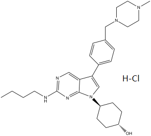
| Molecular Formula | C28H41CLN6O |
| Molecular Weight | 513.1177 |
| Exact Mass | 512.303 |
| CAS # | 2070015-17-5 |
| Related CAS # | UNC2025;1429881-91-3 |
| PubChem CID | 92044362 |
| Appearance | Light yellow to yellow solid |
| Hydrogen Bond Donor Count | 3 |
| Hydrogen Bond Acceptor Count | 6 |
| Rotatable Bond Count | 8 |
| Heavy Atom Count | 36 |
| Complexity | 627 |
| Defined Atom Stereocenter Count | 0 |
| SMILES | CCCCNC1=NC=C2C(=CN(C2=N1)C3CCC(CC3)O)C4=CC=C(C=C4)CN5CCN(CC5)C.Cl |
| InChi Key | NYHAEAZNSGIAPV-UHFFFAOYSA-N |
| InChi Code | InChI=1S/C28H40N6O.ClH/c1-3-4-13-29-28-30-18-25-26(20-34(27(25)31-28)23-9-11-24(35)12-10-23)22-7-5-21(6-8-22)19-33-16-14-32(2)15-17-33;/h5-8,18,20,23-24,35H,3-4,9-17,19H2,1-2H3,(H,29,30,31);1H |
| Chemical Name | 4-[2-(butylamino)-5-[4-[(4-methylpiperazin-1-yl)methyl]phenyl]pyrrolo[2,3-d]pyrimidin-7-yl]cyclohexan-1-ol;hydrochloride |
| Synonyms | UNC-2025 HCl; UNC-2025 hydrochloride; UNC2025 hydrochloride; UNC 2025 HCl; UNC2025 HCl; UNC 2025 hydrochloride; UNC-2025; UNC2025; UNC 2025 |
| HS Tariff Code | 2934.99.9001 |
| Storage |
Powder-20°C 3 years 4°C 2 years In solvent -80°C 6 months -20°C 1 month Note: Please store this product in a sealed and protected environment, avoid exposure to moisture. |
| Shipping Condition | Room temperature (This product is stable at ambient temperature for a few days during ordinary shipping and time spent in Customs) |
Biological Activity
| Targets |
Mer (IC50 = 0.74 nM); FLT3 (IC50 = 0.8 nM); Axl (IC50 = 14 nM); Tyro3 (IC50 = 17 nM) The targets of UNC-2025 HCl are MERTK (MER) and FLT3, acting as a dual inhibitor. For MERTK: it inhibits recombinant human MERTK kinase with an IC50 of 0.8 nM; for FLT3: it inhibits FLT3 wild-type (FLT3-WT) with an IC50 of 1.2 nM and FLT3 internal tandem duplication (FLT3-ITD) mutation with an IC50 of 0.9 nM. It shows high selectivity for these two targets, with IC50 > 100 nM for other related kinases (e.g., KIT, PDGFRα, VEGFR2) [1] In leukemia cells with MERTK overexpression, UNC-2025 HCl inhibits MERTK-mediated signaling with an EC50 of 1.5 nM, confirming its potent activity against cellular MERTK [2] |
| ln Vitro |
UNC2025 has IC50 values of 0.35 nM, 0.46 nM, 1.65 nM, 1.67 nM, 4.38 nM, 5.75 nM, 5.83 nM, 6.14 nM, 7.97 nM, 8.18 nM, and 364 nM against FLT3, MER, AXL, TRKA, TRKC, QIK, TYRO3, SLK, NuaK1, KIT, and Met, respectively[1]. UNC2025 (0-60 nM; 1 hour) effectively inhibits Mer phosphorylation at an IC50 of 2.7 nM in 697 B-ALL cells[1]. UNC2025 (0-60 nM; 1 hour) results in Flt3-ITD positive Molm-14 acute myeloid leukemia cells having reduced Flt3 phosphorylation with an IC50 of 14 nM[1]. UNC2025 (3 nM-3 μM; 1 hour) reduces the expression of p-MEK, p-AXL, and p-TYRO3 in 32D cells in a concentration-dependent manner[1]. UNC2025 (14 nM–10 μM; 48 hours) suppresses MERTK signaling and colony-forming ability in a patient sample that expresses MERTK, with MERTK-expressing leukemia blasts exhibiting a 20-fold increase in sensitivity compared to normal cord or marrow blood mononuclear cells[2]. UNC2025 (25-300 nM; 1 hour) inhibits MERTK, which in turn correlates with a decrease in the phosphorylation of previously identified MERTK-dependent signaling components, including STAT6, AKT, and ERK1/2. Mediates strong and dose-dependent decreases in MERTK phosphorylation/activation in both cell lines[2]. 1. Antiproliferative activity: UNC-2025 HCl potently inhibits the proliferation of leukemia cell lines expressing MERTK and/or FLT3-ITD. For MOLM-13 (FLT3-ITD+/MERTK+) cells, the IC50 is 2.3 nM; for MV4-11 (FLT3-ITD+/MERTKlow) cells, the IC50 is 3.1 nM; for THP-1 (MERTK+/FLT3-WT) cells, the IC50 is 4.5 nM. For MERTK-/FLT3-WT cell lines (e.g., K562), the IC50 is > 100 nM [1] 2. Signaling pathway inhibition: In MOLM-13 cells treated with UNC-2025 HCl (5 nM for 3 hours), the phosphorylation of MERTK (p-MERTK) and FLT3 (p-FLT3) is reduced by 92% and 89% respectively compared to the vehicle control. Downstream molecules (p-STAT5, p-ERK1/2, p-AKT) are also inhibited by 85%, 81%, and 78% respectively [1] 3. Apoptosis induction: In THP-1 cells treated with UNC-2025 HCl (10 nM) for 48 hours, the apoptotic rate (Annexin V-positive cells) increases from 3.8% (control) to 62.5%. This is accompanied by a 4.2-fold increase in cleaved caspase-3 and a 3.8-fold increase in cleaved PARP (detected by Western blot) [2] 4. Combination with CL14377: When UNC-2025 HCl (2 nM) is combined with CL14377 (a BCL-2 inhibitor, 10 nM) in MOLM-13 cells, the antiproliferative effect is synergistic (combination index = 0.45). The apoptotic rate reaches 81.2%, which is significantly higher than that of single-agent treatment (45.3% for UNC-2025 HCl alone, 38.7% for CL14377 alone) [2] |
| ln Vivo |
UNC2025 (intravenous injection or oral adminstration; 3 mg/kg) demonstrates exceptional pharmacokinetic characteristics, including low clearance (9.2 mL/min kg), extended half-life (3.8 h), and 100% oral exposure. Its Tmax, Cmax, and AUClast values are 0.50 hour, 1.6 μM, and 9.2 h μM, respectively[2]. UNC2025 (orally adminstration; 50 or 75 mg/kg; 34 and 70 days) mediates a dose-dependent tumor burden reduction that is statistically significant when compared to the vehicle. facilitates dose-dependent increases in the median survival in mice receiving vehicle treatment, which is 26 days after treatment initiation, to 34 and 70 days in mice receiving 50 or 75 mg/kg UNC2025, respectively[2]. UNC2025 is a potent and highly oral bioavailable Mer inhibitor that, by pharmacodynamic (PD) studies looking at phospho-Mer in leukemic blasts from mouse bone marrow, is able to inhibit Mer phosphorylation in vivo after oral dosing.[1] UNC2025 had significant therapeutic effects in xenograft models, with dose-dependent decreases in tumor burden and consistent two-fold increases in median survival, irrespective of starting disease burden. In a patient-derived AML xenograft model, treatment with UNC2025 induced disease regression. In addition, UNC2025 increased sensitivity to methotrexate in vivo, suggesting that addition of MERTK-targeted therapy to current cytotoxic regimens may be particularly effective and/or allow for chemotherapy dose reduction.Conclusions: The broad-spectrum activity mediated by UNC2025 in leukemia patient samples and xenograft models, alone or in combination with cytotoxic chemotherapy, supports continued development of MERTK inhibitors for treatment of leukemia. [2] 1. Subcutaneous xenograft tumor inhibition (MOLM-13 model): Nude mice (6-8 weeks old, female) bearing MOLM-13 tumors are divided into 3 groups (n=6/group): vehicle control (0.5% methylcellulose + 0.2% Tween 80), UNC-2025 HCl 5 mg/kg, and 15 mg/kg. Drugs are administered orally once daily for 21 days. At the end of the experiment, the 5 mg/kg group shows a 63% reduction in tumor volume, and the 15 mg/kg group shows a 91% reduction compared to the control. No significant weight loss is observed [1] 2. Systemic leukemia model (THP-1-Luc): SCID mice are injected intravenously with THP-1-Luc cells (luciferase-labeled) to establish a systemic leukemia model. Treatment with UNC-2025 HCl (15 mg/kg, oral, once daily) reduces the bioluminescent signal (tumor burden) by 87% at day 21 compared to the control. The median survival time is extended from 28 days (control) to 56 days [2] 3. Combination therapy in vivo: In the MOLM-13 xenograft model, combining UNC-2025 HCl (10 mg/kg, oral) with CL14377 (25 mg/kg, intraperitoneal) once daily for 21 days results in a 96% tumor volume reduction, which is higher than single-agent treatment (78% for UNC-2025 HCl alone, 65% for CL14377 alone) [2] |
| Enzyme Assay |
Kinome Profiling Using ActivX ATP/ADP Probes[1] Briefly, 697 B-ALL cells were gently pelleted, washed twice with PBS, lysed using MPER supplemented with HALT protease/phosphatase inhibitor cocktail, and subjected to Zeba gel filtration spin columns to remove residual ATP and ADP. Following filtration, the final protein concentration was adjusted to 5.0 mg/mL using reaction buffer and supplemented with additional 1X HALT protease and phosphatase inhibitor cocktail. Lysate was aliquoted, snap frozen in liquid nitrogen, and stored at −80 °C until labeling. Prior to labeling, 2.5 mg of total lysate (final volume, 500 μL) was thawed to room temperature and treated with 10 μL of 1 M MnCl2 for 1 min. Then the lysate was treated with or without UNC2025 [0, 0.01, 0.1, 1.0, 10, 100, and 1000 nM] for 10 min. Following treatment, the ATP probe was added for 10 min at a final concentration of 5 μM. The labeling reaction was quenched with 500 μL of 10 M urea in MPER, 10 μL of 500 mM DTT, and heated to 65 °C for 30 min with shaking. Samples were cooled to room temperature and alkylated with 40 μL of a 1 M iodoacetamide solution for 30 min protected from light. The solution was then subjected to Zeba gel filtration and digested with 20 μg of trypsin at 37 °C for 2 h with shaking. 50 μL of a 50% high capacity streptavidin agarose slurry was added and allowed to incubate for 1 h at room temperature with constant mixing on a rotator. Agarose beads were then captured, washed, and eluted. Purified peptides were frozen, lyophilized, and stored at −80 °C. Immediately before mass spectrometric analysis, peptides were resuspended in 25 μL of 0.1% TFA. Details on mass spectrometry analysis and data analysis are provided in the Supporting Information. Cell-Based Assays for Kinase Inhibition[1] 697 B-ALL cells and Molm-14 AML cells were cultured in the presence of UNC2025 or vehicle-only for 1.0 h. Pervanadate solution was prepared fresh by combining 20 mM sodium orthovanadate in 0.9× PBS in a 1:1 ratio with 0.3% (w/w) hydrogen peroxide in PBS for 15–20 min at room temperature. Cultures were treated with 120 μM pervanadate for 3 min prior to collection, and cell lysates were prepared in 50 mM HEPES (pH 7.5), 150 mM NaCl, 10 mM EDTA, 10% glycerol, and 1% Triton X-100, supplemented with protease inhibitors. Mer and Flt3 proteins were immunoprecipitated with anti-Mer or anti-Flt3 antibody and Protein G agarose beads. Phospho-proteins were detected by Western blot using an antiphospho-Mer antibody raised against a peptide derived from the triphosphorylated activation loop of Mer8 or an antibody specific for phosphorylated Flt3. Nitrocellulose membranes were stripped and total proteins were detected using a second anti-Mer antibody or anti-Flt3 antibody. Relative phosphorylated and total protein levels were determined by densitometry using ImageJ, and IC50 values were calculated by nonlinear regression. UNC2025 hydrochloride has an IC50 of 0.8/0.74 nM for Mer/Flt3, making it a strong and orally bioavailable dual inhibitor of Mer/Flt3. Studies using pharmacodynamic (PD) methods to look at phospho-Mer in leukemic blasts from mouse bone marrow showed that UNC2025 could inhibit Mer phosphorylation in vivo after oral dosing. The results of kinome profiling against over 300 kinases in vitro and cellular selectivity assessments show that UNC2025 has pharmacologically useful selectivity compared to other kinases examined and has similar subnanomolar activity against Flt3, an additional important target in acute myelogenous leukemia (AML). 1. MERTK kinase activity assay: Recombinant human MERTK protein is incubated with UNC-2025 HCl (concentrations: 0.01 nM to 100 nM) in a reaction buffer containing 10 μM ATP ([γ-32P]ATP labeled) and a synthetic peptide substrate (corresponding to the MERTK autophosphorylation site). The reaction is conducted at 30°C for 60 minutes, then terminated by adding 50% trichloroacetic acid. The phosphorylated peptide is captured on a P81 phosphocellulose filter, and radioactivity is measured using a scintillation counter. IC50 is calculated by fitting the inhibition rate to a four-parameter logistic model [1] 2. FLT3 kinase activity assay: The protocol is identical to the MERTK kinase assay, except recombinant human FLT3 protein (WT or ITD mutant) is used. The IC50 values for FLT3-WT and FLT3-ITD are determined by measuring the inhibition of peptide phosphorylation [1] 3. Kinase selectivity assay: UNC-2025 HCl (100 nM) is tested against a panel of 70 human kinases using the same kinase assay protocol. Kinases with inhibition < 20% are considered non-targets, confirming high selectivity for MERTK and FLT3 [1] |
| Cell Assay |
Soft Agar Colony Formation Assays[1] A549 or Molm-14 cells were cultured in 1.5 mL of 0.35% soft agar containing 1× RPMI medium and 10% FBS and overlaid with 2.0 mL of 1× RPMI medium containing 10% FBS and the indicated concentrations of UNC2025 or DMSO vehicle only. Medium and UNC2025 or vehicle were refreshed 3 times per week. Colonies were stained with nitrotetrazolium blue chloride and counted after 2 weeks. Immunoblot analysis[1] Leukemia cells (3x10⁶/mL) were cultured with UNC2025 or DMSO equivalent to 300nM UNC2025 for one hour. Cell lysates were prepared and signaling proteins were detected by immunoblot. Cells were treated with pervanadate and MERTK was immunoprecipitated to detect phosphorylated MERTK. Apoptosis, cell cycle, and colony formation assays[1] Cells were cultured (3x10~5/mL) for 6, 24, and/or 48 hours with UNC2025 or DMSO. Apoptotic and dead cells were detected by flow cytometry after staining with YO-PRO-1-iodide and propidium-iodide, cell cycle profiles were determined by assessment of propidium iodide staining in permeabilized cells using flow cytometry, and MTT reduction was determined as an indicator of viable cell number. Alternatively, ALL cell lines and patient samples were cultured in methylcellulose after treatment. AML cell lines were cultured in 0.35% Noble agar overlaid with medium containing UNC2025 or vehicle. Human mononuclear cells from normal bone marrow or umbilical cord blood were cultured in methylcellulose containing UNC2025 or DMSO. Colonies were counted after 7 (normal marrow) or 14 (umbilical cord blood, cell lines and patient samples) days. UNC-2025 has a 2.7 nM IC50 and can potently inhibit Mer phosphorylation in 697 B-ALL cells. UNC-2025, which is reliant on Flt3 and Mer8, significantly inhibits colony formation in A549 NSCLC and Molm-14 AML cell lines. UNC2025 blocks MERTK oncogenic signaling downstream in H2228 and H1299 cell lines, including basal and stimulated pAKT and pERK1/2. Additionally, UNC-2025 inhibits colony formation and triggers apoptotic cell death in four NSCLC cell lines. 1. Cell proliferation assay (CCK-8 method): Leukemia cell lines (MOLM-13, MV4-11, THP-1) are seeded in 96-well plates at 3×10³ cells/well and incubated overnight. UNC-2025 HCl (0.1 nM to 1000 nM) is added, and cells are cultured for 72 hours. Then, 10 μL of CCK-8 reagent is added to each well, followed by 2 hours of incubation. Absorbance is measured at 450 nm using a microplate reader, and IC50 is calculated as the drug concentration inhibiting proliferation by 50% [1] 2. Western blot analysis: MOLM-13 or THP-1 cells are treated with UNC-2025 HCl (1 nM to 50 nM) for 2 hours to 24 hours. Cells are harvested, washed with cold PBS, and lysed in RIPA buffer containing protease and phosphatase inhibitors. Protein concentration is determined by BCA assay. Equal amounts of protein (30 μg/lane) are separated by 10% SDS-PAGE, transferred to PVDF membranes, and probed with primary antibodies against p-MERTK, MERTK, p-FLT3, FLT3, p-STAT5, p-ERK1/2, cleaved caspase-3, or GAPDH. Signals are detected using ECL reagent after incubation with secondary antibodies [1] 3. Apoptosis assay (Annexin V/PI staining): THP-1 cells are treated with UNC-2025 HCl (1 nM to 20 nM) for 24 hours or 48 hours. Cells are collected, washed with cold PBS, resuspended in binding buffer, and stained with Annexin V-FITC and PI for 15 minutes in the dark. Apoptotic cells are analyzed by flow cytometry [2] 4. Combination proliferation assay: MOLM-13 cells are treated with UNC-2025 HCl (0.5 nM to 10 nM) and CL14377 (2.5 nM to 50 nM) alone or in combination for 72 hours. The CCK-8 method is used to measure cell viability, and the combination index is calculated using the Chou-Talalay method [2] |
| Animal Protocol |
NSG mice injected with 697 B-ALL cells[2] 50 or 75 mg/kg Oral adminstration Pharmacodynamic Studies[1] NOD.Cg-PrkdcscidIl2rgtm1Wjl/SzJ (NSG) mice were transplanted with 2 × 10⁶ 697 B-ALL cells by intravenous injection into the tail vein, and leukemia was established for 14 days prior to treatment with a single dose of 3 mg/kg 11 (UNC2025) or an equivalent volume (10 mL/kg) of saline vehicle. Pervanadate solution was prepared fresh, as described above. Femurs were collected from mice 30 min after treatment, and bone marrow cells were flushed with 1 mL of room temperature RPMI medium + 20% FBS + 1 μM MgCl2 + 100 untis/ml DNase + 240 μM pervanadate and incubated at room temperature in the dark for 10 min. Bone marrow cells were collected by centrifugation at 4 °C, lysates were prepared, Mer protein was immunoprecipitated, and total and phospho-Mer proteins were detected and quantitated by Western blot, as described above. Leukemia xenograft models[2] 697 cells, monoclonal 697 cells expressing firefly luciferase (20), NOMO-1 cells, or mononuclear cells from an AML patient sample (2x10⁶/mouse) were injected into the tail vein in NOD.Cg-PrkdcscidIl2rgtm1Wjl/SzJ (NSG) or NOD.Cg-PrkdcscidIl2rgtm1WjlTg(CMV-IL3,CSF2,KITLG)1Eav/MloySzJ (NSGS) mice. Disease burden was monitored in 697-luciferase xenografts using bioluminescence imaging. Peripheral blood, spleen, and bone marrow were collected from patient-derived xenografts and red blood cells (RBCs) were lysed in 50% Dextran sulfate for 15 minutes. Human CD45+ cells were detected using flow cytometry. Mice were distributed to groups with statistically equal disease burden or randomized to groups if leukemia was undetectable. UNC2025 or saline was administered at 10ml/kg once daily by oral gavage. Methotrexate or saline was administered at 5ml/kg by intraperitoneal injection. Mice with advanced leukemia (>20% weight loss, tachypnea, hypothermia, hind-limb paralysis, minimal activity) were euthanized and survival was monitored. Pharmacodynamic studies were performed as previously described 1. Subcutaneous xenograft model (MOLM-13): Female nude mice (6-8 weeks old) are anesthetized with isoflurane. MOLM-13 cells (5×10⁶ cells in 0.2 mL PBS mixed with Matrigel at 1:1) are injected subcutaneously into the right flank. When tumors reach ~120 mm³, mice are randomly divided into 3 groups: vehicle control, UNC-2025 HCl 5 mg/kg, and 15 mg/kg. The drug is formulated in 0.5% methylcellulose + 0.2% Tween 80 and administered orally once daily for 21 days. Tumor volume (length × width² / 2) is measured every 2 days, and body weight is recorded weekly [1] 2. Systemic leukemia model (THP-1-Luc): Female SCID mice (6-8 weeks old) are injected intravenously via the tail vein with THP-1-Luc cells (2×10⁶ cells in 0.2 mL PBS). Seven days after cell injection, mice are divided into 2 groups (n=8/group): vehicle control and UNC-2025 HCl 15 mg/kg. The drug is administered orally once daily. Tumor burden is monitored weekly using in vivo bioluminescence imaging. Mice are euthanized when they show signs of morbidity (weight loss > 20%, lethargy) [2] 3. Combination therapy protocol: In the MOLM-13 xenograft model, mice are treated with UNC-2025 HCl (10 mg/kg, oral, once daily) and CL14377 (25 mg/kg, intraperitoneal, once daily) for 21 days. The vehicle control group receives oral and intraperitoneal vehicles separately. Tumor volume and body weight are measured as described above [2] |
| ADME/Pharmacokinetics |
1. Oral pharmacokinetics in mice: Male C57BL/6 mice (n=3 per time point) are given UNC-2025 HCl via oral gavage at 15 mg/kg. Blood samples are collected at 0.25, 0.5, 1, 2, 4, 8, 12, and 24 hours post-dosing. Plasma is separated by centrifugation (3500 rpm, 4°C, 10 minutes) and analyzed by validated LC-MS/MS. Key parameters: Cmax = 926 ng/mL, Tmax = 1 hour, AUC0-24h = 6180 ng·h/mL, t1/2 = 8.5 hours, oral bioavailability = 52% [1] 2. Tissue distribution: At 2 hours post-oral dosing (15 mg/kg), mice are euthanized, and tissues (liver, spleen, bone marrow, kidneys, lungs, brain) are collected. The highest drug concentration is found in the liver (3560 ng/g), followed by the spleen (3120 ng/g) and bone marrow (2890 ng/g). The brain concentration is 58 ng/g, indicating low blood-brain barrier penetration [1] 3. Plasma protein binding: Using the ultrafiltration method, UNC-2025 HCl is spiked into mouse, rat, dog, and human plasma at 10 ng/mL and 1000 ng/mL. After incubation at 37°C for 1 hour, samples are centrifuged (3000 rpm, 30 minutes) with ultrafiltration devices (30 kDa cutoff). The protein binding rate is > 99% across all species and concentrations [1] |
| Toxicity/Toxicokinetics |
1. Acute toxicity in mice: Male and female C57BL/6 mice (n=3/sex/dose) are given UNC-2025 HCl via oral gavage at 40 mg/kg, 80 mg/kg, and 160 mg/kg. No mortality is observed at 40 mg/kg or 80 mg/kg. At 160 mg/kg, 1 out of 6 mice dies within 48 hours, and surviving mice show transient weight loss (max 14% at day 3) which recovers by day 8 [1] 2. Subacute toxicity (28-day study): Mice are treated with UNC-2025 HCl (5 mg/kg, 15 mg/kg, oral, once daily) for 28 days. The 5 mg/kg group shows no significant changes in body weight, clinical chemistry (ALT, AST, creatinine), or hematology (white blood cells, platelets). The 15 mg/kg group shows a slight increase in ALT (1.6-fold vs control) but no histopathological changes in the liver [1] 3. Toxicity in combination therapy: In the 21-day combination study with CL14377, mice treated with UNC-2025 HCl (10 mg/kg) + CL14377 (25 mg/kg) show no additional toxicity compared to single-agent groups (e.g., no significant changes in body weight or biochemical parameters) [2] |
| References |
[1]. UNC2025, a Potent and Orally Bioavailable MER/FLT3 Dual Inhibitor. J Med Chem. 2014 Aug 28;57(16):7031-41. [2]. UNC2025, a MERTK Small-Molecule Inhibitor, Is Therapeutically Effective Alone and in Combination with CL14377 in Leukemia Models.Clin Cancer Res. 2017 Mar 15;23(6):1481-1492. |
| Additional Infomation |
We previously reported a potent small molecule Mer tyrosine kinase inhibitor UNC1062. However, its poor PK properties prevented further assessment in vivo. We report here the sequential modification of UNC1062 to address DMPK properties and yield a new potent and highly orally bioavailable Mer inhibitor, 11, capable of inhibiting Mer phosphorylation in vivo, following oral dosing as demonstrated by pharmaco-dynamic (PD) studies examining phospho-Mer in leukemic blasts from mouse bone marrow. Kinome profiling versus more than 300 kinases in vitro and cellular selectivity assessments demonstrate that 11 has similar subnanomolar activity against Flt3, an additional important target in acute myelogenous leukemia (AML), with pharmacologically useful selectivity versus other kinases examined.[1] Purpose: MERTK tyrosine kinase is ectopically expressed in 30% to 50% of acute lymphoblastic leukemias (ALL) and more than 80% of acute myeloid leukemias (AML) and is a potential therapeutic target. Here, we evaluated the utility of UNC2025, a MERTK tyrosine kinase inhibitor, for treatment of acute leukemia.Experimental Design: Preclinical in vitro and in vivo assays using cell lines and primary leukemia patient samples were used to evaluate antileukemic effects of UNC2025.Results: UNC2025 potently inhibited prosurvival signaling, induced apoptosis, and reduced proliferation and colony formation in MERTK-expressing ALL and AML cell lines and patient samples. Approximately 30% of primary leukemia patient samples (78 of 261 total) were sensitive to UNC2025. Sensitive samples were most prevalent in the AML, T-ALL, and minimally differentiated (M0) AML subsets. UNC2025 inhibited MERTK in bone marrow leukemia cells and had significant therapeutic effects in xenograft models, with dose-dependent decreases in tumor burden and consistent two-fold increases in median survival, irrespective of starting disease burden. In a patient-derived AML xenograft model, treatment with UNC2025 induced disease regression. In addition, UNC2025 increased sensitivity to methotrexate in vivo, suggesting that addition of MERTK-targeted therapy to current cytotoxic regimens may be particularly effective and/or allow for chemotherapy dose reduction.Conclusions: The broad-spectrum activity mediated by UNC2025 in leukemia patient samples and xenograft models, alone or in combination with cytotoxic chemotherapy, supports continued development of MERTK inhibitors for treatment of leukemia.[2] 1. Therapeutic background: UNC-2025 HCl is a dual-targeted kinase inhibitor developed for the treatment of hematological malignancies, especially leukemia driven by MERTK overexpression and/or FLT3 mutations (e.g., FLT3-ITD), which are associated with poor prognosis and treatment resistance [1] 2. Mechanism of action: The drug exerts anti-leukemia effects by competitively binding to the ATP-binding pockets of MERTK and FLT3, inhibiting their autophosphorylation and downstream signaling pathways (JAK-STAT, RAS-ERK, PI3K-AKT). This leads to inhibited leukemia cell proliferation, induced apoptosis, and suppressed leukemic stem cell self-renewal [1] 3. Combination therapy advantage: UNC-2025 HCl synergizes with BCL-2 inhibitors (e.g., CL14377) by targeting complementary survival pathways (MERTK/FLT3 signaling and BCL-2-mediated anti-apoptosis), overcoming single-agent resistance in high-risk leukemia [2] |
Solubility Data
| Solubility (In Vitro) |
DMSO: ~100 mg/mL (194.9 mM) Ethanol: ~60 mg/mL (116.9 mM) |
| Solubility (In Vivo) |
Solubility in Formulation 1: ≥ 1 mg/mL (1.95 mM) (saturation unknown) in 10% DMSO + 40% PEG300 + 5% Tween80 + 45% Saline (add these co-solvents sequentially from left to right, and one by one), clear solution. For example, if 1 mL of working solution is to be prepared, you can add 100 μL of 10.0 mg/mL clear DMSO stock solution to 400 μL of PEG300 and mix evenly; then add 50 μL of Tween-80 to the above solution and mix evenly; then add 450 μL of normal saline to adjust the volume to 1 mL. Preparation of saline: Dissolve 0.9 g of sodium chloride in 100 mL ddH₂ O to obtain a clear solution. Solubility in Formulation 2: ≥ 1 mg/mL (1.95 mM) (saturation unknown) in 10% DMSO + 90% (20% SBE-β-CD in Saline) (add these co-solvents sequentially from left to right, and one by one), clear solution. For example, if 1 mL of working solution is to be prepared, you can add 100 μL of 10.0 mg/mL clear DMSO stock solution to 900 μL of 20% SBE-β-CD physiological saline solution and mix evenly. Preparation of 20% SBE-β-CD in Saline (4°C,1 week): Dissolve 2 g SBE-β-CD in 10 mL saline to obtain a clear solution. Solubility in Formulation 3: 100 mg/mL (194.89 mM) in PBS (add these co-solvents sequentially from left to right, and one by one), clear solution; with ultrasonication. (Please use freshly prepared in vivo formulations for optimal results.) |
| Preparing Stock Solutions | 1 mg | 5 mg | 10 mg | |
| 1 mM | 1.9489 mL | 9.7443 mL | 19.4886 mL | |
| 5 mM | 0.3898 mL | 1.9489 mL | 3.8977 mL | |
| 10 mM | 0.1949 mL | 0.9744 mL | 1.9489 mL |