Physicochemical Properties
| Molecular Formula | (C14H22O.C2H4O.CH2O)X |
| Molecular Weight | 261.38 (monomer) |
| Exact Mass | 280.203 |
| CAS # | 25301-02-4 |
| Related CAS # | 25301-02-4 |
| PubChem CID | 71388 |
| Appearance | Colorless to light yellow liquids |
| Density | 1.1 |
| Boiling Point | 282.3ºC at 760 mmHg |
| Flash Point | 148.3ºC |
| Vapour Pressure | 0.00198mmHg at 25°C |
| LogP | 4.573 |
| Hydrogen Bond Donor Count | 1 |
| Hydrogen Bond Acceptor Count | 3 |
| Rotatable Bond Count | 3 |
| Heavy Atom Count | 20 |
| Complexity | 204 |
| Defined Atom Stereocenter Count | 0 |
| SMILES | C1=C(O)C=CC(C(C)(C)CC(C)(C)C)=C1.C1OC1.C=O |
| InChi Key | MDYZKJNTKZIUSK-UHFFFAOYSA-N |
| InChi Code | InChI=1S/C14H22O.C2H4O.CH2O/c1-13(2,3)10-14(4,5)11-6-8-12(15)9-7-11;1-2-3-1;1-2/h6-9,15H,10H2,1-5H3;1-2H2;1H2 |
| Chemical Name | formaldehyde;oxirane;4-(2,4,4-trimethylpentan-2-yl)phenol |
| Synonyms | Tyloxapol; 25301-02-4; Macrocyclon; Tyloxypal; Triton WR-1339; |
| HS Tariff Code | 2934.99.9001 |
| Storage |
Powder-20°C 3 years 4°C 2 years In solvent -80°C 6 months -20°C 1 month Note: This product requires protection from light (avoid light exposure) during transportation and storage. |
| Shipping Condition | Room temperature (This product is stable at ambient temperature for a few days during ordinary shipping and time spent in Customs) |
Biological Activity
| Targets | Non-ionic liquid polymer |
| ln Vitro |
Tyloxapol (100 μg/mL) causes HEK293 cell ablation [2]. Tyloxapol promotes nuclear fragmentation and the development of Argentine nuclei [2]. Tyloxapol raises pulmonary venous risk, produces epithelial and erythrocytotoxicity, and promotes human Jurkat T occupancy [2]. #### From Literature [2] - Tyloxapol (Triton WR-1339) exhibited concentration-dependent cytostatic effects on NIH/3T3 fibroblasts and HeLa cells, inhibiting cell proliferation with IC₅₀ values of ~35 μg/mL (NIH/3T3) and ~42 μg/mL (HeLa) (MTT assay) [2] - It suppressed apoptosis in both cell lines, as shown by reduced Annexin V-FITC/PI double-positive cells and decreased caspase-3 activation [2] - Upregulated anti-apoptotic protein Bcl-2 expression and downregulated pro-apoptotic protein Bax expression, as confirmed by Western blot analysis [2] - Inhibited colony formation ability of NIH/3T3 cells, with a 50% reduction in colony number at 50 μg/mL [2] |
| ln Vivo |
Triton WR-1339 increased oxidative stress through an elevation in TBARS associated with depletion in glutathione and the activities of GST, SOD, GSH-Px, and CAT in plasma, liver, and brain. Triton WR-1339 induced DNA fragmentation and inhibited the activities of acetylcholinesterase and mono aminoxidase in the brain. Plasma biochemical parameters including transaminases, phosphatases, lactate dehydrogenase, urea, creatinine, bilirubin, total lipid, cholesterol, triglyceride, and LDL were increased, while total protein, albumin, and high HDL were decreased due to Triton WR-1339-treatment. The histopathological and morphmetric analysis of the liver and dorsal aorta revealed alterations after Triton WR-1339-treatment. The presence of soybean oil with Triton WR-1339 minimized its hepatotoxicity and neurotoxicity via hypolipidemic effects and attenuated the oxidative damage[1]. #### From Literature [1] - In male albino rats, a single intraperitoneal injection of Tyloxapol (Triton WR-1339) (400 mg/kg) induced hyperlipidemia, significantly increasing serum total cholesterol (TC), triglycerides (TG), low-density lipoprotein cholesterol (LDL-C) levels, and decreasing high-density lipoprotein cholesterol (HDL-C) levels at 72 hours post-administration [1] - Caused DNA fragmentation in liver, kidney, and brain tissues, as detected by comet assay (increased tail length and tail moment) [1] - Reduced the levels of neurotransmitters (dopamine, serotonin, norepinephrine) in brain tissue homogenates [1] - Induced oxidative stress: decreased superoxide dismutase (SOD) and catalase (CAT) activities, and increased malondialdehyde (MDA) levels in liver, kidney, and brain tissues [1] - Caused histopathological changes: hepatocellular steatosis, renal tubular epithelial cell damage, and cerebral cortical inflammation in model rats [1] |
| Enzyme Assay |
Plasma and brain acetylcholinesterase and monoamine oxidase estimation[1] Acetylcholinesterase (AChE; EC 3.1.1.7) activity was estimated in plasma and tissue supernatant using acetylcholine iodide as a substrate according to the method of Ellman, Courtney, Anders, and Featherstone (1961). The activity of monoamine oxidase was estimated in plasma and tissue extracts according to the method of Sandler, Reveley, and Glover (1981). Protein estimation[1] The protein content of the tissue extracts mentioned earlier was determined by the method of Lowry, Rosebrough, Farr, and Randall (1951) using bovine serum albumin as a standard. Biochemical parameters[1] Plasma and liver aspartate aminotransaminase (AST; EC 2.6.1.1), alanine aminotransaminase (ALT; EC 2.6.1.2), and lactate dehydrogenase (LDH; EC 1.1.1.27) activities were determined using kits from SENTINEL CH. Stored plasma samples were analyzed for urea and creatinine concentrations using kits from SENTINEL CH. Plasma total cholesterol, high-density lipoproteins (HDL), and triglyceride levels were estimated by using cholesterol and triglyceride kits, span diagnostics Ltd., India. The assays were performed in accordance with the manufacturer’s instructions. |
| Cell Assay |
Solid lipid nanoparticles (SLN) have been praised for their advantageous drug delivery properties such as biocompatibility, controlled release and passive drug targeting. However, the cytotoxicity of SLN and their ingredients, especially over a longer time period, has not been investigated in detail. We examined the critical issues regarding the use of a surface active stabilizer Tyloxapol (Tyl) for the preparation of solid lipid particles (SLP) and their effects on cellular functions and viability. SLP composed of behenate, phospholipids and a stabilizer, Tyloxapol (Tyl) or Lutrol (Lut), were prepared by the lipid melt method, labeled with a fluorescent dye and tested on Jurkat or HEK293 cells. The nano-sized particles were rapidly internalized and exhibited cytoplasmic localization. Incubation of cells with SLP-Tyl resulted in a dose- and time-dependent cytostatic effect, and also caused moderate and delayed cytotoxicity. Tyloxapol solution or SLP-Tyl dispersion caused the detachment of HEK293 cells, a decrease in cell proliferation and alterations in cellular morphology. Cell cycle analysis revealed that, while the unfavourable effects of SLP-Tyl and Tyloxapol solution are similar initially, longer incubation results in partial recovery of cells incubated with the dispersion of SLP-Tyl, whereas the presence ofTyloxapol) solution induces apoptotic cell death. These findings indicate that Tyloxapol is an unfavourable stabilizer of SLP used for intracellular delivery and reinforce the role of stabilizers in a design of SLP with minimal cytotoxic properties. #### From Literature [2] - NIH/3T3 fibroblasts and HeLa cells were cultured in DMEM medium supplemented with 10% fetal bovine serum and antibiotics, maintained at 37°C in a 5% CO₂ incubator [2] - Cell viability assay: Cells were seeded in 96-well plates, treated with Tyloxapol at concentrations of 10, 25, 50, 75, 100 μg/mL for 48 hours; MTT reagent was added, incubated for 4 hours, formazan crystals were dissolved in DMSO, and absorbance was measured at 570 nm [2] - Apoptosis assay: Cells were treated with Tyloxapol (30, 50, 70 μg/mL) for 48 hours, harvested, stained with Annexin V-FITC and PI for 15 minutes in the dark, and apoptotic rate was detected by flow cytometer [2] - Western blot analysis: Cells were lysed in RIPA buffer with protease inhibitors, protein concentrations were determined by BCA assay, equal amounts of protein were separated by SDS-PAGE, transferred to PVDF membranes, incubated with primary antibodies against Bcl-2, Bax, cleaved caspase-3, and β-actin overnight at 4°C, followed by HRP-conjugated secondary antibodies, and protein bands were visualized by ECL detection system [2] - Colony formation assay: Cells were seeded in 6-well plates at low density, treated with Tyloxapol (20, 40, 60 μg/mL) for 10–14 days, colonies were fixed with formaldehyde, stained with crystal violet, and visible colonies (≥50 cells) were counted [2] |
| Animal Protocol |
Animal/Disease Models: 21 adult male Wistar rats, 11-12 weeks old, weighing 180-200 g[1]. Doses: 50 mg/kg. Route of Administration: intraperitoneal (ip) injection, BW, every other day. Experimental Results: TBARS levels were Dramatically increased in rat plasma, liver and brain (P < 0.05), while antioxidant enzymes (GPx, GST, CAT, SOD) were inhibited. Induces DNA fragmentation and inhibits the activity of acetylcholinesterase and monoammonium oxidase in the brain. Animals were treated with Triton WR-1339 (50 mg/kg BW) and soybean oil (50 mg/kg BW) for 28 days. Oxidative stress markers; antioxidant enzymes activity; biochemical parameters, such as acetylcholinesterase and mono aminoxidase, transaminases, phosphatases, lactate dehydrogenase, urea, creatinine, bilirubin, and lipid profile; DNA fragmentation; as well as histopathological and morphmetric analysis of the liver and dorsal aorta were investigated.[1] Twenty-one adult male Wistar rats, aged 11–12 weeks weighing 180–200 g, were used in the present experiments. Animals were caged in groups and given food and water ad libitum. After 2 weeks of acclimation, animals were divided randomly into three groups; seven rats in each group. Group I (control group) rats were treated with a vehicle (injected intraperitoneally with saline and treated orally with corn oil). Group II was injected intraperitoneally with 50 mg/kg BW of Triton WR-1339 every other day. Group III was treated orally with 50 mg/kg BW of soybean oil every day plus 50 mg/kg BW of Triton WR-1339 every other day. The dose of Triton WR-1339 was chosen according to Bhuvaneswari and Sasikumar (2013). The dose of soybean oil was chosen according to Mallo et al. (2013a, b). Rats were administered their respective doses for 28 days.[1] #### From Literature [1] - Male albino rats (150–200 g) were randomly divided into three groups: control group, Tyloxapol model group, and soybean oil treatment group (n=6 per group) [1] - Tyloxapol was dissolved in normal saline, and administered to model group and treatment group rats via intraperitoneal injection at a single dose of 400 mg/kg [1] - Soybean oil was administered to the treatment group via oral gavage (1 mL/kg/day) for 3 consecutive days after Tyloxapol injection; control group received equal volumes of normal saline (intraperitoneal) and distilled water (oral) [1] - At 72 hours post-Tyloxapol administration, rats were sacrificed by cervical dislocation; blood samples were collected for serum lipid analysis (TC, TG, LDL-C, HDL-C) [1] - Liver, kidney, and brain tissues were harvested for DNA fragmentation assay (comet assay), neurotransmitter detection (dopamine, serotonin, norepinephrine), oxidative stress parameter measurement (SOD, CAT, MDA), and histopathological/morphometric analysis (H&E staining) [1] |
| Toxicity/Toxicokinetics |
- In vitro toxicity: Tyloxapol inhibited cell proliferation and suppressed apoptosis in NIH/3T3 and HeLa cells at concentrations ≥10 μg/mL, with no obvious cytotoxicity at concentrations <5 μg/mL [2] - In vivo toxicity: Single intraperitoneal injection of Tyloxapol (400 mg/kg) in rats induced hyperlipidemia, DNA damage, neurotransmitter depletion, oxidative stress, and histopathological lesions in liver, kidney, and brain tissues [1] |
| References |
[1]. Triton WR-1339-induced hyperlipidemia, DNA fragmentation, neurotransmitters inhibition, oxidative damage, histopathological and morphometric changes: the protective role of soybean oil. The Journal of Basic and Applied Zoology volume 79, Article number: 51 (2018). [2]. Surface active stabilizer tyloxapol in colloidal dispersions exerts cytostatic effects and apoptotic dismissal of cells. Toxicol Appl Pharmacol. 2008 Oct 15;232(2):218-25. |
| Additional Infomation |
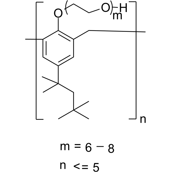
Tyloxapol is a polymeric compound resulting from the reaction of 4-(1,1,3,3-tetramethylbutyl)phenol with formaldehyde to give a chain in which 6-8 molecules are linked together by CH2 groups ortho to the phenolic hydroxy groups, which have then undergone reaction with oxirane to give polyoxyethyleneoxy moieties, Ar(OCH2CH2)xOH, where x = 8-10. A nonionic liquic polymer, it inhibits lipoprotein lipase and hence clearance of triglyceride from the plasma, so is used to induce hyperlipidaemia in test animals. Also used as a surfactant to aid liquefaction and removal of mucus- and pus-containing bronchopulmonary secretions. It has a role as an inhibitor, an excipient, a surfactant and an apoptosis inducer. See also: Tyloxapol (annotation moved to). Tyloxapol (also known as Triton WR-1339) is a non-ionic surfactant commonly used as a research tool to induce hyperlipidemia in animal models [1] It acts as a surface active stabilizer in colloidal dispersions [2] Literature [1] used Tyloxapol to establish a hyperlipidemia model, focusing on the protective effects of soybean oil against its induced toxicity [1] Literature [2] reported its cytostatic and anti-apoptotic effects in vitro, which may be related to the regulation of Bcl-2 family proteins and caspase signaling pathways [2] |
Solubility Data
| Solubility (In Vitro) |
H2O : ~120 mg/mL Ethanol : ~100 mg/mL DMSO : ≥ 38 mg/mL |
| Solubility (In Vivo) |
Solubility in Formulation 1: 100 mg/mL (Infinity mM) in PBS (add these co-solvents sequentially from left to right, and one by one), clear solution; with sonication. (Please use freshly prepared in vivo formulations for optimal results.) |