Physicochemical Properties
| Molecular Formula | C17H14O7 |
| Molecular Weight | 330.2889 |
| Exact Mass | 330.073 |
| CAS # | 520-32-1 |
| Related CAS # | Tricin-d6 |
| PubChem CID | 5281702 |
| Appearance | Off-white to yellow solid powder |
| Density | 1.5±0.1 g/cm3 |
| Boiling Point | 598.5±50.0 °C at 760 mmHg |
| Flash Point | 224.0±23.6 °C |
| Vapour Pressure | 0.0±1.8 mmHg at 25°C |
| Index of Refraction | 1.671 |
| LogP | 1.39 |
| Hydrogen Bond Donor Count | 3 |
| Hydrogen Bond Acceptor Count | 7 |
| Rotatable Bond Count | 3 |
| Heavy Atom Count | 24 |
| Complexity | 491 |
| Defined Atom Stereocenter Count | 0 |
| InChi Key | HRGUSFBJBOKSML-UHFFFAOYSA-N |
| InChi Code | InChI=1S/C17H14O7/c1-22-14-3-8(4-15(23-2)17(14)21)12-7-11(20)16-10(19)5-9(18)6-13(16)24-12/h3-7,18-19,21H,1-2H3 |
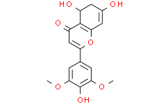
| Chemical Name | 5,7-dihydroxy-2-(4-hydroxy-3,5-dimethoxyphenyl)chromen-4-one |
| Synonyms | Tricin |
| HS Tariff Code | 2934.99.9001 |
| Storage |
Powder-20°C 3 years 4°C 2 years In solvent -80°C 6 months -20°C 1 month Note: This product requires protection from light (avoid light exposure) during transportation and storage. |
| Shipping Condition | Room temperature (This product is stable at ambient temperature for a few days during ordinary shipping and time spent in Customs) |
Biological Activity
| Targets |
- Inhibition of CCL5 induction required for human cytomegalovirus (HCMV) growth [1] - Cyclin-dependent kinase 9 (CDK9): The half-maximal inhibitory concentration (IC50) of Tricin against CDK9/cyclin T1 kinase activity is 0.8 μM [2] - Focal Adhesion Kinase (FAK): Tricin indirectly inhibits FAK by upregulating microRNA-7 (miR-7) that targets FAK [3] |
| ln Vitro |
In Madin-Darby canine kidney (MDCK) cells treated with influenza virus HA and M messenger RNA, tricin inhibits the expression of hemagglutinin (HA) protein and matrix (M) protein [4]. In a dose-dependent manner, tricin (3.3-30 μM; 8 hours) dramatically lowers seasonal A (H1N1), (H3N2), new A (H1N1pdm), and B viruses [4]. Anti-HCMV activity via CCL5 inhibition: Human foreskin fibroblasts (MRC-5 cells) were infected with HCMV (MOI = 0.1). Treatment with Tricin (1 μM, 10 μM) for 72 hours significantly reduced HCMV-induced CCL5 mRNA expression (by ~50% at 1 μM and ~80% at 10 μM, compared to infected controls) and CCL5 protein secretion (by ~40% at 1 μM and ~70% at 10 μM). Additionally, Tricin (10 μM) decreased HCMV viral titer by ~1.5 log10 PFU/mL (measured via plaque assay) [1] Anti-HCMV activity via CDK9 inhibition: In HCMV-infected MRC-5 cells, Tricin (5 μM, 10 μM) inhibited CDK9/cyclin T1 kinase activity, which reduced phosphorylation of RNA polymerase II (RNAP II) at Ser2 (a marker of transcriptional elongation). This led to downregulation of HCMV immediate-early (IE) genes (IE1/72 and IE2/86) mRNA expression (by ~60% at 5 μM and ~90% at 10 μM) and IE1/72 protein levels (by ~50% at 5 μM and ~80% at 10 μM). In vitro kinase assays confirmed Tricin directly inhibited CDK9 with an IC50 of 0.8 μM [2] Anti-glioma activity via miR-7/FAK pathway: For C6 glioma cells: 1) Proliferation inhibition (MTT assay): Tricin (5 μM, 10 μM, 20 μM, 40 μM) treatment for 48 hours reduced cell viability in a dose-dependent manner, with an IC50 of ~25 μM; 2) Invasion inhibition (Transwell assay): Tricin (20 μM, 40 μM) decreased the number of invasive cells by ~40% and ~70%, respectively, compared to controls; 3) Molecular regulation: Tricin (20 μM, 40 μM) upregulated miR-7 expression (by ~2-fold and ~3.5-fold) and downregulated FAK mRNA (by ~30% and ~60%) and protein (by ~25% and ~55%) levels. It also increased apoptotic cell rate (by ~15% at 20 μM and ~30% at 40 μM, detected via flow cytometry) by activating caspase-3 (protein level increased by ~2-fold at 40 μM) [3] |
| ln Vivo |
In mice, tricin (50–250 ppm diet; 15 weeks) dramatically lowers the overall tumor burden and adenocarcinoma count [5]. Anti-glioma efficacy in xenograft models: Nude mice (6-8 weeks old) were subcutaneously inoculated with C6 glioma cells (1×10⁶ cells/mouse) to establish tumor xenografts. When tumors reached ~100 mm³, mice were divided into two groups: 1) Control group (treated with vehicle: 0.1% DMSO in normal saline); 2) Tricin treatment group (50 mg/kg, dissolved in 0.1% DMSO-normal saline). Mice were administered intraperitoneally 3 times per week for 21 days. Tricin significantly inhibited tumor growth: tumor volume in the treatment group was ~40% smaller than that in the control group at day 21, and tumor weight was ~35% lower at the end of the experiment. Immunohistochemical staining of tumor tissues showed FAK protein expression was reduced by ~50% in the Tricin group, and qRT-PCR confirmed miR-7 expression was ~2.5-fold higher than in controls. No significant weight loss or abnormal behavior was observed in Tricin-treated mice [3] |
| Enzyme Assay |
CDK9/cyclin T1 kinase activity assay: Recombinant human CDK9/cyclin T1 complex, ATP (10 μM), and a synthetic carboxy-terminal domain (CTD) peptide of RNAP II (substrate) were mixed in kinase buffer. Different concentrations of Tricin (0.1 μM, 0.5 μM, 1 μM, 5 μM, 10 μM) were added to the reaction system, and the mixture was incubated at 37°C for 60 minutes. The amount of phosphorylated CTD peptide (product) was detected using a specific antibody against phosphorylated Ser2 of CTD via enzyme-linked immunosorbent assay (ELISA). The absorbance at 450 nm was measured, and the IC50 value of Tricin against CDK9 was calculated by fitting the dose-response curve [2] |
| Cell Assay |
HCMV infection and CCL5 detection assay (MRC-5 cells): MRC-5 cells were cultured in DMEM supplemented with 10% fetal bovine serum (FBS) and seeded in 6-well plates (1×10⁵ cells/well). After 24 hours, cells were infected with HCMV (MOI = 0.1) for 2 hours, then Tricin (0.1 μM, 1 μM, 10 μM) was added. After 72 hours of incubation: 1) Total RNA was extracted, and CCL5 mRNA expression was measured via quantitative real-time PCR (qRT-PCR) using specific primers; 2) Culture supernatant was collected, and CCL5 protein concentration was detected via sandwich ELISA; 3) Viral titer was determined via plaque assay on MRC-5 cells (fixed with formaldehyde, stained with crystal violet, and plaques counted) [1] CDK9 and HCMV gene detection assay (MRC-5 cells): MRC-5 cells were infected with HCMV (MOI = 0.1) and treated with Tricin (5 μM, 10 μM) for 48 hours. 1) Total protein was extracted, and phosphorylated RNAP II (Ser2) and HCMV IE1/72 protein levels were detected via Western blot using specific primary antibodies and horseradish peroxidase (HRP)-conjugated secondary antibodies (signal visualized via chemiluminescence); 2) Total RNA was extracted, and IE1/72 and IE2/86 mRNA levels were measured via qRT-PCR [2] C6 glioma cell functional assays: 1) MTT assay: C6 cells were seeded in 96-well plates (5×10³ cells/well) and treated with Tricin (5 μM, 10 μM, 20 μM, 40 μM) for 48 hours. MTT reagent was added, incubated for 4 hours, then dimethyl sulfoxide (DMSO) was added to dissolve formazan crystals; absorbance at 570 nm was measured to calculate cell viability. 2) Transwell invasion assay: C6 cells (1×10⁴ cells/well) treated with Tricin (20 μM, 40 μM) were seeded in the upper chamber of Transwell inserts (coated with Matrigel), and medium containing 10% FBS was added to the lower chamber. After 24 hours, non-invasive cells on the upper surface were removed, invasive cells on the lower surface were fixed and stained, and counted under a microscope. 3) qRT-PCR and Western blot: C6 cells treated with Tricin (20 μM, 40 μM) for 48 hours were used to extract total RNA (for miR-7 and FAK mRNA detection via qRT-PCR) and total protein (for FAK, phosphorylated FAK, and caspase-3 detection via Western blot) [3] |
| Animal Protocol |
C6 glioma xenograft experiment in nude mice: 1) Tumor establishment: 6-8 week-old male nude mice were anesthetized, and 100 μL of cell suspension containing 1×10⁶ C6 glioma cells was subcutaneously injected into the right dorsal flank. 2) Grouping and treatment: When tumors grew to ~100 mm³, mice were randomly divided into two groups (n = 6 per group): Control group (intraperitoneal injection of 0.1% DMSO in normal saline, 0.2 mL/mouse) and Tricin group (intraperitoneal injection of Tricin dissolved in 0.1% DMSO-normal saline at 50 mg/kg, 0.2 mL/mouse). Injections were administered 3 times per week for 21 consecutive days. 3) Sample collection and detection: Every 3 days, tumor volume (calculated as V = length × width² / 2) and mouse body weight were measured. After 21 days, mice were euthanized, tumor tissues were excised and weighed. Part of the tumor tissue was fixed in formalin for immunohistochemical staining (to detect FAK protein), and the remaining tissue was used for total RNA extraction (to detect miR-7 expression via qRT-PCR) [3] |
| Toxicity/Toxicokinetics |
- In the in vivo C6 glioma xenograft experiment, Tricin (50 mg/kg, intraperitoneal injection, 3 times/week for 21 days) did not cause significant changes in mouse body weight (no statistically significant difference compared to the control group) or obvious abnormal behaviors (e.g., reduced activity, poor appetite) [3] |
| References |
[1]. Tricin inhibits the CCL5 induction required for efficient growth of human cytomegalovirus. Microbiol Immunol. 2018 May;62(5):341-347. [2]. The anti-human cytomegalovirus drug tricin inhibits cyclin-dependent kinase 9. FEBS Open Bio. 2018 Feb 20;8(4):646-654. [3]. Inhibition of the Proliferation and Invasion of C6 Glioma Cells by Tricin via the Upregulation of Focal-Adhesion-Kinase-Targeting MicroRNA-7. J Agric Food Chem. 2018 Jul 5;66(26):6708-6716. |
| Additional Infomation |
3',5'-di-O-methyltricetin is the 3',5'-di-O-methyl ether of tricetin. Known commonly as tricin, it is a constituent of rice bran and has been found to potently inhibit colon cancer cell growth. It has a role as an EC 1.14.99.1 (prostaglandin-endoperoxide synthase) inhibitor and a metabolite. It is a trihydroxyflavone, a dimethoxyflavone and a member of 3'-methoxyflavones. It is functionally related to a tricetin. It is a conjugate acid of a 3',5'-di-O-methyltricetin(1-). Tricin has been reported in Trichoderma virens, Crocus heuffelianus, and other organisms with data available. See also: Arnica montana Flower (part of); Elymus repens root (part of). - Tricin exerts anti-HCMV activity by targeting two distinct pathways: inhibiting CCL5 induction (which is essential for efficient HCMV growth) [1] and suppressing CDK9 activity (which is required for HCMV gene transcription) [2]. This dual mechanism suggests Tricin may be a potential candidate for HCMV infection treatment [1,2] - In glioma, FAK overexpression promotes cell proliferation and invasion. Tricin modulates the miR-7/FAK axis: upregulating miR-7 (a tumor suppressor miRNA that directly targets FAK mRNA) to downregulate FAK expression, thereby inhibiting C6 glioma cell proliferation and invasion. This indicates Tricin may have therapeutic potential for glioma [3] |
Solubility Data
| Solubility (In Vitro) | DMSO : ~125 mg/mL (~378.46 mM) |
| Solubility (In Vivo) |
Solubility in Formulation 1: ≥ 2.08 mg/mL (6.30 mM) (saturation unknown) in 10% DMSO + 40% PEG300 +5% Tween-80 + 45% Saline (add these co-solvents sequentially from left to right, and one by one), clear solution. For example, if 1 mL of working solution is to be prepared, you can add 100 μL of 20.8 mg/mL clear DMSO stock solution to 400 μL PEG300 and mix evenly; then add 50 μL Tween-80 + to the above solution and mix evenly; then add 450 μL normal saline to adjust the volume to 1 mL. Preparation of saline: Dissolve 0.9 g of sodium chloride in 100 mL ddH₂ O to obtain a clear solution. (Please use freshly prepared in vivo formulations for optimal results.) |
| Preparing Stock Solutions | 1 mg | 5 mg | 10 mg | |
| 1 mM | 3.0276 mL | 15.1382 mL | 30.2764 mL | |
| 5 mM | 0.6055 mL | 3.0276 mL | 6.0553 mL | |
| 10 mM | 0.3028 mL | 1.5138 mL | 3.0276 mL |