Tirbanibulin dihydrochloride (also known as KXO1 dihydrochloride and KX2-391 dihydrochloride) is a dual Src/tubulin inhibitor approved in 2020 for the treatment of actinic keratosis on the face or scalp.
Physicochemical Properties
| Molecular Formula | C₂₆H₃₁CL₂N₃O₃ | |
| Molecular Weight | 504.45 | |
| Exact Mass | 503.174 | |
| Elemental Analysis | C, 61.23; H, 5.96; Cl, 14.46; N, 8.57; O, 9.79 | |
| CAS # | 1038395-65-1 | |
| Related CAS # | Tirbanibulin;897016-82-9;Tirbanibulin Mesylate;1080645-95-9 | |
| PubChem CID | 24989633 | |
| Appearance | Light yellow to yellow solid powder | |
| LogP | 5.7 | |
| Hydrogen Bond Donor Count | 3 | |
| Hydrogen Bond Acceptor Count | 5 | |
| Rotatable Bond Count | 9 | |
| Heavy Atom Count | 34 | |
| Complexity | 540 | |
| Defined Atom Stereocenter Count | 0 | |
| InChi Key | CPTPOZGQCQXHJO-UHFFFAOYSA-N | |
| InChi Code | InChI=1S/C26H29N3O3.2ClH/c30-26(28-19-21-4-2-1-3-5-21)18-24-9-6-23(20-27-24)22-7-10-25(11-8-22)32-17-14-29-12-15-31-16-13-29;;/h1-11,20H,12-19H2,(H,28,30);2*1H | |
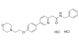
| Chemical Name | N-benzyl-2-[5-[4-(2-morpholin-4-ylethoxy)phenyl]pyridin-2-yl]acetamide;dihydrochloride | |
| Synonyms |
|
|
| HS Tariff Code | 2934.99.9001 | |
| Storage |
Powder-20°C 3 years 4°C 2 years In solvent -80°C 6 months -20°C 1 month Note: Please store this product in a sealed and protected environment (e.g. under nitrogen), avoid exposure to moisture. |
|
| Shipping Condition | Room temperature (This product is stable at ambient temperature for a few days during ordinary shipping and time spent in Customs) |
Biological Activity
| Targets | Src HuH7 (GI50 = 9 nM); Src PLC/PRF/5 (IC50 = 13 nM); Src Hep3B (IC50 = 26 nM); Src HepG2 (IC50 = 60 nM) | |
| ln Vitro |
|
|
| ln Vivo |
|
|
| Enzyme Assay | The Src inhibitor tirbanibulin (KX2-391) targets the Src substrate pocket. The hepatic cell cancer (HCC) cell lines Huh7 (GI50=9 nM), PLC/PRF/5 (GI50=13 nM), Hep3B (GI50=26 nM), and HepG2 (GI50=60 nM) exhibit steep dose-response curves when treated with tirbanibulin (KX2-391). | |
| Cell Assay | Hep3B, HepG2, PLC/PRF/5, Huh7, and other liver cell lines are frequently grown and kept in basal medium with 2% fetal bovine serum (FBS) at 37°C and 5% CO2. In each well of a 96-well plate, cells are seeded at 4.0×10³/190 μL and 8.0×10³/190 μL in basal medium containing 1.5% FBS. Before adding Tirbanibulin (KX2-391) at concentrations ranging from 6,564 to 0.012 nM in triplicates, these are cultured for an additional night at 37°C and 5% CO2. Three days are spent incubating treated cells. On day three, 10 μL of a 5-mg/mL 3-(4,5-dimethylthiazol-2-yl)-2,5-diphenyltetrazolium bromide (MTT) solution is added to each well, and the cells are incubated for four hours. 10% SDS is added to diluted HCl to dissolve the formazan product. The optical density is measured at 570 nm. Parallel experiments are conducted with Tirbanibulin (KX2-391) to compare its potency and activity. With GraphPad Prism 5 statistical software, growth inhibition curves, 50% inhibition concentration (GI50), and 80% inhibition concentration (GI80) are calculated. Both the optical density at wavelength of 570 nm (OD570) signal format and the normalized data representing the percentage of maximum response are reported. | |
| Animal Protocol |
Mouse bearing MDA-MB-231 tumors; Oral gavage; 1, 5mg/kg dose Xenograft procedures and KX-01 oral dosing were as described (11). Briefly, mammary fat pad tumors were established by injecting 5×10⁶ MDA-MB-231 cells in 150μl of PBS-Matrigel mixture (1:2) orthotopically and bilaterally into the mammary fat pads of female NUDE mice (two tumors/mouse). Treatments were started when tumors reached ∼80-100mm3. The first study used MDA-MB-231 xenografts and was performed using vehicle (ultra-pure water) and two doses of KX-01 (1, 5mg/kg) administered twice/day (BID) by oral gavage (using metal 22g feeding needle) for 28 days. A similar experiment was performed with MDA-MB-157 xenografts (another ER/PR/HER2 negative model) to assess KX-01 response. A second study was performed to test combination of KX-01 with paclitaxel on tumor growth. MDA-MD-231 tumor xenograft bearing mice were treated with vehicle or KX-01 (5mg/kg) BID, paclitaxel by intraperitoneal injection (IP) once/week, or combination of KX-0+paclitaxel. Treatments were for 40 days for all groups. A third study used MDA-MB-157 xenografts with the same combination treatment. A fourth study tested the effect of KX-01 or combination with paclitaxel for 24 days on larger MDA-MB-231 tumors (∼300mm3). Tumors were allowed to reach ∼300mm3 before beginning treatments. In this experiment mice were treated with KX-01 at a higher dose of 15mg/kg, and mice were treated once/day instead of twice/day. Paclitaxel was used at a dose of 20mg/kg IP once/week. In all experiments, tumor caliper measurements were taken twice/week and tumor volume was by calculated by the formula: 0.523×LM2 (where L-large diameter, M-small diameter). At the end the experiments animals were sacrificed and tumors and mouse organs removed. Tissues were either stored in 10% neutral buffered formalin for paraffin embedding, or snap frozen for measurement of chromosome-17 by real-time PCR, and embedded for frozen sectioning for CD-31 staining. Immunohistochemistry (IHC) was performed as described on paraffin-embedded tumor tissues [3]. |
|
| References |
[1]. Expression of Src and FAK in hepatocellular carcinoma and the effect of Src inhibitors on hepatocellular carcinoma in vitro. Dig Dis Sci, 2009, 54(7), 1465-1474. [2]. Thiazolyl N-benzyl-substituted acetamide derivatives: synthesis, Src kinase inhibitory and anticancer activities. Eur J Med Chem, 2011, 46(10), 4853-4858. [3]. Peptidomimetic Src/pretubulin inhibitor KX-01 alone and in combination with paclitaxel suppresses growth, metastasis in human ER/PR/HER2-negative tumor xenografts. Mol Cancer Ther. 2012 Sep; 11(9): 1936–1947. |
Solubility Data
| Solubility (In Vitro) |
|
|||
| Solubility (In Vivo) |
Solubility in Formulation 1: ≥ 2.5 mg/mL (4.96 mM) (saturation unknown) in 10% DMSO + 40% PEG300 + 5% Tween80 + 45% Saline (add these co-solvents sequentially from left to right, and one by one), clear solution. For example, if 1 mL of working solution is to be prepared, you can add 100 μL of 25.0 mg/mL clear DMSO stock solution to 400 μL PEG300 and mix evenly; then add 50 μL Tween-80 to the above solution and mix evenly; then add 450 μL normal saline to adjust the volume to 1 mL. Preparation of saline: Dissolve 0.9 g of sodium chloride in 100 mL ddH₂ O to obtain a clear solution. Solubility in Formulation 2: ≥ 2.5 mg/mL (4.96 mM) (saturation unknown) in 10% DMSO + 90% (20% SBE-β-CD in Saline) (add these co-solvents sequentially from left to right, and one by one), clear solution. For example, if 1 mL of working solution is to be prepared, you can add 100 μL of 25.0 mg/mL clear DMSO stock solution to 900 μL of 20% SBE-β-CD physiological saline solution and mix evenly. Preparation of 20% SBE-β-CD in Saline (4°C,1 week): Dissolve 2 g SBE-β-CD in 10 mL saline to obtain a clear solution. Solubility in Formulation 3: ≥ 2.5 mg/mL (4.96 mM) (saturation unknown) in 10% DMSO + 90% Corn Oil (add these co-solvents sequentially from left to right, and one by one), clear solution. For example, if 1 mL of working solution is to be prepared, you can add 100 μL of 25.0 mg/mL clear DMSO stock solution to 900 μL of corn oil and mix evenly. Solubility in Formulation 4: 4% DMSO+30% PEG 300+ddH2O: 5 mg/mL (Please use freshly prepared in vivo formulations for optimal results.) |
| Preparing Stock Solutions | 1 mg | 5 mg | 10 mg | |
| 1 mM | 1.9824 mL | 9.9118 mL | 19.8236 mL | |
| 5 mM | 0.3965 mL | 1.9824 mL | 3.9647 mL | |
| 10 mM | 0.1982 mL | 0.9912 mL | 1.9824 mL |