Tirbanibulin Mesylate (also known as KXO-1 Mesylate and KX2-391 Mesylate) is a tubulin/Src inhibitor that received FDA approval in 2020 to treat scalp or facial actinic keratosis
Physicochemical Properties
| Molecular Formula | C27H33N3O6S |
| Molecular Weight | 527.63 |
| Exact Mass | 527.209 |
| Elemental Analysis | C, 61.46; H, 6.30; N, 7.96; O, 18.19; S, 6.08 |
| CAS # | 1080645-95-9 |
| Related CAS # | 1080645-95-9 (mesylate); 897016-82-9; 1038395-65-1 (HCl) |
| PubChem CID | 25100013 |
| Appearance | White to off-white solid powder |
| LogP | 4.232 |
| Hydrogen Bond Donor Count | 2 |
| Hydrogen Bond Acceptor Count | 8 |
| Rotatable Bond Count | 9 |
| Heavy Atom Count | 37 |
| Complexity | 633 |
| Defined Atom Stereocenter Count | 0 |
| SMILES | S(C([H])([H])[H])(=O)(=O)O[H].O1C([H])([H])C([H])([H])N(C([H])([H])C([H])([H])OC2C([H])=C([H])C(C3=C([H])N=C(C([H])=C3[H])C([H])([H])C(N([H])C([H])([H])C3C([H])=C([H])C([H])=C([H])C=3[H])=O)=C([H])C=2[H])C([H])([H])C1([H])[H] |
| InChi Key | JGSYRKUPDSSTCB-UHFFFAOYSA-N |
| InChi Code | InChI=1S/C26H29N3O3.CH4O3S/c30-26(28-19-21-4-2-1-3-5-21)18-24-9-6-23(20-27-24)22-7-10-25(11-8-22)32-17-14-29-12-15-31-16-13-29;1-5(2,3)4/h1-11,20H,12-19H2,(H,28,30);1H3,(H,2,3,4) |
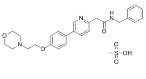
| Chemical Name | N-benzyl-2-[5-[4-(2-morpholin-4-ylethoxy)phenyl]pyridin-2-yl]acetamide;methanesulfonic acid |
| Synonyms | KXO1 HCl; KX-01, KX 01 HCl; KXO1 HCl; Mesylate; KX-01, KX 01 Mesylate; KX2-391; KX-2-391 Mesylate; KX 2-391; KX2391; KX-2391; KX 2391 Mesylate |
| HS Tariff Code | 2934.99.03.00 |
| Storage |
Powder-20°C 3 years 4°C 2 years In solvent -80°C 6 months -20°C 1 month Note: (1). This product requires protection from light (avoid light exposure) during transportation and storage.(2). Please store this product in a sealed and protected environment (e.g. under nitrogen), avoid exposure to moisture. |
| Shipping Condition | Room temperature (This product is stable at ambient temperature for a few days during ordinary shipping and time spent in Customs) |
Biological Activity
| Targets |
Src, in HuH7 cells (GI50 = 9 nM); Src, in PLC/PRF/5 cells (IC50 = 13 nM); Src, in Hep3B cells (IC50 = 26 nM); Src, in HepG2 cells (IC50 = 60 nM) Tirbanibulin Mesylate, also known as KX2-391 Mesylate, is a substrate-specific Src inhibitor. The hepatic cell cancer (HCC) cell lines Huh7 (GI50=9 nM), PLC/PRF/5 (GI50=13 nM), Hep3B (GI50=26 nM), and HepG2 (GI50=60 nM) exhibit steep dose-response curves when exposed to tirbanibulin (KX2-391)[1]. It has been discovered that tirbanibulin mesylate (KX2-391 mesylate) inhibits some leukemia cells, such as those derived from chronic leukemia cells with the T3151 mutation, that are resistant to currently available commercial drugs. In NICH3T3/c-Src527F and SYF/c-Src527F cells, tirbanibulin mesylate (KX2-391 mesylate) is assessed in engineered Src driven cell growth assays and displays GI50 values of 23 nM and 39 nM, respectively[2]. |
| ln Vitro |
Tirbanibulin Mesylate, also known as KX2-391 Mesylate, is a substrate-specific Src inhibitor. The hepatic cell cancer (HCC) cell lines Huh7 (GI50=9 nM), PLC/PRF/5 (GI50=13 nM), Hep3B (GI50=26 nM), and HepG2 (GI50=60 nM) exhibit steep dose-response curves when exposed to tirbanibulin (KX2-391)[1]. It has been discovered that tirbanibulin mesylate (KX2-391 mesylate) inhibits some leukemia cells, such as those derived from chronic leukemia cells with the T3151 mutation, that are resistant to currently available commercial drugs. In NICH3T3/c-Src527F and SYF/c-Src527F cells, tirbanibulin mesylate (KX2-391 mesylate) is assessed in engineered Src driven cell growth assays and displays GI50 values of 23 nM and 39 nM, respectively[2]. KX-01 at varying concentrations (10-250 nmol/L) inhibited the in vitro growth of ER/PR/HER2-negative human breast cancer cell lines MDA-MB-231, MDA-MB-157, and MDA-MB-468 after 48 hours of incubation. [3] In dasatinib-resistant MDA-MB-468 cells, KX-01 (at the same concentration range) inhibited growth by approximately 75%, whereas dasatinib inhibited growth by only about 20%. [3] KX-01 (25 nmol/L) induced significant apoptosis in MDA-MB-468 cells, whereas dasatinib (250 nmol/L) did not. [3] Co-incubation of KX-01 (25 nmol/L) with paclitaxel (5 nmol/L) for 24 hours resulted in synergistic growth inhibition (43.2% inhibition) of MDA-MB-231 cells, compared to modest inhibition by each drug alone (11.2% for KX-01, 14.1% for paclitaxel). Combination Index (CI) values calculated over a concentration range indicated synergism (CI < 1.0). Similar synergistic patterns were observed in MDA-MB-157 and MDA-MB-468 cells. [3] Combination of KX-01 (25 nmol/L) with paclitaxel (5 nmol/L) enhanced apoptosis in MDA-MB-231 cells approximately two-fold compared to single drug treatments after 24 hours. [3] KX-01 (200 nmol/L, 24 hours) markedly inhibited microtubule formation in MDA-MB-231 cells in vitro, as observed by immunofluorescence staining. [3] In a 3D invasion assay, treatment of MDA-MB-231 cells with KX-01 (25 and 50 nmol/L) reduced the formation of invasive stellate structures, with almost complete inhibition at 50 nmol/L. [3] In a Boyden chamber Matrigel invasion assay, KX-01 (50 nmol/L) significantly decreased MDA-MB-231 cell invasion by 52.9% after 24 hours, with data normalized to cell number to account for growth inhibition. [3] In a wound healing/scratch assay, KX-01 at 5, 10, and 25 nmol/L significantly decreased MDA-MB-231 cell migration by 11.7%, 15.6%, and 39.9%, respectively, after 24 hours compared to vehicle. [3] KX-01 (10, 25 nM) synergized with doxorubicin for cell growth inhibition in ER/PR/HER2-negative cell lines, but at higher concentrations (50 nM) the combination resulted in antagonism (CI > 1.0). [3] |
| ln Vivo |
Tirbanibulin Mesylate (KX2-391 Mesylate), when taken orally, has been demonstrated in pre-clinical animal models of cancer to inhibit the growth of primary tumors and to suppress metastasis[2]. In female athymic NUDE mice bearing orthotopic MDA-MB-231 xenograft tumors (~80-100 mm³), oral administration of KX-01 (1 mg/kg and 5 mg/kg, twice daily for 28 days) resulted in a dose-dependent reduction in tumor volume by 35.3% and 79.0%, respectively, compared to vehicle. [3] In the same model, oral administration of KX-01 (5 mg/kg, twice daily) alone for 40 days decreased tumor volume by 61.1%. When combined with paclitaxel (3 mg/kg, intraperitoneal injection once weekly), tumor volume was reduced by 93.0%, with complete regression of tumors observed in two out of five mice. [3] In mice bearing larger, established MDA-MB-231 tumors (~300 mm³), oral administration of a higher dose of KX-01 (15 mg/kg, once daily) alone resulted in significant tumor growth inhibition (48.2% volume decrease) without regression. Combination with a higher dose of paclitaxel (20 mg/kg, once weekly) resulted in 90.6% tumor regression, reducing volume below the starting point. [3] Similar antitumor effects were observed in MDA-MB-157 xenograft models with KX-01 alone and in combination with paclitaxel. [3] Immunohistochemical analysis of MDA-MB-231 tumors from mice treated with KX-01 showed significant inhibition of Src phosphorylation (P-Y416-Src) and its downstream target FAK phosphorylation (P-Y861-FAK), with no significant effect on total Src or FAK expression. [3] TUNEL assay showed that KX-01 treatment significantly enhanced the number of apoptotic cells (3-4 fold) in MDA-MB-231 tumors compared to vehicle. [3] KX-01 treatment reduced the percentage of Ki67-positive proliferating tumor cells (from 58.7% in vehicle to 37.3%) and modestly reduced microvessel density (assessed by CD31 staining) in MDA-MB-231 tumors. [3] Immunofluorescence staining of tumor sections revealed that KX-01 treatment disrupted the microtubule network, resulting in a diffuse staining pattern with reduced intensity, unlike the distinct microtubule fibers seen in vehicle-treated tumors or the intensely stabilized arrays seen in paclitaxel-treated tumors. The combination of KX-01 and paclitaxel resulted in a diffuse tubulin staining pattern with evidence of fragmented microtubules. [3] Quantitative real-time PCR analysis of human chromosome 17 DNA in mouse organs showed that KX-01 treatment alone (5 mg/kg) or in combination with paclitaxel (3 mg/kg) significantly reduced or eliminated detectable micrometastasis of MDA-MB-231 cells to mouse lungs and liver compared to vehicle after 40 days of treatment. No significant effect was observed on bone marrow metastasis. [3] |
| Enzyme Assay |
Tirbanibulin (KX2-391) is a Src inhibitor that targets the substrate pocket of Src. Tirbanibulin (KX2-391) exhibits sharp dose-response curves for four hepatic cell cancer (HCC) cell lines: Hep3B (GI50=26 nM), HepG2 (GI50=60 nM), PLC/PRF/5 (GI50=13 nM), and Huh7 (GI50=9 nM). The study utilized immunohistochemistry (IHC) on paraffin-embedded tumor tissues to assess the activity of Src and its downstream target FAK in vivo. Tumor sections were stained with antibodies specific for the phosphorylated (active) forms of Src (P-Y416-Src) and FAK (P-Y861-FAK), as well as their total protein levels. The staining intensity and distribution were evaluated to determine the inhibitory effect of KX-01 on these kinases. [3] |
| Cell Assay |
Regularly cultured and maintained in basal medium containing 2% fetal bovine serum (FBS) at 37°C and 5% CO2, liver cell lines such as Huh7, PLC/PRF/5, Hep3B, and HepG2 (NutriCyte, Buffalo, NY) are used. 1.5% FBS-containing basal medium is used to seed cells at 4.0×10³/190 μL and 8.0×10³/190 μL per well of a 96-well plate. KX2-391, at concentrations ranging from 6,564 to 0.012 nM in triplicates, is added after these are cultivated for an additional night at 37°C and 5% CO2. Incubation lasts three days for treated cells. Day 3 then sees the addition of 10 microliters of 3-(4,5-dimethylthiazol-2-yl)-2,5-diphenyltetrazolium bromide (MTT) solution (5 mg/mL) to each well, followed by a 4-hour incubation period. With 10% SDS in diluted HCl, the formazan product is dissolved. Using a BioTek Synergy HT multiplatform microplate reader, optical density at 570 nm is discovered. Parallel experiments using KX2-391 are carried out for comparison of potency and activity. A statistical program called GraphPad Prism 5 is used to calculate growth inhibition curves, 50% inhibition concentration (GI50), and 80% inhibition concentration (GI80). The data are presented in the optical density at wavelength of 570 nm (OD570) signal format and normalized to represent the percentage of maximum response. For cell growth inhibition assays (MTT assay), cells (MDA-MB-231, MDA-MB-157, MDA-MB-468) were incubated with vehicle or varying concentrations of KX-01, paclitaxel, doxorubicin, or dasatinib (5, 10, 25, 50, 100, 250 nmol/L) for 48 hours. Cell growth was expressed as a percentage of the vehicle control. [3] Apoptosis was evaluated using an ELISA-based DNA fragmentation assay. Cells were incubated with drugs for 24 hours, and apoptosis was quantified according to the assay protocol. [3] For the 3D invasion ("on-top") assay, cells were seeded on a layer of reconstituted extracellular matrix (Matrigel). KX-01 was added to the culture medium at the time of plating (10, 25, 50 nmol/L). The Matrigel-medium mixture and drug were replaced every 2 days for a total of 4 days. Invasive stellate structure formation was photographed and analyzed. [3] For the Boyden chamber Matrigel invasion assay, cells were incubated with KX-01 (10, 25, 50 nmol/L) for 24 hours in serum-free medium in the upper chamber. The lower chamber contained medium with serum as a chemoattractant. After incubation, non-invaded cells were removed from the upper surface, and invaded cells on the lower surface were fixed, stained, counted using imaging software, and normalized to cell viability. [3] For the scratch (wound healing) assay, a confluent monolayer of cells was scratched with a pipette tip to create a wound. Cells were then incubated with vehicle or KX-01 (5, 10, 25 nmol/L) for 24 hours. Wound closure was photographed at the beginning and end of incubation, and the gap area was measured using imaging software to calculate the percentage of gap closure. [3] For microtubule immunofluorescence in cells, MDA-MB-231 cells were incubated with 200 nmol/L KX-01 for 24 hours, then fixed and immunostained with an anti-alpha-tubulin antibody conjugated to a fluorescent dye. Fluorescence images were captured using a confocal microscope. [3] Synergy calculations for drug combinations were performed using dedicated software. Combination Index (CI) values were determined from dose-response data for single agents and their combinations. CI < 1.0 indicated synergism, CI = 1.0 indicated additivity, and CI > 1.0 indicated antagonism. [3] |
| Animal Protocol |
Mouse bearing MDA-MB-231 tumors; Oral gavage; 1, 5mg/kg dose Xenograft procedures and KX-01 oral dosing were as described (11). Briefly, mammary fat pad tumors were established by injecting 5×10⁶ MDA-MB-231 cells in 150μl of PBS-Matrigel mixture (1:2) orthotopically and bilaterally into the mammary fat pads of female NUDE mice (two tumors/mouse). Treatments were started when tumors reached ∼80-100mm3. The first study used MDA-MB-231 xenografts and was performed using vehicle (ultra-pure water) and two doses of KX-01 (1, 5mg/kg) administered twice/day (BID) by oral gavage (using metal 22g feeding needle) for 28 days. A similar experiment was performed with MDA-MB-157 xenografts (another ER/PR/HER2 negative model) to assess KX-01 response. A second study was performed to test combination of KX-01 with paclitaxel on tumor growth. MDA-MD-231 tumor xenograft bearing mice were treated with vehicle or KX-01 (5mg/kg) BID, paclitaxel by intraperitoneal injection (IP) once/week, or combination of KX-0+paclitaxel. Treatments were for 40 days for all groups. A third study used MDA-MB-157 xenografts with the same combination treatment. A fourth study tested the effect of KX-01 or combination with paclitaxel for 24 days on larger MDA-MB-231 tumors (∼300mm3). Tumors were allowed to reach ∼300mm3 before beginning treatments. In this experiment mice were treated with KX-01 at a higher dose of 15mg/kg, and mice were treated once/day instead of twice/day. Paclitaxel was used at a dose of 20mg/kg IP once/week. In all experiments, tumor caliper measurements were taken twice/week and tumor volume was by calculated by the formula: 0.523×LM2 (where L-large diameter, M-small diameter). At the end the experiments animals were sacrificed and tumors and mouse organs removed. Tissues were either stored in 10% neutral buffered formalin for paraffin embedding, or snap frozen for measurement of chromosome-17 by real-time PCR, and embedded for frozen sectioning for CD-31 staining. Immunohistochemistry (IHC) was performed as described on paraffin-embedded tumor tissues [3]. For xenograft studies, female athymic NUDE mice (BALB/c) aged 4-5 weeks were used. Orthotopic mammary fat pad tumors were established by injecting 5 × 10⁶ MDA-MB-231 or MDA-MB-157 cells suspended in a PBS-Matrigel mixture (1:2 ratio) bilaterally into the mammary fat pads. [3] Treatments were initiated when tumors reached approximately 80-100 mm³. For single-agent efficacy studies, mice were treated with vehicle (ultra-pure water) or KX-01 at 1 mg/kg or 5 mg/kg, administered twice daily (BID) by oral gavage for 28 days. Tumor volumes were measured twice weekly using calipers. [3] For combination studies with paclitaxel, mice bearing MDA-MB-231 or MDA-MB-157 xenografts were treated with: vehicle; KX-01 alone (5 mg/kg, BID, oral gavage); paclitaxel alone (3 mg/kg, intraperitoneal injection once weekly); or the combination of KX-01 and paclitaxel for 40 days. [3] A separate study tested the effect on larger, established tumors (~300 mm³). Mice were treated with: vehicle; KX-01 alone at a higher dose (15 mg/kg, once daily, oral gavage); paclitaxel alone at a higher dose (20 mg/kg, intraperitoneal injection once weekly); or the combination for 24 days. [3] For the assessment of metastasis, at the termination of the 40-day combination study, mouse organs (lungs, liver, bone marrow from femur) were collected, homogenized, and genomic DNA was extracted. Quantitative real-time PCR targeting a human-specific α-satellite DNA sequence on chromosome 17 was performed to detect and quantify human tumor cell DNA (micrometastasis) in mouse tissues. A standard curve was generated by spiking known numbers of human breast cancer cells into control mouse organs prior to DNA isolation. [3] KX-01 was provided as a methanesulfonic acid (MSA) salt powder, which was water-soluble. It was administered orally in ultra-pure water. Paclitaxel was administered intraperitoneally. [3] |
| Toxicity/Toxicokinetics |
In mice bearing MDA-MB-231 or MDA-MB-157 xenografts treated with KX-01 alone (5 mg/kg, BID) or in combination with paclitaxel (3 mg/kg, weekly) for up to 40 days, no noticeable toxicity or significant reduction in mouse body weight was observed compared to vehicle controls. [3] In the study with larger tumors using higher doses (KX-01 at 15 mg/kg once daily + paclitaxel at 20 mg/kg weekly), mice in the combination group showed approximately a 10% reduction in body weight by the end of the 24-day treatment (from a mean of 19.4 g to 17.6 g) but without other noticeable signs of toxicity. [3] |
| References |
[1]. Expression of Src and FAK in hepatocellular carcinoma and the effect of Src inhibitors on hepatocellular carcinoma in vitro. Dig Dis Sci, 2009, 54(7), 1465-1474. [2]. Thiazolyl N-benzyl-substituted acetamide derivatives: synthesis, Src kinase inhibitory and anticancer activities. Eur J Med Chem, 2011, 46(10), 4853-4858. [3]. Peptidomimetic Src/pretubulin inhibitor KX-01 alone and in combination with paclitaxel suppresses growth, metastasis in human ER/PR/HER2-negative tumor xenografts. Mol Cancer Ther. 2012 Sep; 11(9): 1936–1947. |
| Additional Infomation |
KX-01 (also referred to as KX2-391) is described as a 'first-in-class' peptidomimetic Src kinase inhibitor with an additional, distinct mechanism of action involving inhibition of microtubule polymerization (pretubulin inhibition). [3] It is orally bioavailable and had completed phase 1 clinical testing at the time of the study, with phase 2 trials ongoing for prostate cancer and a phase 1b trial for Acute Myeloid Leukemia. [3] The study proposes that the dual mechanism of action of KX-01 (Src inhibition + microtubule disruption) may provide superior antitumor activity compared to agents that inhibit Src alone, particularly for aggressive ER/PR/HER2-negative breast cancer. [3] The synergy observed with paclitaxel is suggested to stem from targeting microtubules via two different mechanisms: inhibition of assembly (by KX-01) and stabilization (by paclitaxel), potentially leading to fragmented and dysfunctional microtubules. [3] KX-01 was effective in a dasatinib-resistant cell line (MDA-MB-468), suggesting potential utility in tumors resistant to ATP-competitive Src inhibitors. [3] |
Solubility Data
| Solubility (In Vitro) |
|
|||
| Solubility (In Vivo) |
Solubility in Formulation 1: ≥ 2.5 mg/mL (4.74 mM) (saturation unknown) in 10% DMSO + 40% PEG300 + 5% Tween80 + 45% Saline (add these co-solvents sequentially from left to right, and one by one), clear solution. For example, if 1 mL of working solution is to be prepared, you can add 100 μL of 25.0 mg/mL clear DMSO stock solution to 400 μL PEG300 and mix evenly; then add 50 μL Tween-80 to the above solution and mix evenly; then add 450 μL normal saline to adjust the volume to 1 mL. Preparation of saline: Dissolve 0.9 g of sodium chloride in 100 mL ddH₂ O to obtain a clear solution. Solubility in Formulation 2: ≥ 2.5 mg/mL (4.74 mM) (saturation unknown) in 10% DMSO + 90% (20% SBE-β-CD in Saline) (add these co-solvents sequentially from left to right, and one by one), clear solution. For example, if 1 mL of working solution is to be prepared, you can add 100 μL of 25.0 mg/mL clear DMSO stock solution to 900 μL of 20% SBE-β-CD physiological saline solution and mix evenly. Preparation of 20% SBE-β-CD in Saline (4°C,1 week): Dissolve 2 g SBE-β-CD in 10 mL saline to obtain a clear solution. Solubility in Formulation 3: ≥ 2.5 mg/mL (4.74 mM) (saturation unknown) in 10% DMSO + 90% Corn Oil (add these co-solvents sequentially from left to right, and one by one), clear solution. For example, if 1 mL of working solution is to be prepared, you can add 100 μL of 25.0 mg/mL clear DMSO stock solution to 900 μL of corn oil and mix evenly. Solubility in Formulation 4: Formulation 1: ≥ 5 mg/mL (9.5 mM) in 4% DMSO+30% PEG 300+ddH2O Formulation 2: ≥ 2.5 mg/mL (4.7 mM) in 10% DMSO + 40% PEG300 + 5% Tween80 + + 45% Saline For example, if 1 mL of working solution is to be prepared, you can take 100 μL of 25 mg/mL of DMSO stock solution and add tO + 400 μL of PEG300, mix well (clear solution); Then add 50 μL of Tween 80 to the above solution, mix well (clear solution); Finally, add 450 μL of saline to the above solution, mix well (clear solution). Preparation of saline: Dissolve 0.9 g sodium chloride in 100 mL ddH ₂ O to obtain a clear solution. Formulation 3: ≥ 2.5 mg/mL (4.7 mM) in 10% DMSO + 90% (20% SBE-β-CD in saline) For example, if 1 mL of working solution is to be prepared, you can take 100 μL of 25 mg/mL of DMSO stock solution and add to 900 μL of 20% SBE-β-CD in saline, mix well (clear solution). Preparation of 20% SBE-β-CD in Saline (4°C,1 week): Dissolve 2 g SBE-β-CD in 10 mL saline to obtain a clear solution. Formulation 4: ≥ 2.5 mg/mL (4.7 mM) in 10% DMSO + 90% Corn oil For example, if 1 mL of working solution is to be prepared, you can take 100 μL of 25 mg/mL of DMSO stock solution and add to 900 μL of corn oil, mix well (clear solution). (Please use freshly prepared in vivo formulations for optimal results.) |
| Preparing Stock Solutions | 1 mg | 5 mg | 10 mg | |
| 1 mM | 1.8953 mL | 9.4763 mL | 18.9527 mL | |
| 5 mM | 0.3791 mL | 1.8953 mL | 3.7905 mL | |
| 10 mM | 0.1895 mL | 0.9476 mL | 1.8953 mL |