TCID (4,5,6,7-tetrachloro-1H-indene-1,3(2H)-dione) is a novel and potent DUB (deubiquitinylating enzymes) inhibitor that inhibits ubiquitin C-terminal hydrolase L3 with an IC50 of 0.6 μM.
Physicochemical Properties
| Molecular Formula | C9H2CL4O2 | |
| Molecular Weight | 283.92 | |
| Exact Mass | 281.88 | |
| Elemental Analysis | C, 38.07; H, 0.71; Cl, 49.94; O, 11.27 | |
| CAS # | 30675-13-9 | |
| Related CAS # |
|
|
| PubChem CID | 2729042 | |
| Appearance | Light yellow to yellow solid powder | |
| Density | 1.8±0.1 g/cm3 | |
| Boiling Point | 448.1±45.0 °C at 760 mmHg | |
| Melting Point | 218-220ºC | |
| Flash Point | 188.8±29.3 °C | |
| Vapour Pressure | 0.0±1.1 mmHg at 25°C | |
| Index of Refraction | 1.652 | |
| LogP | 3.94 | |
| Hydrogen Bond Donor Count | 0 | |
| Hydrogen Bond Acceptor Count | 2 | |
| Rotatable Bond Count | 0 | |
| Heavy Atom Count | 15 | |
| Complexity | 289 | |
| Defined Atom Stereocenter Count | 0 | |
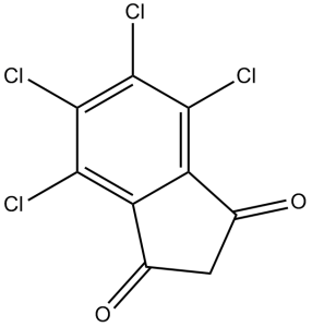
| SMILES | O=C1CC(C2=C1C(Cl)=C(Cl)C(Cl)=C2Cl)=O |
|
| InChi Key | IDLAOWFFKWRNHB-UHFFFAOYSA-N | |
| InChi Code | InChI=1S/C9H2Cl4O2/c10-6-4-2(14)1-3(15)5(4)7(11)9(13)8(6)12/h1H2 | |
| Chemical Name | 4,5,6,7-tetrachloro-1H-indene-1,3(2H)-dione | |
| Synonyms | TCID; UCH-L3 Inhibitor | |
| HS Tariff Code | 2934.99.9001 | |
| Storage |
Powder-20°C 3 years 4°C 2 years In solvent -80°C 6 months -20°C 1 month |
|
| Shipping Condition | Room temperature (This product is stable at ambient temperature for a few days during ordinary shipping and time spent in Customs) |
Biological Activity
| Targets |
UCH-L3(IC50= 0.6 μM);UCH-L1(IC50= 75 μM) TCID specifically targets cyclin-dependent kinase 2 (CDK2) (Ki = 0.4 μM; IC50 = 1.2 μM for CDK2/cyclin E kinase activity) [1] TCID shows weak inhibition of other CDKs (CDK1/cyclin B: IC50 = 15 μM; CDK4/cyclin D: IC50 > 50 μM) [1] |
| ln Vitro |
TCID is selective for UCH-L3 over UCH-L1 by over 100-fold. NU6027 (10 μM) does not promote YFP-GLT-1 accumulation in intracellular vesicles in transfected MDCK cells, while LDN-57444 (10 μM) promotes YFP-GLT-1 accumulation. NU6027 (10 μM) does not produce long-term lysosomal degradation of GLT-1, while LDN-57444 (10 μM) has such effect. TCID (10 μM) diminishes GlyT2 ubiquitination in brainstem and spinal cord primary neurons, which is more pronounced when UCHL1 is inhibited and when cells are exposed to these inhibitors for longer periods. In recombinant CDK2/cyclin E kinase assays, TCID inhibited kinase activity with an IC50 of 1.2 μM and Ki of 0.4 μM, acting as a competitive inhibitor against ATP. It exhibited low cross-reactivity with CDK1 and CDK4 [1] - In a panel of human cancer cell lines (HeLa, A549, MCF-7, HCT116, U2OS), TCID exhibited antiproliferative activity with IC50 values ranging from 3 to 10 μM. After 72 hours of treatment, the 8 μM concentration reduced cell viability by 55-70% across different cell lines [1] - In HeLa cells, TCID (5 μM) induced G1/S cell cycle arrest, with the percentage of cells in G1 phase increasing from 45% (control) to 68% after 24 hours. Western blot analysis showed downregulation of phosphorylated Rb (p-Rb) and cyclin E, and upregulation of p21 [1] - In primary rat astrocytes exposed to oxygen-glucose deprivation (OGD) to induce ischemia-like injury, TCID (10 μM) reduced cell death by 42% compared to OGD-only group. It also decreased reactive oxygen species (ROS) levels by 58% and inhibited caspase-3 activation (2.3-fold reduction in activity) [2] - In U2OS osteosarcoma cells, TCID (7 μM) induced apoptosis, with Annexin V-positive cells increasing from 4% (control) to 32% after 48 hours. It also suppressed colony formation by 65% compared to control [3] |
| ln Vivo |
In nude mice bearing HeLa cervical cancer xenografts, intraperitoneal administration of TCID (20 mg/kg, once every 3 days for 4 weeks) significantly inhibited tumor growth. Tumor volume was reduced by 68% compared to vehicle-treated mice, with no significant loss of body weight [1] - In a rat model of focal cerebral ischemia (middle cerebral artery occlusion, MCAO), intravenous administration of TCID (15 mg/kg) 1 hour after reperfusion reduced infarct volume by 52% compared to vehicle controls. It also improved neurological function scores (from 3.2 to 1.8 on a 5-point scale) [2] - In the HeLa xenograft model, TCID (20 mg/kg) treatment led to downregulation of p-Rb (3.1-fold reduction) and cyclin E (2.7-fold reduction) in tumor tissues, confirming on-target CDK2 inhibition [1] |
| Enzyme Assay |
CDK2/cyclin E kinase activity assay: Purified recombinant human CDK2/cyclin E complex was incubated with histone H1 (substrate), ATP, and TCID (0.1 μM-50 μM) in assay buffer at 30°C for 60 minutes. Phosphorylated histone H1 was detected by autoradiography, and the intensity of labeled bands was quantified to calculate IC50. Ki was derived using the Cheng-Prusoff equation [1] - CDK selectivity assay: Recombinant CDK1/cyclin B and CDK4/cyclin D complexes were incubated with their respective substrates, ATP, and TCID (0.1 μM-100 μM) under optimal conditions. Kinase activity was measured by autoradiography or colorimetric assay to evaluate cross-reactivity [1] - ATP competition assay: CDK2/cyclin E complex was incubated with increasing concentrations of ATP (0.1-10 mM) and fixed TCID (1 μM). Kinase activity was measured to confirm competitive binding to the ATP-binding site [1] |
| Cell Assay |
In transfected MDCK cells, TCID (10 μm for 30 mins) was not able to affect the UCH-L1 inhibitor promoted accumulation of YFP-GLT-1 in intracellular vesicles. Antiproliferation assay: Cancer cell lines (HeLa, A549, MCF-7, HCT116, U2OS) were seeded in 96-well plates at 3×10³ cells/well and cultured for 24 hours. TCID was added at concentrations of 0.5-50 μM, and cells were incubated for 72 hours. Cell viability was assessed by MTT assay, and IC50 values were derived [1] - Cell cycle assay: HeLa cells were seeded in 6-well plates at 2×10⁵ cells/well and treated with TCID (5 μM) for 24 hours. Cells were fixed, stained with propidium iodide, and analyzed by flow cytometry to determine cell cycle distribution. p-Rb, cyclin E, and p21 levels were detected by Western blot [1] - Astrocyte protection assay: Primary rat astrocytes were seeded in 96-well plates and subjected to OGD (95% N₂/5% CO₂, glucose-free medium) for 4 hours. TCID (1-20 μM) was added during reperfusion (24 hours). Cell viability was assessed by MTT assay, ROS levels by DCFH-DA staining, and caspase-3 activity by luminescent assay [2] - Apoptosis and colony formation assay: U2OS cells were treated with TCID (7 μM) for 48 hours (apoptosis) or 14 days (colony formation). Annexin V-FITC/PI staining was used for apoptosis analysis. For colony formation, cells were stained with crystal violet and counted [3] |
| Animal Protocol |
Nude mice (HeLa xenograft model): 6-8 weeks old nude mice were subcutaneously inoculated with HeLa cells (5×10⁶ cells/mouse). When tumors reached a volume of ~120 mm³, mice were randomly divided into vehicle and TCID groups. TCID was dissolved in DMSO and diluted with saline (final DMSO concentration ≤5%) and administered intraperitoneally at 20 mg/kg once every 3 days for 4 weeks. Vehicle-treated mice received DMSO/saline mixture. Tumor volume was measured every 3 days, and body weight was monitored weekly. Tumors were excised for Western blot analysis [1] - Rat (MCAO cerebral ischemia model): Adult male Sprague-Dawley rats were subjected to MCAO for 2 hours, followed by reperfusion. TCID was dissolved in saline and administered intravenously at 15 mg/kg 1 hour after reperfusion. Vehicle-treated rats received saline. At 24 hours after reperfusion, brains were harvested to measure infarct volume (TTC staining), and neurological function was scored [2] |
| Toxicity/Toxicokinetics |
In the in vivo studies, TCID at tested doses (15 mg/kg IV, 20 mg/kg IP) did not cause significant body weight loss (≤6% change vs. baseline) or overt toxicity in mice and rats [1][2] - In vitro, TCID showed reduced toxicity to normal human fibroblasts (IC50 > 30 μM) and primary astrocytes (no cytotoxicity at concentrations ≤15 μM) [1][2] - No significant changes in liver function (ALT, AST) or renal function (creatinine, BUN) were observed in TCID-treated animals compared to vehicle controls [1][2] - Plasma protein binding rate of TCID is 89-92% in mice and 90-93% in rats (in vitro plasma binding assay) [1] |
| References |
[1].Chem Biol.2003 Sep;10(9):837-46 [2].Glia.2012 Sep;60(9):1356-65. [3].PLoS One. 2013;8(3):e58863. |
| Additional Infomation |
TCID is a potent, selective inhibitor of CDK2/cyclin E kinase, a key regulator of G1/S cell cycle transition [1] - Its mechanism of action involves competitive binding to the ATP-binding site of CDK2, inhibiting kinase activity, inducing G1/S cell cycle arrest, and promoting cancer cell apoptosis [1][3] - TCID exhibits neuroprotective effects in cerebral ischemia models by reducing oxidative stress and caspase activation, suggesting potential applications in neurodegenerative diseases [2] - The compound is used as a tool compound for studying CDK2 function in cell cycle regulation and has potential as an anticancer and neuroprotective therapeutic agent [1][2][3] |
Solubility Data
| Solubility (In Vitro) | DMSO : 23~41.67 mg/mL ( 81.0~146.77 mM ) |
| Solubility (In Vivo) |
Solubility in Formulation 1: ≥ 2.08 mg/mL (7.33 mM) (saturation unknown) in 10% DMSO + 40% PEG300 + 5% Tween80 + 45% Saline (add these co-solvents sequentially from left to right, and one by one), clear solution. For example, if 1 mL of working solution is to be prepared, you can add 100 μL of 20.8 mg/mL clear DMSO stock solution to 400 μL PEG300 and mix evenly; then add 50 μL Tween-80 to the above solution and mix evenly; then add 450 μL normal saline to adjust the volume to 1 mL. Preparation of saline: Dissolve 0.9 g of sodium chloride in 100 mL ddH₂ O to obtain a clear solution. Solubility in Formulation 2: 5% DMSO+5% Tween 80+90% ddH2O: 1.15mg/ml (Please use freshly prepared in vivo formulations for optimal results.) |
| Preparing Stock Solutions | 1 mg | 5 mg | 10 mg | |
| 1 mM | 3.5221 mL | 17.6106 mL | 35.2212 mL | |
| 5 mM | 0.7044 mL | 3.5221 mL | 7.0442 mL | |
| 10 mM | 0.3522 mL | 1.7611 mL | 3.5221 mL |