TAS-301 is a novel, potent and selective inhibitor of smooth muscle cell migration and proliferation and inhibits intimal thickening after balloon injury to rat carotid arteries. TAS-103 inhibits smooth muscle cell migration and proliferation. It also inhibits intimal thickening after balloon injury to rat carotid arteries. TAS-301 blocks receptor-operated calcium influx and inhibits rat vascular smooth muscle cell proliferation induced by basic fibroblast growth factor and platelet-derived growth factor. TAS-103 inhibits calcium-dependent signal transduction and cytoskeletal reorganization.
Physicochemical Properties
| Molecular Formula | C23H19NO3 |
| Molecular Weight | 357.4019 |
| Exact Mass | 357.136 |
| CAS # | 193620-69-8 |
| PubChem CID | 9885087 |
| Appearance | Light yellow to yellow solid powder |
| Density | 1.2±0.1 g/cm3 |
| Boiling Point | 573.2±50.0 °C at 760 mmHg |
| Flash Point | 300.5±30.1 °C |
| Vapour Pressure | 0.0±1.6 mmHg at 25°C |
| Index of Refraction | 1.630 |
| LogP | 4.72 |
| Hydrogen Bond Donor Count | 1 |
| Hydrogen Bond Acceptor Count | 3 |
| Rotatable Bond Count | 4 |
| Heavy Atom Count | 27 |
| Complexity | 530 |
| Defined Atom Stereocenter Count | 0 |
| InChi Key | KUEYYIJXBRWZIB-UHFFFAOYSA-N |
| InChi Code | InChI=1S/C23H19NO3/c1-26-17-11-7-15(8-12-17)21(16-9-13-18(27-2)14-10-16)22-19-5-3-4-6-20(19)24-23(22)25/h3-14H,1-2H3,(H,24,25) |
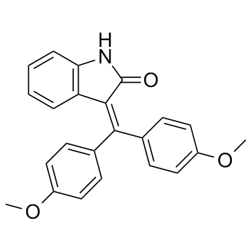
| Chemical Name | 3-[bis(4-methoxyphenyl)methylidene]-1H-indol-2-one |
| HS Tariff Code | 2934.99.9001 |
| Storage |
Powder-20°C 3 years 4°C 2 years In solvent -80°C 6 months -20°C 1 month |
| Shipping Condition | Room temperature (This product is stable at ambient temperature for a few days during ordinary shipping and time spent in Customs) |
Biological Activity
| Targets | PDGF-induced PKC activation and Ca2+ influx were both concentration-dependently decreased by TAS-301 (1–10 μM), which reduced PMA-induced AP-1 by 38 and inhibited PKC activation by 62.7% at 10 μM. % and 67.6% inhibition, respectively, were 3 and 10 μM[1]. The growth factor-induced cell migration (PDGF-BB, IGF-1, HB-EGF) is dose-dependently reduced by TAS-301 (0.3-3 μM). Additionally, TAS-301 (1–10 μM), particularly at 3 and 10 μM, decreases bFGF-induced BrdU incorporation [2]. |
| ln Vitro |
PDGF-induced PKC activation and Ca2+ influx were both concentration-dependently decreased by TAS-301 (1–10 μM), which reduced PMA-induced AP-1 by 38 and inhibited PKC activation by 62.7% at 10 μM. % and 67.6% inhibition, respectively, were 3 and 10 μM[1]. The growth factor-induced cell migration (PDGF-BB, IGF-1, HB-EGF) is dose-dependently reduced by TAS-301 (0.3-3 μM). Additionally, TAS-301 (1–10 μM), particularly at 3 and 10 μM, decreases bFGF-induced BrdU incorporation [2]. TAS-301 inhibited the proliferation of rat vascular smooth muscle cells stimulated by platelet-derived growth factor-BB, basic fibroblast growth factor, or 2% fetal bovine serum in a concentration-dependent manner (1-10 µM), as measured by bromodeoxyuridine incorporation assay. At 10 µM, it significantly inhibited DNA synthesis induced by PDGF-BB (53.3% inhibition), bFGF (72.2% inhibition), and 2% FBS (17.6% inhibition). [2] TAS-301 (1-10 µM) did not affect the PDGF-BB-induced activation of extracellular signal-regulated kinases in rat vascular smooth muscle cells. [2] TAS-301 (1-10 µM) inhibited PDGF-BB-induced activation of protein kinase C in a concentration-dependent manner in rat vascular smooth muscle cells, with significant inhibition (62.7%) at 10 µM. [2] TAS-301 (1-10 µM) inhibited phorbol 12-myristate 13-acetate-mediated induction of activator protein 1 transcriptional activity in a concentration-dependent manner in transfected rat vascular smooth muscle cells, with significant inhibition at 3 µM (38.0%) and 10 µM (67.6%). [2] TAS-301 (3-10 µM) significantly inhibited the PDGF-BB-induced unidirectional influx of calcium (⁴⁵Ca²⁺) into rat vascular smooth muscle cells. [2] The inhibitory effects of TAS-301 on cell proliferation were not due to nonspecific cytotoxicity, as assessed by cell viability (WST assay) and lactate dehydrogenase release assays at concentrations up to 10 µM. [2] |
| ln Vivo | In rats, TAS-301 (3–100 mg/kg, oral) decreases intimal cell numbers and I/M ratio in a dose-dependent manner, as well as neointimal thickening 14 days following balloon damage [2]. |
| Cell Assay |
For the cell proliferation assay, rat vascular smooth muscle cells were seeded in 96-well plates and growth-arrested in serum-free medium. Cells were pretreated with TAS-301 for 2 hours before stimulation with growth factors or serum. After 24 hours of stimulation, bromodeoxyuridine was added to the culture for another 24 hours. Cells were then fixed, and the incorporated bromodeoxyuridine was quantified using a specific enzyme-linked immunosorbent assay. [2] For measuring ERK activity, growth-arrested cells in 24-well plates were pretreated with compounds and stimulated with PDGF-BB. Cells were lysed, and ERK activity in the supernatant was measured using a commercial assay kit that quantifies the incorporation of radioactive phosphate from [γ-³³P]ATP into a synthetic ERK-specific peptide substrate. Radioactivity was measured by scintillation counting. [2] For measuring PKC activity, growth-arrested cells in 6-well plates were pretreated and stimulated. Cells were lysed, and cytosolic and particulate fractions were separated by centrifugation. The particulate fraction was solubilized, and PKC activity was measured using a commercial assay kit that quantifies the transfer of radioactive phosphate from [γ-³²P]ATP to a PKC-specific peptide substrate. [2] For the AP-1 luciferase reporter assay, rat vascular smooth muscle cells were transiently transfected with a luciferase reporter plasmid containing multiple AP-1 response elements. After transfection and recovery, cells were pretreated with TAS-301 for 2 hours and then stimulated with phorbol ester. Cells were lysed after a specific time, and luciferase activity was measured using a luminometer. Activity was expressed as fold increase over unstimulated cells. [2] For measuring unidirectional calcium influx, confluent, serum-starved cells in 6-well plates were pretreated with inhibitors and then stimulated with PDGF-BB. During the stimulation, radioactive calcium (⁴⁵Ca²⁺) was added to the medium for the final 5 minutes. Cells were then rapidly washed with ice-cold buffer, solubilized, and the incorporated radioactivity was measured by scintillation counting. The calcium influx rate was calculated based on the amount of radiolabel taken up per milligram of protein. [2] |
| Toxicity/Toxicokinetics |
In vitro, TAS-301 at concentrations up to 10 µM showed no significant effects on cell viability or lactate dehydrogenase release in rat vascular smooth muscle cells, indicating a lack of acute cytotoxicity at the tested concentrations. [2] The literature does not provide information on in vivo toxicity, median lethal dose, organ toxicity, drug-drug interactions, or plasma protein binding for TAS-301. [2] |
| References |
[1]. TAS-301, an inhibitor of smooth muscle cell migration and proliferation, inhibits intimal thickening after balloon injury to rat carotid arteries. J Pharmacol Exp Ther. 1998 Jun;285(3):1280-6. [2]. TAS-301 blocks receptor-operated calcium influx and inhibits rat vascular smooth muscle cell proliferation induced by basic fibroblast growth factor and platelet-derived growth factor. Jpn J Pharmacol. 2000 Nov;84(3):252-8. |
| Additional Infomation |
3-[bis(4-methoxyphenyl)methylidene]-1H-indol-2-one is a diarylmethane. TAS-301 (3-bis(4-methoxyphenyl)methylene-2-indolinone) is a synthetically derived compound. [2] The proposed mechanism of action for its anti-proliferative effect on vascular smooth muscle cells involves the blockade of receptor-operated (non-voltage-dependent) calcium influx triggered by growth factors like PDGF. This inhibition of calcium entry leads to the suppression of downstream signaling pathways, including the calcium/protein kinase C pathway, and ultimately inhibits the induction of the transcription factor complex activator protein 1, which is involved in cell cycle progression. [2] The study suggests that by inhibiting common mitogenic signals activated by multiple growth factors, TAS-301 has potential as a therapeutic agent for preventing vascular disorders characterized by smooth muscle cell proliferation, such as atherosclerosis and restenosis after angioplasty. [2] |
Solubility Data
| Solubility (In Vitro) | DMSO : ≥ 38 mg/mL (~106.32 mM) |
| Solubility (In Vivo) |
Note: Listed below are some common formulations that may be used to formulate products with low water solubility (e.g. < 1 mg/mL), you may test these formulations using a minute amount of products to avoid loss of samples. Injection Formulations (e.g. IP/IV/IM/SC) Injection Formulation 1: DMSO : Tween 80: Saline = 10 : 5 : 85 (i.e. 100 μL DMSO stock solution → 50 μL Tween 80 → 850 μL Saline) *Preparation of saline: Dissolve 0.9 g of sodium chloride in 100 mL ddH ₂ O to obtain a clear solution. Injection Formulation 2: DMSO : PEG300 :Tween 80 : Saline = 10 : 40 : 5 : 45 (i.e. 100 μL DMSO → 400 μLPEG300 → 50 μL Tween 80 → 450 μL Saline) Injection Formulation 3: DMSO : Corn oil = 10 : 90 (i.e. 100 μL DMSO → 900 μL Corn oil) Example: Take the Injection Formulation 3 (DMSO : Corn oil = 10 : 90) as an example, if 1 mL of 2.5 mg/mL working solution is to be prepared, you can take 100 μL 25 mg/mL DMSO stock solution and add to 900 μL corn oil, mix well to obtain a clear or suspension solution (2.5 mg/mL, ready for use in animals). Injection Formulation 4: DMSO : 20% SBE-β-CD in saline = 10 : 90 [i.e. 100 μL DMSO → 900 μL (20% SBE-β-CD in saline)] *Preparation of 20% SBE-β-CD in Saline (4°C,1 week): Dissolve 2 g SBE-β-CD in 10 mL saline to obtain a clear solution. Injection Formulation 5: 2-Hydroxypropyl-β-cyclodextrin : Saline = 50 : 50 (i.e. 500 μL 2-Hydroxypropyl-β-cyclodextrin → 500 μL Saline) Injection Formulation 6: DMSO : PEG300 : castor oil : Saline = 5 : 10 : 20 : 65 (i.e. 50 μL DMSO → 100 μLPEG300 → 200 μL castor oil → 650 μL Saline) Injection Formulation 7: Ethanol : Cremophor : Saline = 10: 10 : 80 (i.e. 100 μL Ethanol → 100 μL Cremophor → 800 μL Saline) Injection Formulation 8: Dissolve in Cremophor/Ethanol (50 : 50), then diluted by Saline Injection Formulation 9: EtOH : Corn oil = 10 : 90 (i.e. 100 μL EtOH → 900 μL Corn oil) Injection Formulation 10: EtOH : PEG300:Tween 80 : Saline = 10 : 40 : 5 : 45 (i.e. 100 μL EtOH → 400 μLPEG300 → 50 μL Tween 80 → 450 μL Saline) Oral Formulations Oral Formulation 1: Suspend in 0.5% CMC Na (carboxymethylcellulose sodium) Oral Formulation 2: Suspend in 0.5% Carboxymethyl cellulose Example: Take the Oral Formulation 1 (Suspend in 0.5% CMC Na) as an example, if 100 mL of 2.5 mg/mL working solution is to be prepared, you can first prepare 0.5% CMC Na solution by measuring 0.5 g CMC Na and dissolve it in 100 mL ddH2O to obtain a clear solution; then add 250 mg of the product to 100 mL 0.5% CMC Na solution, to make the suspension solution (2.5 mg/mL, ready for use in animals). Oral Formulation 3: Dissolved in PEG400 Oral Formulation 4: Suspend in 0.2% Carboxymethyl cellulose Oral Formulation 5: Dissolve in 0.25% Tween 80 and 0.5% Carboxymethyl cellulose Oral Formulation 6: Mixing with food powders Note: Please be aware that the above formulations are for reference only. InvivoChem strongly recommends customers to read literature methods/protocols carefully before determining which formulation you should use for in vivo studies, as different compounds have different solubility properties and have to be formulated differently. (Please use freshly prepared in vivo formulations for optimal results.) |
| Preparing Stock Solutions | 1 mg | 5 mg | 10 mg | |
| 1 mM | 2.7980 mL | 13.9899 mL | 27.9799 mL | |
| 5 mM | 0.5596 mL | 2.7980 mL | 5.5960 mL | |
| 10 mM | 0.2798 mL | 1.3990 mL | 2.7980 mL |