TAK-593 is a novel, orally bioavailable and potent small-molecule receptor of VEGFR and PDGFR with IC50s of 3.2, 0.95, 1.1, 4.3 and 13 nM for VEGFR1, VEGFR2, VEGFR3, PDFGRα and PDFGRβ, respectively. TAK-593 has potential antineoplastic activity. TAK-593 inhibits VEGFR and PDGFR through specific binding, which may stop angiogenesis and the growth of tumor cells.
Physicochemical Properties
| Molecular Formula | C23H23N7O3 |
| Molecular Weight | 445.4738 |
| Exact Mass | 445.186 |
| Elemental Analysis | C, 62.01; H, 5.20; N, 22.01; O, 10.77 |
| CAS # | 1005780-62-0 |
| Related CAS # | 1005780-62-0 |
| PubChem CID | 24767976 |
| Appearance | White solid powder |
| LogP | 3.618 |
| Hydrogen Bond Donor Count | 2 |
| Hydrogen Bond Acceptor Count | 6 |
| Rotatable Bond Count | 6 |
| Heavy Atom Count | 33 |
| Complexity | 735 |
| Defined Atom Stereocenter Count | 0 |
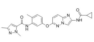
| SMILES | O=C(C1([H])C([H])([H])C1([H])[H])N([H])C1=C([H])N2C(C([H])=C([H])C(=N2)OC2C([H])=C([H])C(C([H])([H])[H])=C(C=2[H])N([H])C(C2=C([H])C(C([H])([H])[H])=NN2C([H])([H])[H])=O)=N1 |
| InChi Key | DZFZXPPHBWCXPQ-UHFFFAOYSA-N |
| InChi Code | InChI=1S/C23H23N7O3/c1-13-4-7-16(11-17(13)24-23(32)18-10-14(2)27-29(18)3)33-21-9-8-20-25-19(12-30(20)28-21)26-22(31)15-5-6-15/h4,7-12,15H,5-6H2,1-3H3,(H,24,32)(H,26,31) |
| Chemical Name | N-[5-[2-(cyclopropanecarbonylamino)imidazo[1,2-b]pyridazin-6-yl]oxy-2-methylphenyl]-2,5-dimethylpyrazole-3-carboxamide |
| Synonyms | TAK-593; TAK593; TAK 593 |
| HS Tariff Code | 2934.99.9001 |
| Storage |
Powder-20°C 3 years 4°C 2 years In solvent -80°C 6 months -20°C 1 month |
| Shipping Condition | Room temperature (This product is stable at ambient temperature for a few days during ordinary shipping and time spent in Customs) |
Biological Activity
| Targets |
VEGFR1 (IC50 = 3.2 nM); VEGFR2 (IC50 = 0.95 nM); VEGFR3 (IC50 = 1.1 nM); PDGFRα (IC50 = 4.3 nM); PDGFRβ (IC50 = 13 nM); PDGFRαV561D (IC50 = 1 nM) TAK-593 suppresses HUVEC growth with an IC50 of 0.30 nM. It exhibits strong inhibitory action against the PDGFR (PDGFRα, β: IC50=4.3, 13 nM) and VEGFR (VEGFR1-3: IC50=3.2, 0.95, 1.1 nM) families. TAK-593 exhibits IC50 values greater than 100 nM against all kinases, with the exception of Fms (IC50 = 10 nM) and Ret (IC50 = 18 nM) kinases [1]. Human umbilical vein endothelial cells and human coronary artery smooth muscle cells' cellular phosphorylation and proliferation are both potently inhibited by TAK-593 in response to VEGF and PDGF stimulation. Additionally, TAK-593 strongly suppresses the formation of tubes in co-cultured endothelial cells and fibroblasts caused by VEGF[2]. |
| ln Vitro |
TAK-593 suppresses HUVEC growth with an IC50 of 0.30 nM. It exhibits strong inhibitory action against the PDGFR (PDGFRα, β: IC50=4.3, 13 nM) and VEGFR (VEGFR1-3: IC50=3.2, 0.95, 1.1 nM) families. TAK-593 exhibits IC50 values greater than 100 nM against all kinases, with the exception of Fms (IC50 = 10 nM) and Ret (IC50 = 18 nM) kinases [1]. Human umbilical vein endothelial cells and human coronary artery smooth muscle cells' cellular phosphorylation and proliferation are both potently inhibited by TAK-593 in response to VEGF and PDGF stimulation. Additionally, TAK-593 strongly suppresses the formation of tubes in co-cultured endothelial cells and fibroblasts caused by VEGF[2]. TAK-593 potently inhibited VEGF-induced VEGFR2 phosphorylation in human umbilical vein endothelial cells (HUVECs) with an IC₅₀ of 0.34 nM. It also inhibited PDGF-BB-induced PDGFRβ phosphorylation in coronary artery smooth muscle cells (CASMCs) with an IC₅₀ of 2.1 nM. Consistent with this, TAK-593 inhibited VEGF-stimulated proliferation of HUVECs (IC₅₀ = 0.30 nM) and PDGF-BB-stimulated proliferation of CASMCs (IC₅₀ = 3.5 nM). In contrast, it showed much weaker inhibition of proliferation in various human cancer cell lines (A549, CFPAC-1, DU-145, HT-29, MDA-MB-231) and MRC-5 human fibroblasts, with IC₅₀ values ranging from 8.6 to 30 µM. In a tube formation assay mimicking angiogenesis, TAK-593 strongly inhibited VEGF-induced endothelial tube formation with an IC₅₀ of 0.32 nM.[2] |
| ln Vivo |
TAK-593 suppresses HUVEC growth with an IC50 of 0.30 nM. It exhibits strong inhibitory action against the PDGFR (PDGFRα, β: IC50=4.3, 13 nM) and VEGFR (VEGFR1-3: IC50=3.2, 0.95, 1.1 nM) families. TAK-593 exhibits IC50 values greater than 100 nM against all kinases, with the exception of Fms (IC50 = 10 nM) and Ret (IC50 = 18 nM) kinases [1]. Human umbilical vein endothelial cells and human coronary artery smooth muscle cells' cellular phosphorylation and proliferation are both potently inhibited by TAK-593 in response to VEGF and PDGF stimulation. Additionally, TAK-593 strongly suppresses the formation of tubes in co-cultured endothelial cells and fibroblasts caused by VEGF[2]. Oral administration of TAK-593 (twice daily) exhibited broad-spectrum anti-tumor activity in subcutaneous xenograft models of lung (A549), colon (HT-29), breast (MDA-MB-231), prostate (DU145), pancreas (CFPAC-1), renal (RCC-02-JCK), thyroid (TT), glioblastoma (U87MG), ovary (SK-OV-3), and gastric (MKN45) cancer. Doses as low as 0.25 mg/kg showed statistically significant tumor growth inhibition in most models. In an established A549 lung cancer model (larger tumors), treatment with TAK-593 at 0.125 and 0.25 mg/kg (BID) strongly inhibited growth, while doses of 1.5 and 3 mg/kg (BID) induced tumor regression. Treatment cessation led to regrowth, but re-initiation re-established efficacy. In an orthotopic U87MG glioblastoma model, TAK-593 (1 and 4 mg/kg, BID) significantly prolonged median survival time. Immunohistochemical analysis of A549 tumors showed that TAK-593 treatment significantly decreased tumor microvessel density (CD31 staining) and pericyte coverage (αSMA staining), while increasing tumor cell apoptosis (TUNEL assay) and decreasing proliferation (Ki-67 staining). Dynamic contrast-enhanced MRI (DCE-MRI) in HT-29 tumor-bearing mice revealed that TAK-593 significantly reduced tumor vascular permeability (Ktrans value) after 4 days of treatment.[2] |
| Enzyme Assay | In 50 mM TrisHCl pH 7.5, 5 mM MnCl2, 5 mM MgCl2, 0.01% Tween-20, and 2 mM DTT, with 10 μM ATP, 0.1 μg/mL biotinylated polyGluTyr (4:1), and 0.1 nM of VEGFR2, enzyme reactions are carried out. Preincubation involves incubating the compound (TAK-593) and enzyme for five minutes at room temperature before catalytic initiation with ATP. The reactions are stopped by adding 25 μL of 100 mM EDTA, 10 μg/mL donor and acceptor streptavidine beads in 62.5 mM HEPES pH 7.4, 250 mM NaCl, and 0.1% BSA. Plates are read by a plate reader after being incubated in the dark for the entire night[1]. |
| Cell Assay |
HUVECs are cultured at 37 C for an entire night in an incubator with 5% CO2 after being seeded at a density of 3000 cells/well in a 96-well plate using Human Endothelial-SFM Growth Medium (Invitrogen) supplemented with 3% fetal bovine serum (FBS). After adding the test compounds (TAK-593) at different concentrations and adding 60 ng/mL VEGF, the cells are cultured for an additional five days. The WST-8 formazan assay, which uses the Cell Counting Kit-8, is used to measure cellular proliferation[1]. For the receptor phosphorylation assay, HUVECs or CASMCs were treated with TAK-593 for 2 hours prior to stimulation with VEGF or PDGF-BB for 5 minutes. Cell lysates were then analyzed by western blotting using antibodies against phospho-VEGFR2, total VEGFR2, or PDGFRβ. Phospho-PDGFRβ was detected using an anti-phosphotyrosine antibody after immunoprecipitation with an anti-PDGFRβ antibody. For cell proliferation assays, HUVECs were treated with TAK-593 and VEGF for 5 days, CASMCs were starved and then treated with TAK-593 and PDGF-BB for 6 days, and cancer cell lines/fibroblasts were treated with TAK-593 for 3 days. Cell viability was determined using a colorimetric cell counting kit. For the tube formation assay, HUVECs were co-cultured with normal human dermal fibroblasts in the presence of VEGF and TAK-593 for 7 days. Endothelial cells were then stained with an anti-CD31 antibody and visualized by fluorescence imaging. The tube area was quantified using image analysis software.[2] |
| Animal Protocol |
Rats: Diethyl ether is used to induce anesthesia during the intravenous administration of medication to rats. Blood is drawn from the femoral vein in monkeys, and the tail vein in rats, at 5, 10, (only for IV dosing), 15, 30 min, and 1, 2, 3, 4, 6, 8, 12, 24, 32, and 48 hours (only for monkeys) following dosing. After that, the plasma fraction is extracted from the blood by centrifugation. Until analysis, the plasma is maintained frozen at 20°C. Utilizing a fluorescence detector and high-performance liquid chromatography, the concentration of TAK-593 in plasma is ascertained. The wavelengths of the excitation and emission are 346 and 420 nm, respectively. Mice: Test compounds are given to female BALB/cAJcl mice that have not been fasted at a dose of 10 mg/kg via cassette dosing. Blood samples are taken following oral administration. To extract the plasma fraction, the blood samples are centrifuged. Acetonitrile with an internal standard is used to deproteinize the plasma samples. Following centrifugation, the supernatant is diluted and then centrifuged once more using a mixture of acetonitrile (9:1, v/v) and 0.01 M ammonium formate solution. By using LC/MS/MS, the compound concentrations in the supernatant are determined[1]. TAK-593 was prepared as a 0.5% (w/v) methylcellulose suspension for oral administration. For subcutaneous xenograft efficacy studies, tumor cells or tissue fragments were implanted into the flank of athymic nude or SCID mice. When tumors reached a predetermined size (e.g., ~120-200 mm³), mice were randomized and orally administered TAK-593 or vehicle twice daily (BID) for 10-21 days. Tumor volumes and body weights were monitored regularly. For pharmacodynamic (PD) and pharmacokinetic (PK) analysis, A549 tumor-bearing nude mice were orally administered a single dose of TAK-593. Blood and tissue samples were collected at various time points for drug concentration measurement by HPLC-MS/MS or HPLC-fluorescence. For PD analysis, VEGF was injected intravenously 5 minutes before euthanasia, and lung tissues were collected for phospho-VEGFR2 analysis by western blot. For immunohistochemistry studies, A549 tumor-bearing mice were treated with TAK-593 BID for specified durations. Tumors were harvested, embedded, sectioned, and stained for CD31 (vessels), Ki-67 (proliferation), TUNEL (apoptosis), and αSMA (pericytes). For the orthotopic glioblastoma model, U87MG cells were injected intracranially. Mice were treated with TAK-593 BID starting 4 days post-inoculation and monitored for survival. For DCE-MRI studies, HT-29 tumor-bearing mice were treated with TAK-593 BID for 3 days. MRI scans were performed before and after treatment to assess changes in vascular permeability using a gadolinium-based contrast agent.[2] |
| ADME/Pharmacokinetics |
Following a single oral dose of 0.125 mg/kg in mice, TAK-593 achieved a maximum plasma concentration (Cmax) of 0.069 µg/mL at 15 minutes and an area under the plasma concentration-time curve from 0 to 24 hours (AUC₀₋₂₄) of 0.078 µg·h/mL. At a dose of 1 mg/kg, plasma Cmax was 0.451 µg/mL (at 1 hour) and AUC₀₋₂₄ was 0.883 µg·h/mL. In lung tissue, Cmax was 0.242 µg/mL and AUC₀₋₂₄ was 0.556 µg·h/mL. TAK-593 exhibited rapid absorption and clearance; it was undetectable in plasma or lung tissue of most mice by 8 hours after a 1 mg/kg dose. Despite rapid clearance from plasma, a single oral dose of 1 mg/kg led to almost complete suppression of VEGF-induced VEGFR2 phosphorylation in lung tissue for up to 8 hours, with recovery by 16 hours, indicating a long duration of target inhibition.[2] |
| Toxicity/Toxicokinetics |
In multiple xenograft studies, oral administration of TAK-593 at efficacious doses (up to 4 mg/kg twice daily) did not cause significant body weight loss or obvious adverse effects in mice, indicating good tolerability.[2] |
| References |
[1]. Discovery of N-[5-({2-[(cyclopropylcarbonyl)amino]imidazo[1,2-b]pyridazin-6-yl}oxy)-2-methylphenyl]-1,3-dimethyl-1H-pyrazole-5-carboxamide (TAK-593), a highly potent VEGFR2 kinase inhibitor. Bioorg Med Chem. 2013 Apr 15;21(8):2333-2345. [2]. Anti-angiogenic and anti-tumor effects of TAK-593, a potent and selective inhibitor of vascular endothelial growth factor and platelet-derived growth factor receptor tyrosine kinase. Cancer Sci. 2013 Apr;104(4):486-94. |
| Additional Infomation |
TAK-593 has been used in trials studying the treatment of Solid Tumors. VEGFR/PDGFR Tyrosine Kinase Inhibitor TAK-593 is an oral formulation containing a small-molecule receptor tyrosine kinase inhibitor of both vascular endothelial growth factor receptor (VEGFR) and platelet-derived growth factor receptor (PDGFR) with potential antineoplastic activity. TAK-593 selectively binds to and inhibits VEGFR and PDGFR, which may result in the inhibition of angiogenesis and tumor cell proliferation. TAK-593 is a novel imidazo[1,2-b]pyridazine derivative with a "pseudo-irreversible" or long-residence time mechanism of binding to VEGFR2 and PDGFRβ, contributing to its prolonged pharmacodynamic effect despite short systemic exposure. Its anti-tumor efficacy is primarily mediated through an anti-angiogenic mechanism, involving inhibition of endothelial cell signaling (VEGFR2), pericyte recruitment (PDGFRβ), leading to vessel regression, reduced vascular permeability, and subsequent tumor cell death. The study suggests that the dual inhibition of VEGF and PDGF signaling by TAK-593 may be more effective than VEGF inhibition alone in disrupting tumor vasculature.[2] |
Solubility Data
| Solubility (In Vitro) | DMSO: ≥ 48.5 mg/mL (~108.9 mM) |
| Solubility (In Vivo) |
Solubility in Formulation 1: ≥ 2.67 mg/mL (5.99 mM) (saturation unknown) in 10% DMSO + 40% PEG300 + 5% Tween80 + 45% Saline (add these co-solvents sequentially from left to right, and one by one), clear solution. For example, if 1 mL of working solution is to be prepared, you can add 100 μL of 26.7 mg/mL clear DMSO stock solution to 400 μL PEG300 and mix evenly; then add 50 μL Tween-80 to the above solution and mix evenly; then add 450 μL normal saline to adjust the volume to 1 mL. Preparation of saline: Dissolve 0.9 g of sodium chloride in 100 mL ddH₂ O to obtain a clear solution. Solubility in Formulation 2: ≥ 2.67 mg/mL (5.99 mM) (saturation unknown) in 10% DMSO + 90% (20% SBE-β-CD in Saline) (add these co-solvents sequentially from left to right, and one by one), clear solution. For example, if 1 mL of working solution is to be prepared, you can add 100 μL of 26.7 mg/mL clear DMSO stock solution to 900 μL of 20% SBE-β-CD physiological saline solution and mix evenly. Preparation of 20% SBE-β-CD in Saline (4°C,1 week): Dissolve 2 g SBE-β-CD in 10 mL saline to obtain a clear solution. Solubility in Formulation 3: ≥ 2.67 mg/mL (5.99 mM) (saturation unknown) in 10% DMSO + 90% Corn Oil (add these co-solvents sequentially from left to right, and one by one), clear solution. For example, if 1 mL of working solution is to be prepared, you can add 100 μL of 26.7 mg/mL clear DMSO stock solution to 900 μL of corn oil and mix evenly. (Please use freshly prepared in vivo formulations for optimal results.) |
| Preparing Stock Solutions | 1 mg | 5 mg | 10 mg | |
| 1 mM | 2.2448 mL | 11.2241 mL | 22.4482 mL | |
| 5 mM | 0.4490 mL | 2.2448 mL | 4.4896 mL | |
| 10 mM | 0.2245 mL | 1.1224 mL | 2.2448 mL |