Physicochemical Properties
| Molecular Formula | C7H14NO2 |
| Molecular Weight | 144.1916 |
| Exact Mass | 143.094 |
| CAS # | 471-87-4 |
| Related CAS # | Stachydrine hydrochloride;4136-37-2 |
| PubChem CID | 115244 |
| Appearance | White to off-white solid powder |
| Density | 1.1095 (rough estimate) |
| Boiling Point | 261.28°C (rough estimate) |
| Melting Point | 235°C |
| Index of Refraction | 1.4150 (estimate) |
| LogP | -2.97 |
| Hydrogen Bond Donor Count | 0 |
| Hydrogen Bond Acceptor Count | 2 |
| Rotatable Bond Count | 0 |
| Heavy Atom Count | 10 |
| Complexity | 148 |
| Defined Atom Stereocenter Count | 1 |
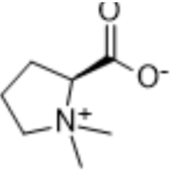
| SMILES | C[N+]1(CCC[C@H]1C(=O)[O-])C |
| InChi Key | CMUNUTVVOOHQPW-LURJTMIESA-N |
| InChi Code | InChI=1S/C7H13NO2/c1-8(2)5-3-4-6(8)7(9)10/h6H,3-5H2,1-2H3/t6-/m0/s1 |
| Chemical Name | (2S)-1,1-dimethylpyrrolidin-1-ium-2-carboxylate |
| HS Tariff Code | 2934.99.9001 |
| Storage |
Powder-20°C 3 years 4°C 2 years In solvent -80°C 6 months -20°C 1 month |
| Shipping Condition | Room temperature (This product is stable at ambient temperature for a few days during ordinary shipping and time spent in Customs) |
Biological Activity
| Targets |
- Stachydrine acts on signaling pathways related to myocardial cell hypertrophy (specific targets not specified) [1] - Stachydrine acts on pathways associated with anoxia-reoxygenation-induced human umbilical vein endothelial cell (HUVEC) injury (specific targets not specified) [2] - Stachydrine acts on antiproliferative-related pathways in prostate cancer cells (PC-3, DU-145; specific targets not specified) [3] - Stachydrine acts on pathways involved in norepinephrine-induced myocardial cell hypertrophy and intracellular calcium transients (specific targets not specified) [4] - Stachydrine acts on endoplasmic reticulum (ER) stress-related apoptotic pathways in rat kidney cells (specific targets not specified) [5] |
| ln Vitro |
Stachyine's anti-hypertrophic action may be associated with its ability to suppress the NF-κB signaling pathway. The levels of NF-κB protein in the nucleus and p-IκB protein in the cytoplasm are both markedly inhibited by strepthydrine intervention [1]. After stachydrine treatment, human umbilical vein endothelial cells exhibit a decrease in tissue factor mRNA. Stachyrine has the ability to mitigate the effects of hypoxia and reoxygenation-induced increases in LDH activity and decreases in human umbilical vein endothelial cell viability [2]. Stachydrine-treated human prostate cancer cells (PC-3 and LNcaP) showed a dose-dependent reduction in both protein levels and mRNA expression [3]. - For neonatal rat myocardial cells: Stachydrine (concentrations: 10, 50, 100 μM) inhibited norepinephrine-induced myocardial hypertrophy, as evidenced by downregulated mRNA expression of atrial natriuretic peptide (ANP) and brain natriuretic peptide (BNP) (detected via RT-PCR) and reduced hypertrophy-related morphological changes (observed via immunocytochemistry) [1] - For HUVECs: Stachydrine (concentrations: 10, 50, 100 μM) ameliorated anoxia-reoxygenation-induced injury, including reduced lactate dehydrogenase (LDH) release, increased superoxide dismutase (SOD) activity, decreased malondialdehyde (MDA) content, and improved cell viability (detected via MTT assay) [2] - For prostate cancer cells (PC-3, DU-145): Stachydrine (concentrations: 25, 50, 100, 200 μg/mL) inhibited cell proliferation in a dose-dependent manner, with IC50 values of 85 μg/mL (PC-3) and 92 μg/mL (DU-145) (detected via MTT assay); it also induced cell cycle arrest at G0/G1 phase (detected via flow cytometry) [3] - For neonatal rat myocardial cells: Stachydrine (concentrations: 10, 50, 100 μM) suppressed norepinephrine-induced hypertrophy (downregulated ANP, α-smooth muscle actin (α-SMA) mRNA via RT-PCR) and reduced the amplitude of intracellular calcium transients (detected via fluorescent calcium probe) [4] - For rat renal proximal tubule cells: Stachydrine (concentrations: 10, 50, 100 μM) inhibited ER stress-induced apoptosis, as shown by decreased apoptotic rate (flow cytometry) and downregulated protein expression of ER stress markers GRP78 and CHOP (detected via western blot) [5] |
| ln Vivo |
Stachyhydrine treatment can decrease the expression of PERK, CHOP, and caspase-3 in the endoplasmic reticulum stress-related apoptotic pathway [5]. Stachyhydrine can also potentially protect against β-adrenergic receptor-induced Ca2+ mishandling [4]. - For unilateral ureteral obstruction (UUO)-induced rat renal injury model: Stachydrine was administered via gavage at doses of 10, 20, 40 mg/kg once daily for 14 days. It reduced renal tissue apoptosis (detected via TUNEL staining) and downregulated renal GRP78, CHOP protein expression (western blot), thereby alleviating ER stress-related renal damage [5] |
| Cell Assay |
- Neonatal rat myocardial cell assay (for hypertrophy): Neonatal rat myocardial cells were isolated and cultured, then pretreated with norepinephrine to induce hypertrophy, followed by Stachydrine (10, 50, 100 μM) treatment for 48 hours. mRNA expression of ANP and BNP was detected via RT-PCR (RNA extraction, reverse transcription, and PCR amplification), and α-actin distribution (hypertrophy marker) was observed via immunocytochemistry [1] - HUVEC assay (for anoxia-reoxygenation injury): HUVECs were cultured to confluence, subjected to anoxia (hypoxic environment) followed by reoxygenation, and co-treated with Stachydrine (10, 50, 100 μM) for 24 hours. Cell viability was measured via MTT assay (adding MTT reagent, incubating, and detecting absorbance at 490 nm); LDH release, SOD activity, and MDA content were determined via colorimetric assays [2] - Prostate cancer cell assay (PC-3, DU-145): Cancer cells were seeded in culture plates, treated with Stachydrine (25, 50, 100, 200 μg/mL) for 48 hours. Proliferation was assessed via MTT assay; cell cycle distribution was analyzed via flow cytometry after PI staining (fixing cells, staining with PI, and detecting via flow cytometer) [3] - Neonatal rat myocardial cell assay (for hypertrophy and calcium transients): Myocardial cells were cultured, induced with norepinephrine, and treated with Stachydrine (10, 50, 100 μM) for 48 hours. ANP and α-SMA mRNA were detected via RT-PCR; intracellular calcium transients were measured by loading cells with fluorescent calcium probe and recording fluorescence intensity changes [4] - Rat renal proximal tubule cell assay (for ER stress-induced apoptosis): Renal proximal tubule cells were cultured, treated with ER stress inducer to trigger apoptosis, then co-incubated with Stachydrine (10, 50, 100 μM) for 24 hours. Apoptotic rate was detected via flow cytometry (Annexin V-FITC/PI double staining); GRP78 and CHOP proteins were analyzed via western blot (protein extraction, SDS-PAGE, membrane transfer, antibody incubation, and chemiluminescence detection) [5] |
| References |
[1]. Effect of Leonurus stachydrine on myocardial cell hypertrophy. Zhong Yao Cai. 2012 Jun;35(6):940-3. [2]. Stachydrine, a major constituent of the Chinese herb leonurus heterophyllus sweet, ameliorates human umbilical vein endothelial cells injury induced by anoxia-reoxygenation. Am J Chin Med. 2010;38(1):157-71. [3]. In vitro anticancer activity of stachydrine isolated from Capparis decidua on prostate cancer cell lines. Nat Prod Res. 2012;26(18):1737-40. [4]. Effects of stachydrine on norepinephrine-induced neonatal rat cardiac myocytes hypertrophy and intracellular calcium transients. BMC Complement Altern Med. 2014 Dec 8;14:474. [5]. Effect of stachydrine on endoplasmic reticulum stress-induced apoptosis in rat kidney after unilateral ureteral obstruction. J Asian Nat Prod Res. 2013;15(4):373-81. |
| Additional Infomation |
L-proline betaine is an amino acid betaine that is L-proline zwitterion in which both of the hydrogens attached to the nitrogen are replaced by methyl groups. It has a role as a food component, a plant metabolite and a human blood serum metabolite. It is a N-methyl-L-alpha-amino acid, an alkaloid and an amino-acid betaine. It is functionally related to a L-prolinium. It is a conjugate base of a N,N-dimethyl-L-prolinium. It is an enantiomer of a D-proline betaine. Stachydrine is a metabolite found in or produced by Escherichia coli (strain K12, MG1655). Stachydrine has been reported in Leonurus japonicus, Achillea setacea, and other organisms with data available. - Stachydrine is a major constituent of the Chinese herb Leonurus heterophyllus Sweet [2] - Stachydrine was isolated from the plant Capparis decidua for its in vitro anticancer activity against prostate cancer [3] - The protective effect of Stachydrine on UUO-induced renal injury is mainly mediated by inhibiting ER stress, which reduces downstream apoptotic pathways in renal tissue [5] |
Solubility Data
| Solubility (In Vitro) |
H2O : ~100 mg/mL (~698.42 mM) DMSO : ~100 mg/mL (~698.42 mM) |
| Solubility (In Vivo) |
Solubility in Formulation 1: ≥ 2.5 mg/mL (17.46 mM) (saturation unknown) in 10% DMSO + 40% PEG300 + 5% Tween80 + 45% Saline (add these co-solvents sequentially from left to right, and one by one), clear solution. For example, if 1 mL of working solution is to be prepared, you can add 100 μL of 25.0 mg/mL clear DMSO stock solution to 400 μL PEG300 and mix evenly; then add 50 μL Tween-80 to the above solution and mix evenly; then add 450 μL normal saline to adjust the volume to 1 mL. Preparation of saline: Dissolve 0.9 g of sodium chloride in 100 mL ddH₂ O to obtain a clear solution. Solubility in Formulation 2: ≥ 2.5 mg/mL (17.46 mM) (saturation unknown) in 10% DMSO + 90% (20% SBE-β-CD in Saline) (add these co-solvents sequentially from left to right, and one by one), clear solution. For example, if 1 mL of working solution is to be prepared, you can add 100 μL of 25.0 mg/mL clear DMSO stock solution to 900 μL of 20% SBE-β-CD physiological saline solution and mix evenly. Preparation of 20% SBE-β-CD in Saline (4°C,1 week): Dissolve 2 g SBE-β-CD in 10 mL saline to obtain a clear solution. Solubility in Formulation 3: ≥ 2.5 mg/mL (17.46 mM) (saturation unknown) in 10% DMSO + 90% Corn Oil (add these co-solvents sequentially from left to right, and one by one), clear solution. For example, if 1 mL of working solution is to be prepared, you can add 100 μL of 25.0 mg/mL clear DMSO stock solution to 900 μL of corn oil and mix evenly. Solubility in Formulation 4: 110 mg/mL (768.26 mM) in PBS (add these co-solvents sequentially from left to right, and one by one), clear solution; with ultrasonication. (Please use freshly prepared in vivo formulations for optimal results.) |
| Preparing Stock Solutions | 1 mg | 5 mg | 10 mg | |
| 1 mM | 6.9353 mL | 34.6765 mL | 69.3529 mL | |
| 5 mM | 1.3871 mL | 6.9353 mL | 13.8706 mL | |
| 10 mM | 0.6935 mL | 3.4676 mL | 6.9353 mL |