Sodium salicylate (Magsalyl; Kerasalicyl; Kerosal), the sodium salt of salicylic acid and a metabolite of acetylsalicylic acid, is an inhibitor of NF-kB with potential anti-inflammatory activity. It has been used as a nonsteroidal anti-inflammatory drug (NSAID). It works by permanently acetylating COX I/II (cyclooxygenases I and II), which prevents the production of prostaglandins, which is a key factor in inflammation and pain.
Physicochemical Properties
| Molecular Formula | C7H6O3.NA | |
| Molecular Weight | 161.11 | |
| Exact Mass | 160.013 | |
| Elemental Analysis | C, 52.51; H, 3.15; Na, 14.36; O, 29.98 | |
| CAS # | 54-21-7 | |
| Related CAS # | Salicylic acid;69-72-7 | |
| PubChem CID | 16760658 | |
| Appearance | White to off-white solid powder | |
| Boiling Point | 336.3ºC at 760mmHg | |
| Melting Point | >300 °C(lit.) | |
| Flash Point | 144.5ºC | |
| Index of Refraction | 1.435 | |
| Hydrogen Bond Donor Count | 1 | |
| Hydrogen Bond Acceptor Count | 3 | |
| Rotatable Bond Count | 1 | |
| Heavy Atom Count | 11 | |
| Complexity | 138 | |
| Defined Atom Stereocenter Count | 0 | |
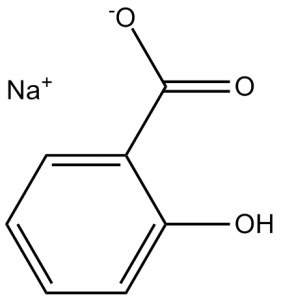
| SMILES | [Na+].O([H])C1=C([H])C([H])=C([H])C([H])=C1C(=O)[O-] |
|
| InChi Key | ABBQHOQBGMUPJH-UHFFFAOYSA-M | |
| InChi Code | InChI=1S/C7H6O3.Na/c8-6-4-2-1-3-5(6)7(9)10;/h1-4,8H,(H,9,10);/q;+1/p-1 | |
| Chemical Name | sodium;2-hydroxybenzoate | |
| Synonyms |
|
|
| HS Tariff Code | 2934.99.03.00 | |
| Storage |
Powder-20°C 3 years 4°C 2 years In solvent -80°C 6 months -20°C 1 month Note: Please store this product in a sealed and protected environment, avoid exposure to moisture. |
|
| Shipping Condition | Room temperature (This product is stable at ambient temperature for a few days during ordinary shipping and time spent in Customs) |
Biological Activity
| Targets |
COX-2; Microbial Metabolite; Autophagy; S6K Cyclooxygenase-2 (COX-2): Sodium salicylate inhibits sheep seminal vesicle-derived COX-2 activity, with an IC50 value of 2.5 ± 0.3 mM (determined by measuring prostaglandin E2 (PGE2) production) [1] - 11β-Hydroxysteroid Dehydrogenase Type 1 (11β-HSD1): Sodium salicylate downregulates the expression and activity of 11β-HSD1 in adipose tissue [3] |
| ln Vitro |
At concentrations much lower than those necessary to prevent the activation of NF-κB (20 mg/mL), sodium salicylate effectively inhibits COX-2 activity. When combined with interleukin 1β for 24 hours, sodium salicylate inhibits prostaglandin E2 release with an IC50 value of 5 μg/mL, an effect that is not influenced by NF-κB activation, COX-2 transcription, or COX-2 translation. Additionally, sodium salicylate acutely (30 min) inhibits COX-2 activity as measured in the presence of 0, 1, or 10 μM exogenous arachidonic acid in a concentration-dependent manner. In contrast, sodium salicylate is a very weak inhibitor of COX-2 activity with an IC50 of >100 μg/mL when exogenous arachidonic acid is increased to 30 μM. With an apparent IC50 value of about 5 μg/mL when combined with IL-1 for 24 hours, sodium salicylate inhibits PGE2 release in a concentration-dependent manner. After a 30-min exposure period, different concentrations of exogenous arachidonic acid (1, 10, and 30 μM) are added, and then the ability of sodium salicylate to directly inhibit COX-2 activity in A549 cells is tested. In the absence of additional arachidonic acid or in the presence of 1 or 10 μM exogenous substrate, sodium salicylate inhibits COX-2 activity in a concentration-dependent manner, with an apparent IC50 value of roughly 5 μg/mL. With an apparent IC50 value of more than 100 g/mL and a maximal inhibition of less than 50%, sodium salicylate is an ineffective inhibitor of COX-2 activity when the same experiments are carried out using 30 μM arachidonic acid[1]. COX-2 Inhibition (Independent of NF-κB): Incubation of purified sheep seminal vesicle COX-2 with Sodium salicylate (0.5–10 mM) resulted in concentration-dependent inhibition of PGE2 synthesis. At 2 mM, PGE2 production was reduced by 42% compared to the control; at 5 mM, inhibition reached 78%; and at 10 mM, inhibition was ~90%. To test NF-κB dependence, RAW264.7 macrophages were pre-treated with Sodium salicylate (5 mM) for 1 hour, then stimulated with LPS (1 μg/mL) for 6 hours. Western blot and RT-PCR showed that Sodium salicylate did not affect LPS-induced NF-κB p65 nuclear translocation or COX-2 mRNA expression, indicating that its COX-2 inhibitory activity is independent of NF-κB activation [1] - 11β-HSD1 Downregulation and Insulin Sensitization in Adipocytes: 3T3-L1 adipocytes (differentiated) were treated with Sodium salicylate (1–5 mM) for 24 hours. RT-PCR analysis revealed a dose-dependent decrease in 11β-HSD1 mRNA levels: 1 mM reduced expression by 28%, 3 mM by 55%, and 5 mM by 72%. Western blot confirmed a corresponding reduction in 11β-HSD1 protein (by ~68% at 5 mM). Additionally, Sodium salicylate (5 mM) increased insulin-stimulated 2-deoxyglucose uptake by 45% compared to insulin-only controls, as measured by a radioactive glucose uptake assay. In primary human subcutaneous adipocytes (isolated from obese donors), 5 mM Sodium salicylate reduced 11β-HSD1 mRNA by 60% and protein by 55% [3] |
| ln Vivo |
Salicylate lowers plasma glucose levels in C57Bl/6 DIO mice during fasting and after meals. Additionally, after Salicylate treatment in C57Bl/6 DIO mice, there is a tendency to decrease plasma triglyceride levels (P=0.059). Salicylate significantly lowers 11β-HSD1 mRNA in the omental adipose tissue of C57Bl/6 DIO mice, with a trend toward the same result in the mesenteric adipose (P=0.057). Salicylate also lowers 11β-HSD1 enzyme activity in the mesenteric adipose of C57Bl/6 DIO mice[2]. 11β-HSD1 Downregulation and Metabolic Improvement in Obese Mice: Male C57BL/6 mice were fed a high-fat diet (HFD, 45% fat content) for 12 weeks to induce obesity, then randomly divided into two groups (n=10 per group): HFD control group and Sodium salicylate treatment group. The treatment group received Sodium salicylate via drinking water at a dose of 200 mg/kg/day for 4 weeks, while the control group received plain drinking water. Compared to the control group, Sodium salicylate treatment significantly reduced 11β-HSD1 mRNA (by 58%) and protein (by 52%) in epididymal white adipose tissue (eWAT), and 11β-HSD1 mRNA (by 45%) in liver. Metabolically, Sodium salicylate decreased fasting blood glucose (from 12.8 ± 1.2 mmol/L to 8.3 ± 0.9 mmol/L), fasting insulin (from 42.5 ± 4.1 μU/mL to 21.3 ± 3.2 μU/mL), and HOMA-IR (from 14.6 ± 1.4 to 6.8 ± 0.8). Glucose tolerance test (GTT) showed that Sodium salicylate reduced the area under the curve (AUC) by 35% [3] - 11β-HSD1 Reduction in Human Obese Subjects: A pilot clinical study included 8 obese volunteers (BMI 30–35 kg/m²) who received oral Sodium salicylate at a dose of 3 g/day (divided into 3 doses) for 2 weeks. Before and after treatment, subcutaneous adipose tissue biopsies were collected. RT-PCR and Western blot showed that Sodium salicylate reduced 11β-HSD1 mRNA by 48% and protein by 42% in adipose tissue. Fasting insulin levels decreased by 32%, and insulin sensitivity (measured by hyperinsulinemic-euglycemic clamp) increased by 28% [3] |
| Enzyme Assay |
The cofactors Glutathione (5 mM), Adrenaline (5 mM), and Hematin (1 M) are dissolved in 50 mM Tris buffer (pH 7.5), along with human purified COX-2. Prior to further diluting in Tris buffer, hematin is first dissolved in a concentrated stock of 100 mM in 1 M NaOH. 96-well plates with individual wells are used to conduct enzyme reactions, with a final reaction volume of 200 μL. The plate is first filled with various sodium salicylate concentrations, then 10 enzyme units (180 μL) are added. Arachidonic acid (10 nM to 30 μM) is then added to the plates and incubated at 37 ° for an additional 15 min. The reaction is stopped by heating the plate to 100°C for five minutes. The 96-well plate is then centrifuged at 10,000 g for 10 min, after which the appropriate samples are taken out and added to the radioimmunoassay[1]. COX-2 Activity Assay: Purified COX-2 (from sheep seminal vesicles) was resuspended in assay buffer (50 mM Tris-HCl pH 8.0, 1 mM EDTA, 10 μM hemoglobin). The reaction mixture contained 0.1 μg COX-2, 10 μM arachidonic acid (substrate), and Sodium salicylate (0.5–10 mM). The mixture was incubated at 37°C for 10 minutes, then the reaction was stopped by adding 0.5 M HCl. PGE2 concentration in the mixture was measured by radioimmunoassay (RIA) using a specific anti-PGE2 antibody. COX-2 activity was calculated as pmol of PGE2 produced per minute per μg of enzyme, and inhibition rate was relative to the vehicle control [1] - 11β-HSD1 Activity Assay: eWAT homogenates (from obese mice) or primary human adipocyte lysates were prepared in activity buffer (20 mM HEPES pH 7.4, 1 mM EDTA, 100 mM NaCl). The reaction mixture contained 50 μg homogenate/lysate, 100 nM cortisone (substrate), 1 mM NADPH (cofactor), and Sodium salicylate (1–5 mM). After incubation at 37°C for 60 minutes, the reaction was stopped by adding methanol. The product (cortisol) was quantified by high-performance liquid chromatography (HPLC) with UV detection at 240 nm. 11β-HSD1 activity was expressed as pmol of cortisol formed per hour per mg of protein [3] |
| Cell Assay |
A549 cells are first treated with IL-1β for 24 hours, and the culture medium is replaced with DMEM containing different concentrations of sodium salicylate (10, 100, and 1000 μg/mL) in order to assess the direct effect of sodium salicylate on COX-2 activity after induction. For 30 minutes, cells are incubated at 37°C. Following a 15-minute addition of arachide acid (1–30 μM), the medium is removed to measure PGE2[1]. RAW264.7 Macrophage COX-2 Expression Assay: RAW264.7 cells were seeded in 6-well plates at 2×10⁵ cells/well and cultured overnight. Cells were pre-treated with Sodium salicylate (1–5 mM) for 1 hour, then stimulated with LPS (1 μg/mL) for 6 hours (for mRNA) or 12 hours (for protein). Total RNA was extracted and reverse-transcribed to cDNA; RT-PCR was performed with COX-2 and GAPDH (internal control) primers. For protein, cells were lysed, and Western blot was conducted with anti-COX-2 and anti-β-actin antibodies. Band intensity was quantified using ImageJ, and COX-2 expression was normalized to GAPDH/β-actin [1] - 3T3-L1 Adipocyte 11β-HSD1 Expression and Glucose Uptake Assay: 3T3-L1 cells were differentiated into adipocytes over 8 days. Differentiated adipocytes were treated with Sodium salicylate (1–5 mM) for 24 hours. RNA and protein were extracted for RT-PCR (11β-HSD1 primers) and Western blot (anti-11β-HSD1 antibody). For glucose uptake, adipocytes were treated with Sodium salicylate (5 mM) for 24 hours, then incubated with insulin (100 nM) for 30 minutes, followed by 2-deoxy-[³H]-glucose (1 μCi/mL) for 10 minutes. Cells were lysed, and radioactivity was measured with a scintillation counter. Glucose uptake was normalized to total protein content [3] |
| Animal Protocol |
Mice: Male C57Bl/6 adult mice are 12 weeks old. Prior to treatment, 10 weeks of a high-fat diet (58% fat, 12% sucrose) are given to C57Bl/6 mice that have developed diet-induced obesity (C57Bl/6 DIO). To groups of n=8, osmotic minipumps implanted subcutaneously between the scapulae are used to administer sodium salicylate (120 mg/kg/day) or distilled water (vehicle) starting one week after arrival (C57Bl/6 Lean), after ten weeks of high-fat feeding (C57Bl/6 DIO), or after reaching the target weight (HSD1KO-DIO) for four weeks. HFD-Induced Obese Mouse Model for Metabolic Study: Male C57BL/6 mice (6 weeks old) were fed HFD (45% fat) for 12 weeks to establish obesity. Mice were then assigned to HFD control (n=10) or Sodium salicylate treatment (n=10) groups. Sodium salicylate was dissolved in drinking water to a concentration of 0.2% (w/v), providing a daily dose of ~200 mg/kg (adjusted based on average daily water intake). During the 4-week treatment, body weight, water intake, and food intake were recorded weekly. At the end of treatment, mice were fasted for 12 hours: blood was collected for glucose and insulin measurement; eWAT, liver, and skeletal muscle were harvested. eWAT and liver were divided into portions: one fixed in 10% neutral buffered formalin for histology, another stored at -80°C for RNA/protein extraction and enzyme activity assay [3] |
| Toxicity/Toxicokinetics |
In Vitro Cytotoxicity: MTT assay was performed in RAW264.7 cells and 3T3-L1 adipocytes treated with Sodium salicylate (0.5–10 mM) for 48 hours. Cell viability remained >90% at concentrations ≤5 mM; at 10 mM, viability decreased by ~12% in RAW264.7 cells and ~10% in 3T3-L1 adipocytes [1,3] - In Vivo Safety (Mice): In the HFD-induced obese mouse model, Sodium salicylate (200 mg/kg/day for 4 weeks) did not cause significant changes in body weight (no difference vs. control), organ weights (liver, kidney, spleen), or serum levels of alanine transaminase (ALT), aspartate transaminase (AST), blood urea nitrogen (BUN), or creatinine. Histological examination of liver and kidney showed no obvious damage [3] - Human Safety (Pilot Study): In 8 obese subjects receiving Sodium salicylate (3 g/day for 2 weeks), 2 subjects reported mild gastrointestinal discomfort (nausea), which resolved without treatment. No significant changes in serum ALT, AST, BUN, creatinine, or platelet count were observed [3] |
| References |
[1]. Sodium salicylate inhibits cyclo-oxygenase-2 activity independently of transcription factor (nuclear factor kappaB) activation: role of arachidonic acid. Mol Pharmacol. 1997 Jun;51(6):907-12. [2]. Palmitic acid negatively regulates tumor suppressor PTEN through T366 phosphorylation and protein degradation. Cancer Lett. 2020 Oct 8;S0304-3835(20)30506-1. [3]. Salicylate downregulates 11β-HSD1 expression in adipose tissue in obese mice and in humans, mediating insulin sensitization. Diabetes. 2012 Apr;61(4):790-6. [4]. Age-associated NF-κB signaling in myofibers alters the satellite cell niche and re-strains muscle stem cell function. Aging (Albany NY). 2016 Nov; 8(11): 2871–2884. |
| Additional Infomation |
Sodium salicylate is an organic molecular entity. Sodium Salicylate is the sodium salt of salicylic acid. As a nonsteroidal anti-inflammatory drug (NSAID), sodium salicylate irreversibly acetylates cyclooxygenases I and II, thereby inhibiting prostaglandin synthesis and associated inflammation and pain. This agent may also activate mitogen-activated protein kinase (p38MAPK), thereby inducing apoptosis in cancer cells. (NCI04) A non-steroidal anti-inflammatory agent that is less effective than equal doses of ASPIRIN in relieving pain and reducing fever. However, individuals who are hypersensitive to ASPIRIN may tolerate sodium salicylate. In general, this salicylate produces the same adverse reactions as ASPIRIN, but there is less occult gastrointestinal bleeding. (From AMA Drug Evaluations Annual, 1992, p120) See also: Salicylic Acid (has active moiety); Methenamine; Sodium Salicylate (component of) ... View More ... Mechanism of COX-2 Inhibition: Unlike non-steroidal anti-inflammatory drugs (NSAIDs) that inhibit COX-2 by acetylating the active site, Sodium salicylate acts by competing with arachidonic acid (the COX-2 substrate) for binding to the enzyme’s active site, which explains its NF-κB-independent activity [1] - Link Between 11β-HSD1 and Insulin Sensitivity: 11β-HSD1 converts inactive cortisone to active cortisol in adipose tissue; excess cortisol promotes insulin resistance. Sodium salicylate downregulates 11β-HSD1 to reduce local cortisol levels, thereby improving insulin sensitivity—this mechanism supports its potential use in metabolic disorders like type 2 diabetes and obesity-related insulin resistance [3] - Clinical Relevance: Sodium salicylate is a classic anti-inflammatory and antipyretic drug. The findings in literature [3] expand its potential therapeutic scope to metabolic diseases, though further large-scale clinical trials are needed to confirm its efficacy in insulin-resistant patients [3] |
Solubility Data
| Solubility (In Vitro) |
|
|||
| Solubility (In Vivo) |
Solubility in Formulation 1: ≥ 2.5 mg/mL (15.62 mM) (saturation unknown) in 10% DMSO + 40% PEG300 + 5% Tween80 + 45% Saline (add these co-solvents sequentially from left to right, and one by one), clear solution. For example, if 1 mL of working solution is to be prepared, you can add 100 μL of 25.0 mg/mL clear DMSO stock solution to 400 μL PEG300 and mix evenly; then add 50 μL Tween-80 to the above solution and mix evenly; then add 450 μL normal saline to adjust the volume to 1 mL. Preparation of saline: Dissolve 0.9 g of sodium chloride in 100 mL ddH₂ O to obtain a clear solution. Solubility in Formulation 2: ≥ 2.5 mg/mL (15.62 mM) (saturation unknown) in 10% DMSO + 90% (20% SBE-β-CD in Saline) (add these co-solvents sequentially from left to right, and one by one), clear solution. For example, if 1 mL of working solution is to be prepared, you can add 100 μL of 25.0 mg/mL clear DMSO stock solution to 900 μL of 20% SBE-β-CD physiological saline solution and mix evenly. Preparation of 20% SBE-β-CD in Saline (4°C,1 week): Dissolve 2 g SBE-β-CD in 10 mL saline to obtain a clear solution. Solubility in Formulation 3: ≥ 2.5 mg/mL (15.62 mM) (saturation unknown) in 10% DMSO + 90% Corn Oil (add these co-solvents sequentially from left to right, and one by one), clear solution. For example, if 1 mL of working solution is to be prepared, you can add 100 μL of 25.0 mg/mL clear DMSO stock solution to 900 μL of corn oil and mix evenly. Solubility in Formulation 4: 110 mg/mL (687.07 mM) in PBS (add these co-solvents sequentially from left to right, and one by one), clear solution; with ultrasonication. (Please use freshly prepared in vivo formulations for optimal results.) |
| Preparing Stock Solutions | 1 mg | 5 mg | 10 mg | |
| 1 mM | 6.2069 mL | 31.0347 mL | 62.0694 mL | |
| 5 mM | 1.2414 mL | 6.2069 mL | 12.4139 mL | |
| 10 mM | 0.6207 mL | 3.1035 mL | 6.2069 mL |