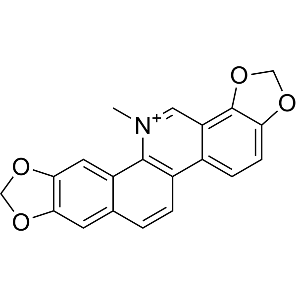
Sanguinarine chloride is a naturally occurring benzophenanthridine alkaloid isolated from the root of Sanguinaria Canadensis. It stimulates apoptosis via activating the production of reactive oxygen species (ROS).
Physicochemical Properties
| Molecular Formula | C20H14NO4 |
| Molecular Weight | 332.3295 |
| Exact Mass | 332.091 |
| CAS # | 2447-54-3 |
| Related CAS # | Sanguinarine chloride;5578-73-4;Sanguinarine (gluconate);132210-34-5 |
| PubChem CID | 5154 |
| Appearance | Typically exists as solid at room temperature |
| Density | 1.3463 (rough estimate) |
| Boiling Point | 483.53°C (rough estimate) |
| Melting Point | 205-215ºC |
| Index of Refraction | 1.5180 (estimate) |
| LogP | 3.428 |
| Hydrogen Bond Donor Count | 0 |
| Hydrogen Bond Acceptor Count | 4 |
| Rotatable Bond Count | 0 |
| Heavy Atom Count | 25 |
| Complexity | 530 |
| Defined Atom Stereocenter Count | 0 |
| SMILES | O1C([H])([H])OC2C([H])=C([H])C3C(C1=2)=C([H])[N+](C([H])([H])[H])=C1C2=C([H])C4=C(C([H])=C2C([H])=C([H])C1=3)OC([H])([H])O4 |
| InChi Key | INVGWHRKADIJHF-UHFFFAOYSA-N |
| InChi Code | InChI=1S/C20H14NO4/c1-21-8-15-12(4-5-16-20(15)25-10-22-16)13-3-2-11-6-17-18(24-9-23-17)7-14(11)19(13)21/h2-8H,9-10H2,1H3/q+1 |
| Chemical Name | 24-methyl-5,7,18,20-tetraoxa-24-azoniahexacyclo[11.11.0.02,10.04,8.014,22.017,21]tetracosa-1(24),2,4(8),9,11,13,15,17(21),22-nonaene |
| HS Tariff Code | 2934.99.9001 |
| Storage |
Powder-20°C 3 years 4°C 2 years In solvent -80°C 6 months -20°C 1 month |
| Shipping Condition | Room temperature (This product is stable at ambient temperature for a few days during ordinary shipping and time spent in Customs) |
Biological Activity
| ln Vitro |
The activation of JNK and NF-κB signaling pathways is linked to apoptosis inducing sanguinarine (SANG). 22B-cFluc cells were stimulated with varying concentrations of sanguinarine for 24 hours in order to ascertain the impact of sanguinarine on cell viability. Following this, the CKK-8 test was carried out. The administration of sanguinarine exhibited a dose-dependent reduction in the proliferation of 22B cells. In parallel, cellular caspase-3 activity was assessed using the validated caspase-3 substrate Ac-DEVD-pNA in the cytoplasmic extracts of 22B-cFluc cells treated with various dosages of sanguinarine. Greater Sanguinarine-stimulated caspase-3 activity is indicated by a dose-dependent rise in absorbance at 450 nm [1]. Sanguinarine exhibits concentration-dependent antiproliferative and pro-apoptotic activity in multiple cancer cell lines. In human hepatocellular carcinoma HepG2 cells, it inhibited cell viability with an IC50 of 2.3 μM (MTT assay) after 48 hours of treatment. At 5 μM, the apoptotic rate reached 58% (Annexin V-FITC/PI double staining), accompanied by a 3.6-fold increase in reactive oxygen species (ROS) production (DCFH-DA staining) compared to the control [1] Western blot analysis showed that Sanguinarine (1-5 μM) upregulated pro-apoptotic proteins: cleaved caspase-3 (3.2-fold), cleaved caspase-9 (2.8-fold), and Bax (2.1-fold), while downregulating anti-apoptotic protein Bcl-2 (0.4-fold) in HepG2 cells. It also increased the ratio of Bax/Bcl-2 (5.2-fold at 5 μM) and induced cytochrome c release from mitochondria to cytoplasm [1] In human breast cancer MCF-7 cells and colon cancer HCT116 cells, Sanguinarine (1-4 μM) similarly inhibited cell proliferation (IC50 = 1.8 μM for MCF-7, 2.1 μM for HCT116) and induced apoptosis (45% and 52% apoptotic rate at 4 μM, respectively), with consistent ROS overproduction (3.1-fold and 3.4-fold increase) [1] |
| ln Vivo |
To examine the apoptosis produced by sanguinarine (SANG) in vivo, 22B-cFluc cells were subcutaneously implanted into one side of nude mice and a xenograft model was constructed. Mice were treated with 10 mg/kg of sanguinarine intravenously. At 24, 48 and 72 hours following treatment, mice were intraperitoneally injected with 150 mg/kg D-luciferin substrate and then bioluminescence imaging was conducted. Treatment with sanguinarine generated a considerable increase in luminescence signal as early as 48 hours after initial treatment. A constant increase in bioluminescence imaging (BLI) intensity was seen throughout the experiment. 72 hours after therapy, tumors were collected and TUNEL staining was conducted to determine apoptosis. Compared with control tumors, the sanguinarine-treated group displayed much more apoptosis, as shown by enhanced green signal of sporadic apoptotic cells [1]. In nude mice bearing HepG2 xenograft tumors (n=6 per group), intraperitoneal administration of Sanguinarine (5 mg/kg/day or 10 mg/kg/day) for 21 days dose-dependently inhibited tumor growth. The 10 mg/kg dose reduced tumor volume by 68% and tumor weight by 63% compared to the vehicle control group. Bioluminescence imaging (using caspase-3-sensitive luciferase reporter) showed that the 10 mg/kg dose enhanced apoptotic signal in tumors by 4.7-fold, confirming in vivo apoptotic induction [1] Histopathological examination of tumor tissues revealed that Sanguinarine (10 mg/kg) increased the number of TUNEL-positive apoptotic cells (6.2-fold vs. control) and reduced Ki-67-positive proliferative cells (by 59%). Immunohistochemical staining showed upregulated cleaved caspase-3 and downregulated Bcl-2 expression in tumor tissues [1] |
| Cell Assay |
HepG2/MCF-7/HCT116 cell viability assay: Cells were cultured in DMEM/RPMI 1640 medium supplemented with fetal bovine serum, seeded in 96-well plates (1×10⁴ cells/well), and treated with Sanguinarine (0.5-10 μM) for 48 hours. Cell viability was measured by MTT assay, and IC50 values were calculated from concentration-response curves [1] Apoptosis and ROS detection assay: HepG2 cells were seeded in 6-well plates (2×10⁵ cells/well), treated with Sanguinarine (1-5 μM) for 24 hours. Apoptosis was detected by Annexin V-FITC/PI double staining and flow cytometry; ROS production was measured by DCFH-DA staining and fluorescence microscopy [1] Western blot and cytochrome c release assay: HepG2 cells were treated with Sanguinarine (1-5 μM) for 24 hours, then lysed to extract total proteins or mitochondrial/cytoplasmic fractions. Protein expression (cleaved caspase-3/9, Bax, Bcl-2) was detected by western blot; cytochrome c release was analyzed by comparing mitochondrial and cytoplasmic fractions [1] Bioluminescence imaging of apoptotic cells: HepG2 cells stably transfected with caspase-3-sensitive luciferase reporter plasmid were treated with Sanguinarine (1-5 μM) for 12 hours. Luciferase activity was measured using a luminometer to assess caspase-3 activation and apoptotic progression [1] |
| Animal Protocol | Nude mouse HepG2 xenograft model: 4-6-week-old female BALB/c nude mice were subcutaneously inoculated with HepG2 cells (5×10⁶ cells/mouse). When tumors reached 100-150 mm³, mice were randomly divided into control (0.1% DMSO in saline), 5 mg/kg, and 10 mg/kg Sanguinarine groups (n=6 per group). Sanguinarine was dissolved in 0.1% DMSO and diluted with saline to the desired concentration, then administered via intraperitoneal injection once daily for 21 days. Tumor volume was measured every 3 days; bioluminescence imaging was performed on days 7, 14, and 21 to monitor apoptotic activity. Mice were euthanized at the end of treatment, and tumor tissues were collected for histopathological and immunohistochemical analysis [1] |
| Toxicity/Toxicokinetics |
In vitro toxicity: Sanguinarine (up to 5 μM) showed minimal cytotoxicity to normal human liver LO2 cells (viability >85% at 5 μM), while exerting potent toxicity to cancer cells (IC50 1.8-2.3 μM) [1] In nude mice, 21-day intraperitoneal administration of Sanguinarine (up to 10 mg/kg/day) did not cause significant changes in body weight, food intake, or serum levels of ALT, AST, creatinine, and BUN. No histopathological abnormalities were observed in the liver, kidney, heart, or spleen [1] |
| References |
[1]. Wang Y, Noninvasive bioluminescence imaging of the dynamics of sanguinarine induced apoptosis via activation of reactive oxygen species. Oncotarget. 2016 Apr 19;7(16):22355-67. |
| Additional Infomation |
Sanguinarine is a benzophenanthridine alkaloid, an alkaloid antibiotic and a botanical anti-fungal agent. Sanguinarine has been reported in Corydalis ophiocarpa, Glaucium squamigerum, and other organisms with data available. Sanguinarine is found in opium poppy. Consumption of Sanguinarine, present in poppy seeds and in the oil of Argemone mexicana which has been used as an adulterant for mustard oil in India, has been linked to development of glaucoma. Sanguinarine is banned by FDA. Sanguinarine is a quaternary ammonium salt from the group of benzylisoquinoline alkaloids. It is extracted from some plants, including bloodroot (Sanguinaria canadensis), Mexican prickly poppy Argemone mexicana, Chelidonium majus and Macleaya cordata. It is also found in the root, stem and leaves of the opium poppy but not in the capsule. Sanguinarine is a toxin that kills animal cells through its action on the Na+-K+-ATPase transmembrane protein. Epidemic dropsy is a disease that results from ingesting sanguinarine. Sanguinarine has been shown to exhibit antibiotic, anti-apoptotic, anti-fungal, anti-inflammatory and anti-angiogenic functions Sanguinarine belongs to the family of Benzoquinolines. These are organic compounds containing a benzene fused to a quinoline ring system. (A3208, A3209, A3208, A3208, A3208). See also: Sanguinaria canadensis root (part of); Chelidonium majus flowering top (part of). Sanguinarine is a natural benzophenanthridine alkaloid isolated from plants of the Papaveraceae family (e.g., Sanguinaria canadensis, Macleaya cordata) [1] Its anti-cancer mechanism is mediated by ROS overproduction, which triggers mitochondrial-dependent apoptotic pathway. ROS accumulation disrupts mitochondrial membrane potential, promotes cytochrome c release, activates caspases-9 and -3, and modulates the Bax/Bcl-2 balance to induce apoptosis [1] It exhibits broad-spectrum anti-proliferative activity against multiple cancer cell lines (hepatocellular, breast, colon cancer) and inhibits tumor growth in vivo without obvious systemic toxicity, supporting its potential as an anti-cancer agent [1] Noninvasive bioluminescence imaging confirms that Sanguinarine induces apoptosis in tumor tissues, providing a tool for real-time monitoring of its in vivo therapeutic effect [1] |
Solubility Data
| Solubility (In Vitro) | May dissolve in DMSO (in most cases), if not, try other solvents such as H2O, Ethanol, or DMF with a minute amount of products to avoid loss of samples |
| Solubility (In Vivo) |
Note: Listed below are some common formulations that may be used to formulate products with low water solubility (e.g. < 1 mg/mL), you may test these formulations using a minute amount of products to avoid loss of samples. Injection Formulations (e.g. IP/IV/IM/SC) Injection Formulation 1: DMSO : Tween 80: Saline = 10 : 5 : 85 (i.e. 100 μL DMSO stock solution → 50 μL Tween 80 → 850 μL Saline) *Preparation of saline: Dissolve 0.9 g of sodium chloride in 100 mL ddH ₂ O to obtain a clear solution. Injection Formulation 2: DMSO : PEG300 :Tween 80 : Saline = 10 : 40 : 5 : 45 (i.e. 100 μL DMSO → 400 μLPEG300 → 50 μL Tween 80 → 450 μL Saline) Injection Formulation 3: DMSO : Corn oil = 10 : 90 (i.e. 100 μL DMSO → 900 μL Corn oil) Example: Take the Injection Formulation 3 (DMSO : Corn oil = 10 : 90) as an example, if 1 mL of 2.5 mg/mL working solution is to be prepared, you can take 100 μL 25 mg/mL DMSO stock solution and add to 900 μL corn oil, mix well to obtain a clear or suspension solution (2.5 mg/mL, ready for use in animals). Injection Formulation 4: DMSO : 20% SBE-β-CD in saline = 10 : 90 [i.e. 100 μL DMSO → 900 μL (20% SBE-β-CD in saline)] *Preparation of 20% SBE-β-CD in Saline (4°C,1 week): Dissolve 2 g SBE-β-CD in 10 mL saline to obtain a clear solution. Injection Formulation 5: 2-Hydroxypropyl-β-cyclodextrin : Saline = 50 : 50 (i.e. 500 μL 2-Hydroxypropyl-β-cyclodextrin → 500 μL Saline) Injection Formulation 6: DMSO : PEG300 : castor oil : Saline = 5 : 10 : 20 : 65 (i.e. 50 μL DMSO → 100 μLPEG300 → 200 μL castor oil → 650 μL Saline) Injection Formulation 7: Ethanol : Cremophor : Saline = 10: 10 : 80 (i.e. 100 μL Ethanol → 100 μL Cremophor → 800 μL Saline) Injection Formulation 8: Dissolve in Cremophor/Ethanol (50 : 50), then diluted by Saline Injection Formulation 9: EtOH : Corn oil = 10 : 90 (i.e. 100 μL EtOH → 900 μL Corn oil) Injection Formulation 10: EtOH : PEG300:Tween 80 : Saline = 10 : 40 : 5 : 45 (i.e. 100 μL EtOH → 400 μLPEG300 → 50 μL Tween 80 → 450 μL Saline) Oral Formulations Oral Formulation 1: Suspend in 0.5% CMC Na (carboxymethylcellulose sodium) Oral Formulation 2: Suspend in 0.5% Carboxymethyl cellulose Example: Take the Oral Formulation 1 (Suspend in 0.5% CMC Na) as an example, if 100 mL of 2.5 mg/mL working solution is to be prepared, you can first prepare 0.5% CMC Na solution by measuring 0.5 g CMC Na and dissolve it in 100 mL ddH2O to obtain a clear solution; then add 250 mg of the product to 100 mL 0.5% CMC Na solution, to make the suspension solution (2.5 mg/mL, ready for use in animals). Oral Formulation 3: Dissolved in PEG400 Oral Formulation 4: Suspend in 0.2% Carboxymethyl cellulose Oral Formulation 5: Dissolve in 0.25% Tween 80 and 0.5% Carboxymethyl cellulose Oral Formulation 6: Mixing with food powders Note: Please be aware that the above formulations are for reference only. InvivoChem strongly recommends customers to read literature methods/protocols carefully before determining which formulation you should use for in vivo studies, as different compounds have different solubility properties and have to be formulated differently. (Please use freshly prepared in vivo formulations for optimal results.) |
| Preparing Stock Solutions | 1 mg | 5 mg | 10 mg | |
| 1 mM | 3.0091 mL | 15.0453 mL | 30.0906 mL | |
| 5 mM | 0.6018 mL | 3.0091 mL | 6.0181 mL | |
| 10 mM | 0.3009 mL | 1.5045 mL | 3.0091 mL |