Physicochemical Properties
| Molecular Formula | C42H68O13 |
| Molecular Weight | 780.98 |
| Exact Mass | 780.466 |
| CAS # | 20874-52-6 |
| PubChem CID | 107793 |
| Appearance | White to off-white solid powder |
| Density | 1.4±0.1 g/cm3 |
| Boiling Point | 893.7±65.0 °C at 760 mmHg |
| Melting Point | 256- 259ºC |
| Flash Point | 494.3±34.3 °C |
| Vapour Pressure | 0.0±0.6 mmHg at 25°C |
| Index of Refraction | 1.617 |
| LogP | 3.65 |
| Hydrogen Bond Donor Count | 8 |
| Hydrogen Bond Acceptor Count | 13 |
| Rotatable Bond Count | 6 |
| Heavy Atom Count | 55 |
| Complexity | 1490 |
| Defined Atom Stereocenter Count | 21 |
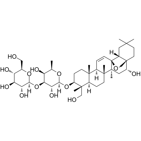
| SMILES | C[C@@H]1[C@@H]([C@@H]([C@H]([C@@H](O1)O[C@H]2CC[C@]3([C@H]([C@]2(C)CO)CC[C@@]4([C@@H]3C=C[C@@]56[C@]4(C[C@H]([C@@]7([C@H]5CC(CC7)(C)C)CO6)O)C)C)C)O)O[C@H]8[C@@H]([C@H]([C@@H]([C@H](O8)CO)O)O)O)O |
| InChi Key | KYWSCMDFVARMPN-LCSVLAELSA-N |
| InChi Code | InChI=1S/C42H68O13/c1-21-28(46)33(55-34-31(49)30(48)29(47)22(18-43)53-34)32(50)35(52-21)54-27-10-11-37(4)23(38(27,5)19-44)8-12-39(6)24(37)9-13-42-25-16-36(2,3)14-15-41(25,20-51-42)26(45)17-40(39,42)7/h9,13,21-35,43-50H,8,10-12,14-20H2,1-7H3/t21-,22-,23-,24-,25-,26-,27+,28+,29-,30+,31-,32-,33+,34+,35+,37+,38+,39-,40+,41-,42+/m1/s1 |
| Chemical Name | (2S,3R,4S,5S,6R)-2-[(2R,3R,4S,5S,6R)-3,5-dihydroxy-2-[[(1S,2R,4S,5R,8R,9R,10S,13S,14R,17S,18R)-2-hydroxy-9-(hydroxymethyl)-4,5,9,13,20,20-hexamethyl-24-oxahexacyclo[15.5.2.01,18.04,17.05,14.08,13]tetracos-15-en-10-yl]oxy]-6-methyloxan-4-yl]oxy-6-(hydroxymethyl)oxane-3,4,5-triol |
| HS Tariff Code | 2934.99.9001 |
| Storage |
Powder-20°C 3 years 4°C 2 years In solvent -80°C 6 months -20°C 1 month |
| Shipping Condition | Room temperature (This product is stable at ambient temperature for a few days during ordinary shipping and time spent in Customs) |
Biological Activity
| Targets |
- Selectin (E-selectin, P-selectin) [1] - Nuclear Factor-κB (NF-κB) [2] - Signal Transducer and Activator of Transcription 3 (STAT3) [2] - Estrogen Receptor-β (ER-β) [3] |
| ln Vitro |
Saikosaponin D (Compound 3) is a triterpene saponin that, at IC50 values of 1.8 µM, 3.0 µM, and 4.3 µM, respectively, inhibits the binding of E-, L-, and P-selectin to THP-1 cells. The cause of this effect is not cytotoxicity. Saikosaponin D (1, 5, 10 µM) inhibits THP-1 adhesion to HUVEC monolayers activated by TNF-α in a dose-dependent manner. In THP-1 cells, saikosaponin D (30 μM) also suppresses P-selectin ligand (CD162) expression [1]. Saikosaponin D (5 μM) has effects similar to those of estradiol (E2) in that it inhibits HSC-T6 cell proliferation induced by H2O2 treatment, decreases the expression levels of α-SMA, TGF-β1, Hyp, COL1, and TIMP-1, and increases the expression of MMP-1. These effects are blocked by ER antagonists. Additionally, saikosaponin D has the ability to suppress the MAPK signaling pathway and prevent ROS generation brought on by oxidative stress; however, ER antagonists can also counteract this inhibitory effect [3]. 1. Inhibition of selectin-mediated cell adhesion: - HL-60 cells (human promyelocytic leukemia) were pretreated with Saikosaponin D (1–20 μM) for 30 minutes, then co-incubated with TNF-α-activated HUVECs (human umbilical vein endothelial cells) or thrombin-activated platelets. The compound dose-dependently inhibited HL-60 cell adhesion to HUVECs (inhibition rates: 32% at 5 μM, 58% at 10 μM, 75% at 20 μM) and to platelets (inhibition rates: 28% at 5 μM, 55% at 10 μM, 72% at 20 μM) [1] 2. Protection against acetaminophen-induced hepatotoxicity: - Human hepatoma HepG2 cells were pretreated with Saikosaponin D (5–20 μM) for 2 hours, then exposed to acetaminophen (10 mM) for 24 hours. The compound dose-dependently increased cell viability (MTT assay): 20 μM Saikosaponin D restored viability from 45% (acetaminophen alone) to 82% [2] - Western blot showed reduced phosphorylation of NF-κB p65 and STAT3, decreased expression of pro-inflammatory cytokines (TNF-α, IL-6) (RT-PCR), and reduced intracellular reactive oxygen species (ROS) levels (DCFH-DA staining) [2] 3. Inhibition of oxidative stress-induced hepatic stellate cell activation: - Rat hepatic stellate cells (HSCs) were pretreated with Saikosaponin D (1–10 μM) for 2 hours, then stimulated with H₂O₂ (200 μM) for 24 hours. The compound dose-dependently suppressed HSC activation, as evidenced by reduced α-SMA and collagen I/III expression (Western blot and RT-PCR) [3] - ER-β knockdown (siRNA) abolished the inhibitory effect, confirming ER-β-dependent mechanism. Saikosaponin D increased SOD and GSH-Px activities, reduced MDA levels, and inhibited ROS production [3] |
| ln Vivo |
When administered intraperitoneally, saikosaponin D (2 mg/kg/day) protects mice from liver damage caused by acetaminophen overdose (APAP). Saikosaponin D does not change PPARα activation, however it does impact GSH levels and APAP metabolism. Additionally, pro-inflammatory cytokines and STAT3 target gene expression can be inhibited by saikosaponin D (2 mg/kg/day, intraperitoneal injection) as well as the activation of NF-kB and STAT3 caused by APAP [2]. 1. Protection against acetaminophen-induced liver injury in mice: - Male ICR mice were randomly divided into 4 groups (n=8/group): normal control, acetaminophen (300 mg/kg, i.p.), Saikosaponin D 20 mg/kg + acetaminophen, Saikosaponin D 40 mg/kg + acetaminophen [2] - Saikosaponin D was administered intragastrically once daily for 3 consecutive days, with acetaminophen injected intraperitoneally on the 3rd day. Serum ALT and AST levels were reduced by 48% and 52% (40 mg/kg group), respectively, compared to acetaminophen alone [2] - Liver tissue analysis: reduced hepatic necrosis (H&E staining), decreased TNF-α/IL-6 mRNA levels (RT-PCR), and inhibited NF-κB/STAT3 phosphorylation (Western blot) [2] |
| Cell Assay |
1. Selectin-mediated cell adhesion assay: - HUVECs were seeded in 96-well plates and activated with TNF-α for 4 hours; platelets were activated with thrombin for 30 minutes. HL-60 cells were labeled with fluorescent dye, pretreated with Saikosaponin D (1–20 μM) for 30 minutes, then added to HUVEC/platelet-coated wells [1] - After 1 hour of incubation at 37°C, unbound cells were washed away, and fluorescence intensity (reflecting adherent cells) was measured. Inhibition rates were calculated relative to vehicle control [1] 2. Hepatocyte protection and HSC activation assays: - HepG2 cell viability assay: Cells were seeded in 96-well plates (5×10³ cells/well), pretreated with Saikosaponin D (5–20 μM) for 2 hours, exposed to acetaminophen (10 mM) for 24 hours. MTT reagent was added, and absorbance was measured at 570 nm [2] - ROS detection: HepG2/HSC cells were loaded with DCFH-DA dye for 30 minutes, treated as above, and fluorescence intensity was measured at 488 nm (excitation)/525 nm (emission) [2,3] - Western blot/RT-PCR: Cells were lysed, protein/RNA was extracted, and target proteins (NF-κB p65, p-STAT3, α-SMA, collagen) or genes (TNF-α, IL-6, α-SMA) were detected with specific antibodies/primers [2,3] - ER-β siRNA experiment: HSCs were transfected with ER-β siRNA or scramble siRNA for 48 hours, then treated with Saikosaponin D and H₂O₂, followed by α-SMA/collagen detection [3] |
| Animal Protocol |
1. Acetaminophen-induced liver injury model: - Male ICR mice (6–8 weeks old) were acclimated for 1 week. Saikosaponin D was dissolved in 0.5% carboxymethylcellulose to prepare 20 mg/kg and 40 mg/kg suspensions [2] - Administration: Saikosaponin D was given intragastrically once daily for 3 days. On the 3rd day, acetaminophen (300 mg/kg) was injected intraperitoneally 1 hour after the last Saikosaponin D dose [2] - Mice were euthanized 24 hours after acetaminophen injection. Serum was collected for ALT/AST detection, and liver tissues were harvested for H&E staining, Western blot, and RT-PCR analysis [2] |
| Toxicity/Toxicokinetics |
1. In vitro toxicity: - HepG2 cells and normal HUVECs treated with Saikosaponin D (up to 20 μM) for 24 hours showed no significant cytotoxicity (viability > 90%), indicating low direct cell toxicity [2] 2. In vivo toxicity: - In the acetaminophen-induced liver injury study (40 mg/kg, i.g., 3 days), mice showed no significant body weight loss, abnormal behavior, or gross pathological changes in major organs (liver, kidney, heart) [2] |
| References |
[1]. Saikosaponin D isolated from Bupleurum falcatum inhibits selectin-mediated cell adhesion. Molecules. 2014 Dec 4;19(12):20340-9. [2]. Saikosaponin d protects against acetaminophen-induced hepatotoxicity by inhibiting NF-κB and STAT3 signaling. Chem Biol Interact. 2014 Nov 5;223:80-6. [3]. Estrogen receptor-β-dependent effects of saikosaponin‑d on the suppression of oxidative stress-induced rat hepatic stellate cell activation. Int J Mol Med. 2018 Mar;41(3):1357-1364. |
| Additional Infomation |
Saikosaponin D has been reported in Bupleurum chinense, Bupleurum rockii, and other organisms with data available. 1. Source and structural background: Saikosaponin D is a triterpenoid saponin isolated from the roots of Bupleurum falcatum (Chinese herbal medicine). It has a characteristic oleanane-type triterpenoid scaffold with sugar moieties, which contributes to its biological activities [1,2,3] 2. Mechanisms of action: - Anti-adhesive: Inhibits selectin-mediated adhesion between leukocytes and endothelial cells/platelets [1] - Hepatoprotective: Inhibits NF-κB/STAT3 signaling, reduces inflammation and oxidative stress to protect against acetaminophen-induced hepatotoxicity [2] - Anti-fibrotic: ER-β-dependent suppression of oxidative stress-induced HSC activation, reducing extracellular matrix deposition [3] 3. Therapeutic potential: Saikosaponin D exhibits hepatoprotective, anti-inflammatory, anti-adhesive, and anti-fibrotic activities, making it a promising candidate for treating liver injury, liver fibrosis, and inflammatory diseases [1,2,3] |
Solubility Data
| Solubility (In Vitro) |
DMSO : ~50 mg/mL (~64.02 mM) H2O : < 0.1 mg/mL |
| Solubility (In Vivo) |
Solubility in Formulation 1: ≥ 2.5 mg/mL (3.20 mM) (saturation unknown) in 10% DMSO + 90% (20% SBE-β-CD in Saline) (add these co-solvents sequentially from left to right, and one by one), clear solution. For example, if 1 mL of working solution is to be prepared, you can add 100 μL of 25.0 mg/mL clear DMSO stock solution to 900 μL of 20% SBE-β-CD physiological saline solution and mix evenly. Preparation of 20% SBE-β-CD in Saline (4°C,1 week): Dissolve 2 g SBE-β-CD in 10 mL saline to obtain a clear solution. Solubility in Formulation 2: ≥ 2.5 mg/mL (3.20 mM) (saturation unknown) in 10% DMSO + 90% Corn Oil (add these co-solvents sequentially from left to right, and one by one), clear solution. For example, if 1 mL of working solution is to be prepared, you can add 100 μL of 25.0 mg/mL clear DMSO stock solution to 900 μL of corn oil and mix evenly. (Please use freshly prepared in vivo formulations for optimal results.) |
| Preparing Stock Solutions | 1 mg | 5 mg | 10 mg | |
| 1 mM | 1.2804 mL | 6.4022 mL | 12.8044 mL | |
| 5 mM | 0.2561 mL | 1.2804 mL | 2.5609 mL | |
| 10 mM | 0.1280 mL | 0.6402 mL | 1.2804 mL |