Physicochemical Properties
| Molecular Formula | C18H16O3S |
| Molecular Weight | 312.3828 |
| Exact Mass | 312.082 |
| Elemental Analysis | C, 69.21; H, 5.16; O, 15.36; S, 10.26 |
| CAS # | 1384268-04-5 |
| Related CAS # | 1384268-04-5; |
| PubChem CID | 123131800 |
| Appearance | Yellow to orange solid powder |
| Density | 1.3±0.1 g/cm3 |
| Boiling Point | 502.0±50.0 °C at 760 mmHg |
| Flash Point | 257.4±30.1 °C |
| Vapour Pressure | 0.0±1.3 mmHg at 25°C |
| Index of Refraction | 1.649 |
| LogP | 6.34 |
| Hydrogen Bond Donor Count | 1 |
| Hydrogen Bond Acceptor Count | 4 |
| Rotatable Bond Count | 3 |
| Heavy Atom Count | 22 |
| Complexity | 483 |
| Defined Atom Stereocenter Count | 0 |
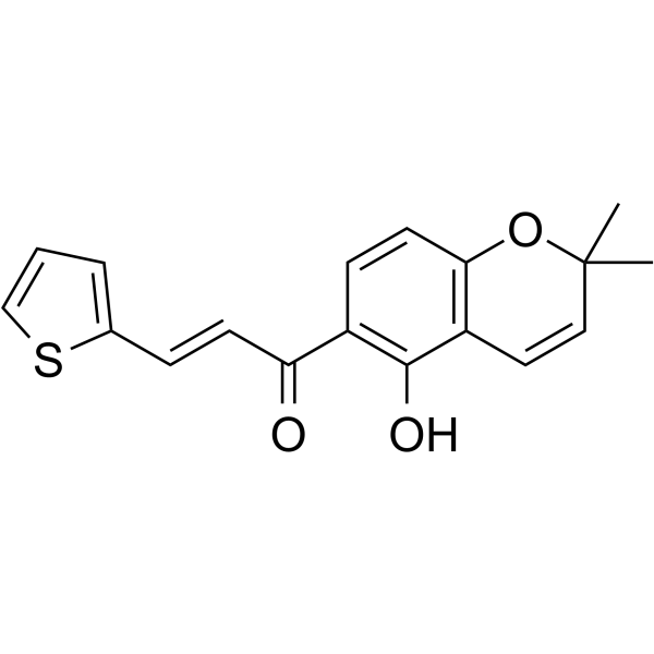
| SMILES | S1C([H])=C([H])C([H])=C1C([H])=C([H])C(C1C([H])=C([H])C2=C(C=1O[H])C([H])=C([H])C(C([H])([H])[H])(C([H])([H])[H])O2)=O |
| InChi Key | OMVURDVBYIJCOI-FNORWQNLSA-N |
| InChi Code | InChI=1S/C18H16O3S/c1-18(2)10-9-14-16(21-18)8-6-13(17(14)20)15(19)7-5-12-4-3-11-22-12/h3-11,20H,1-2H3/b7-5+ |
| Chemical Name | (E)-1-(5-hydroxy-2,2-dimethylchromen-6-yl)-3-thiophen-2-ylprop-2-en-1-one |
| Synonyms | SYP-5; SYP5; SYP 5; |
| HS Tariff Code | 2934.99.9001 |
| Storage |
Powder-20°C 3 years 4°C 2 years In solvent -80°C 6 months -20°C 1 month |
| Shipping Condition | Room temperature (This product is stable at ambient temperature for a few days during ordinary shipping and time spent in Customs) |
Biological Activity
| Targets |
Hypoxia-inducible factor-1 (HIF-1). [1] |
| ln Vitro |
SYP-5 prevents HIF-1 upregulation brought on by hypoxia. In Hep3B and Bcap37 cells, SYP-5 inhibits HIF-1 and downstream gene expression. SYP-5 blocks the PI3K/AKT and MAPK/ERK-dependent HIF-1 pathways, which in turn prevents tumor cell migration, invasion, and angiogenesis. SYP-5 downregulates two proteins that are targets of HIF-1: matrix metalloproteinase (MMP)-2 and vascular endothelial growth factor (VEGF). In both cell lines, hypoxia-induced overexpression of VEGF and MMP2 was significantly inhibited by SYP-5. SYP-5 inhibited hypoxia- and VEGF-induced angiogenesis in vitro in tube formation experiments. Additionally, SYP-5 can postpone the invasion and migration of Bcap37 and Hep3B cells brought on by FBS and hypoxia. The hypoxic induction of luciferase expression in U251-HRE is specifically inhibited by SYP-5, whereas U251-pGL3 is not affected [1]. SYP-5 specifically inhibited hypoxia-induced luciferase expression in U251-HRE reporter cells (a model for HIF-1 activity) in a concentration-dependent manner, while it did not affect the control U251-pGL3 cells. [1] In Hep3B (hepatocellular carcinoma) and Beap37 (breast cancer) cells under hypoxic conditions, SYP-5 (2, 10, 50 μM) treatment for 48 hours downregulated the protein levels of HIF-1α and HIF-1β, as well as the downstream HIF-1 target proteins vascular endothelial growth factor (VEGF) and matrix metalloproteinase-2 (MMP-2), in a concentration-dependent manner. [1] In human umbilical vein endothelial cells (HUVECs), SYP-5 (2, 10, 50 μM) significantly inhibited capillary-like tube formation induced by either hypoxia or exogenous VEGF (20 ng/ml) in a concentration-dependent manner. [1] SYP-5 (2, 10, 50 μM) suppressed the migration and invasion of HUVECs induced by VEGF (20 ng/ml), as measured by Real-Time Cell Analysis (RTCA) and Transwell assays. [1] SYP-5 (2, 10, 50 μM) inhibited hypoxia-induced migration and invasion of Hep3B and Beap37 cells in Transwell assays. It also suppressed fetal bovine serum (FBS)-induced migration (RTCA) and invasion (Transwell) of these cancer cell lines. [1] In Beap37 cells under hypoxia, SYP-5 (2, 10, 50 μM) treatment for 48 hours inhibited the activation (phosphorylation) of epidermal growth factor receptor (EGFR), protein kinase B (AKT), extracellular signal-regulated kinase (ERK1/2), and eukaryotic translation initiation factor 4E-binding protein 1 (4E-BP1). [1] Combination of SYP-5 (10 μM) with the PI3K/AKT pathway inhibitor LY294002 (10 μM) resulted in a dramatic synergistic suppression of hypoxia-induced invasion in Beap37 cells, greater than the effect of either agent alone or the combination with the MAPK/ERK inhibitor PD98059 (10 μM). Western blot analysis confirmed that the combination of SYP-5 with either LY294002 or PD98059 enhanced the inhibition of MMP-2 expression compared to single agent treatments. [1] MTT assays showed that at concentrations up to 50 μM, SYP-5 did not significantly reduce cell viability (<80% viability) in HUVEC, Hep3B, and Beap37 cells after 24-hour treatment, indicating that the observed anti-angiogenic and anti-invasive effects at these concentrations were not due to general cytotoxicity. [1] |
| Cell Assay |
Luciferase Reporter Assay: U251-HRE cells (stably expressing a hypoxia-responsive element-luciferase construct) and control U251-pGL3 cells were seeded in 96-well plates. After overnight incubation, cells were pretreated with various concentrations of SYP-5 for 1 hour and then exposed to hypoxic conditions (1% O2) for 16 hours. Luciferase activity was measured using a commercial luciferase assay reagent. [1] Cell Viability Assay (MTT): Cells (HUVEC, Hep3B, Beap37) were seeded in 96-well plates. After overnight incubation, cells were treated with various concentrations of SYP-5 for 24 hours. MTT solution was added to each well and incubated for 4 hours. The formed formazan crystals were dissolved in dimethyl sulfoxide, and the optical density was measured at 570 nm. [1] Western Blot Analysis: Hep3B and Beap37 cells were treated with SYP-5 under hypoxic conditions. For inhibitor studies, cells were pretreated with pathway inhibitors for 1 hour before SYP-5 addition. Cells were lysed, and proteins were separated by SDS-PAGE, transferred to membranes, and probed with specific primary antibodies against target proteins (e.g., HIF-1α, VEGF, MMP-2, p-EGFR, p-AKT, p-ERK) and corresponding secondary antibodies conjugated to horseradish peroxidase. Protein bands were visualized using enhanced chemiluminescence detection reagents. β-actin was used as a loading control. [1] Tube Formation Assay: Matrigel was polymerized in 96-well plates. HUVECs were seeded onto the Matrigel layer with or without SYP-5 and then exposed to hypoxia or treated with VEGF (20 ng/ml). After 12 hours, tubular structures were stained with Calcein-AM and imaged. The number of branches per field was quantified. [1] RTCA Migration Assay: Cell migration was monitored in real-time using a system with modified Boyden chambers. Cells treated with SYP-5 were placed in serum-free medium in the upper chamber. The lower chamber contained a chemoattractant (15% FBS or 20 ng/ml VEGF). Electrical impedance (Cell Index) across microelectrodes on the underside of the membrane, which changes as cells migrate through pores and adhere, was monitored every 5 minutes. [1] Transwell Migration/Invasion Assay: For migration assay, cells were plated in the upper chamber of a Transwell insert (8 μm pore). For invasion assay, the insert membrane was pre-coated with Matrigel. SYP-5 was added to the lower chamber. Cells were incubated under normoxia or hypoxia for 16 hours. Non-migrating/invading cells on the upper surface were removed. Cells that migrated/invaded to the lower surface were stained with Calcein-AM, photographed, and counted. [1] |
| References |
[1]. SYP-5, a novel HIF-1 inhibitor, suppresses tumor cells invasion and angiogenesis. Eur J Pharmacol. 2016 Nov 15;791:560-568. |
| Additional Infomation |
SYP-5 is a novel chalcone-based compound identified as a potential inhibitor of HIF-1. HIF-1 is a key transcription factor overexpressed in many solid tumors under hypoxia and promotes tumor progression, angiogenesis, and metastasis. [1] The proposed mechanism of action involves SYP-5 inhibiting the hypoxia-induced upregulation of HIF-1α and HIF-1β protein levels. This leads to downregulation of HIF-1 target genes such as VEGF and MMP-2. The anti-angiogenic (inhibition of tube formation) and anti-invasive/migratory effects observed in vitro are attributed to this inhibition of the HIF-1 pathway. [1] Further mechanistic studies suggest that SYP-5 exerts its effects by inhibiting the activation of the EGFR and its downstream PI3K/AKT and MAPK/ERK signaling pathways, which are known to regulate HIF-1α expression. The PI3K/AKT pathway appears to play a particularly important synergistic role in the anti-invasive effect of SYP-5. [1] The study suggests that SYP-5 might be a potential anticancer agent targeting the HIF-1 pathway. [1] |
Solubility Data
| Solubility (In Vitro) | DMSO : ~33.33 mg/mL (~106.70 mM) |
| Solubility (In Vivo) |
Solubility in Formulation 1: ≥ 2.5 mg/mL (8.00 mM) (saturation unknown) in 10% DMSO + 40% PEG300 + 5% Tween80 + 45% Saline (add these co-solvents sequentially from left to right, and one by one), clear solution. For example, if 1 mL of working solution is to be prepared, you can add 100 μL of 25.0 mg/mL clear DMSO stock solution to 400 μL PEG300 and mix evenly; then add 50 μL Tween-80 to the above solution and mix evenly; then add 450 μL normal saline to adjust the volume to 1 mL. Preparation of saline: Dissolve 0.9 g of sodium chloride in 100 mL ddH₂ O to obtain a clear solution. Solubility in Formulation 2: ≥ 2.5 mg/mL (8.00 mM) (saturation unknown) in 10% DMSO + 90% Corn Oil (add these co-solvents sequentially from left to right, and one by one), clear solution. For example, if 1 mL of working solution is to be prepared, you can add 100 μL of 25.0 mg/mL clear DMSO stock solution to 900 μL of corn oil and mix evenly. (Please use freshly prepared in vivo formulations for optimal results.) |
| Preparing Stock Solutions | 1 mg | 5 mg | 10 mg | |
| 1 mM | 3.2012 mL | 16.0061 mL | 32.0123 mL | |
| 5 mM | 0.6402 mL | 3.2012 mL | 6.4025 mL | |
| 10 mM | 0.3201 mL | 1.6006 mL | 3.2012 mL |