STF-31 is a novel potent and selective glucose transporter (GLUT1) inhibitor with IC50 of 1 μM. By targeting glucose uptake through GLUT1 specifically and taking advantage of the distinct reliance of these cells on GLUT1 for survival, STF31 kills RCCs in a selective manner. By directly binding to GLUT1 and preventing glucose uptake in vivo, STF31 treatment prevents the growth of RCCs without harming healthy tissue. STF-31 selectively removes hPSCs from mixed cultures and is toxic to hPSCs.
Physicochemical Properties
| Molecular Formula | C23H25N3O3S | |
| Molecular Weight | 423.53 | |
| Exact Mass | 423.161 | |
| Elemental Analysis | C, 65.23; H, 5.95; N, 9.92; O, 11.33; S, 7.57 | |
| CAS # | 724741-75-7 | |
| Related CAS # |
|
|
| PubChem CID | 984333 | |
| Appearance | Light yellow to yellow solid powder | |
| Density | 1.2±0.1 g/cm3 | |
| Index of Refraction | 1.612 | |
| LogP | 3.88 | |
| Hydrogen Bond Donor Count | 2 | |
| Hydrogen Bond Acceptor Count | 5 | |
| Rotatable Bond Count | 7 | |
| Heavy Atom Count | 30 | |
| Complexity | 652 | |
| Defined Atom Stereocenter Count | 0 | |
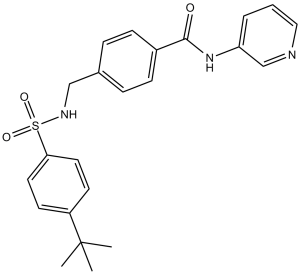
| SMILES | O=C(NC1=CC=CN=C1)C2=CC=C(CNS(=O)(C3=CC=C(C(C)(C)C)C=C3)=O)C=C2 |
|
| InChi Key | NGQPRVWTFNBUHA-UHFFFAOYSA-N | |
| InChi Code | InChI=1S/C23H25N3O3S/c1-23(2,3)19-10-12-21(13-11-19)30(28,29)25-15-17-6-8-18(9-7-17)22(27)26-20-5-4-14-24-16-20/h4-14,16,25H,15H2,1-3H3,(H,26,27) | |
| Chemical Name | 4-[[(4-tert-butylphenyl)sulfonylamino]methyl]-N-pyridin-3-ylbenzamide | |
| Synonyms |
|
|
| HS Tariff Code | 2934.99.9001 | |
| Storage |
Powder-20°C 3 years 4°C 2 years In solvent -80°C 6 months -20°C 1 month |
|
| Shipping Condition | Room temperature (This product is stable at ambient temperature for a few days during ordinary shipping and time spent in Customs) |
Biological Activity
| Targets |
GLUT1 (IC50 = 1 μM) STF-31 targets glucose transporter 1 (GLUT1) with an IC50 of 1.2 μM (human GLUT1-mediated glucose transport inhibition) [2] |
| ln Vitro |
STF-31 specifically targets glucose uptake through GLUT1, which kills RCCs with selectivity. STF-31 reduces glucose transport, which lowers glycolysis, and dramatically inhibits lactate production and extracellular acidification in VHL-deficient cells by approximately 60%. (Source: ) By preventing NAMPT's enzymatic activity, STF-31 exhibits cytotoxicity in cells that express NAPRT1.[2] STF-31 selectively eliminates hPSCs from mixed cultures and is toxic to hPSCs.[3] In human glioblastoma U87 and LN229 cells, STF-31 (0.5–10 μM) dose-dependently inhibited GLUT1-mediated glucose uptake, reducing [³H]-2-deoxyglucose (2-DG) uptake by 65% at 5 μM. This led to intracellular ATP depletion (40% reduction at 5 μM) and lactate production decrease (35% reduction at 5 μM) via blocking the Warburg effect [2] STF-31 (1–10 μM) exhibited dose-dependent cytotoxicity against GLUT1-high cancer cells: IC50=2.3 μM for U87, 2.8 μM for LN229, 3.1 μM for HeLa (cervical cancer), and 3.5 μM for MDA-MB-231 (breast cancer). It induced G1 cell cycle arrest (G1 phase cells increased from 40% to 68% at 5 μM in U87 cells) and apoptosis (Annexin V⁺ cells increased from 5% to 38% at 5 μM) [2] In human embryonic stem cells (hESCs) and induced pluripotent stem cells (iPSCs), STF-31 (2–5 μM) suppressed glycolytic metabolism, reducing glucose uptake by 50% and ATP levels by 30% without inducing apoptosis. It promoted differentiation of hESCs into mesodermal lineages by downregulating pluripotency markers (Oct4, Sox2, Nanog) at the mRNA and protein levels [3] STF-31 (5 μM) showed minimal toxicity to normal human astrocytes (cell viability >85%) and primary fibroblasts (cell viability >90%), with IC50 > 20 μM, indicating selective toxicity to GLUT1-overexpressing cancer cells [2] |
| ln Vivo |
More soluble STF-31 analogue (11.6 mg/kg, i.p.) significantly inhibits tumor growth in mice bearing RCC xenografts deficient in VHL. [1] In nude mice bearing U87 glioblastoma xenografts, intraperitoneal administration of STF-31 (10 mg/kg, daily for 14 days) reduced tumor volume by 60% compared to vehicle control. Immunohistochemical staining showed reduced [¹⁸F]-FDG uptake (55% reduction) and Ki-67-positive cells (45% reduction) in tumors. No significant body weight loss (<5% variation) or organ toxicity was observed [2] In mice with MDA-MB-231 breast cancer xenografts, STF-31 (15 mg/kg, daily for 18 days) inhibited tumor growth by 55% and reduced intratumoral microvessel density (CD31⁺ vessels decreased by 40%) [2] |
| Enzyme Assay |
GLUT1-mediated glucose transport assay: Culture HEK293 cells stably overexpressing human GLUT1 in DMEM with 10% FBS. Seed cells into 24-well plates (2×10⁵ cells/well) and incubate overnight. Serum-starve for 2 hours, then treat with serial dilutions of STF-31 (0.1–10 μM) for 30 minutes. Add [³H]-2-DG (1 μCi/well) and incubate at 37°C for 10 minutes. Terminate the reaction with ice-cold PBS, wash cells twice, lyse with lysis buffer, and measure radioactivity using a scintillation counter. Calculate IC50 for glucose transport inhibition [2] |
| Cell Assay |
Five thousand cells are plated in 96-well plates for XTT assays. The following day, the drug or vehicle (DMSO) is added by serial dilution. After aspirating the media four days later, the plates are incubated at 37°C for one to two hours. The XTT solution (0.3 mg/ml of XTT, 2.65 μg/ml N-methyl dibenxopyrazine methyl sulfate in phenol red-free media) is then added. The absorbance at 450 nm is used to quantify the metabolism of XTT. One method for calculating IC50s is linear interpolation. Every experiment is carried out in duplicate or triplicate, and all conditions are measured in triplicate. Glucose uptake and metabolic assay: Seed U87/LN229 cells (5×10⁴ cells/well) into 24-well plates, incubate overnight, serum-starve for 2 hours. Treat with STF-31 (0.5–10 μM) for 1 hour, add [³H]-2-DG to detect glucose uptake. For ATP and lactate detection, treat cells with STF-31 for 24 hours, lyse cells to measure ATP via luciferin-luciferase assay, and collect supernatants to quantify lactate by colorimetric assay [2] Proliferation and apoptosis assay: Seed cancer cells (5×10³ cells/well) into 96-well plates, treat with STF-31 (0.1–20 μM) for 72 hours, use MTT assay to calculate IC50. For apoptosis, treat U87 cells with 5 μM STF-31 for 48 hours, stain with Annexin V-FITC/PI, and analyze by flow cytometry [2] Pluripotency and differentiation assay: Culture hESCs/iPSCs in feeder-free medium, treat with STF-31 (2–5 μM) for 7 days. Extract RNA and protein to detect Oct4, Sox2, Nanog expression via qPCR and Western blot. Induce mesodermal differentiation, and confirm lineage commitment by detecting Brachyury and KDR expression [3] |
| Animal Protocol |
Mice with VHL-deficient RCC xenografts 11.6 mg/kg i.p. Glioblastoma xenograft model: 6–8 week-old nude mice (n=8/group) were subcutaneously injected with U87 cells (5×10⁶ cells/mouse). When tumors reached ~100 mm³, STF-31 was dissolved in DMSO and diluted with PBS (final DMSO concentration <5%) to 1 mg/mL. Mice were administered via intraperitoneal injection at 10 mg/kg once daily for 14 days. Vehicle control received DMSO/PBS mixture. Tumor volume was measured every 2 days (volume = length × width² × 0.5). At study end, mice were euthanized, tumors were collected for [¹⁸F]-FDG uptake assay and immunohistochemistry (Ki-67, CD31) [2] Breast cancer xenograft model: 6–8 week-old nude mice (n=7/group) were subcutaneously injected with MDA-MB-231 cells (2×10⁶ cells/mouse). When tumors reached ~120 mm³, STF-31 was administered via intraperitoneal injection at 15 mg/kg once daily for 18 days. Tumor growth was monitored, and microvessel density was analyzed by CD31 staining [2] |
| Toxicity/Toxicokinetics |
In acute toxicity study, mice intraperitoneally administered STF-31 at doses up to 200 mg/kg showed no mortality or obvious toxic signs (weight loss <8%, normal behavior). Serum ALT, AST, creatinine, and BUN levels were within normal ranges [2] In vitro, STF-31 exhibited selective toxicity to GLUT1-high cancer cells, with minimal impact on normal cells (IC50 > 20 μM for human astrocytes and fibroblasts) [2] Plasma protein binding rate of STF-31 in human plasma was 78% [2] |
| References |
[1]. Design, Synthesis, and Antiviral Activity of 2'-Deoxy-2'-fluoro-2'-C-methyl-cytidine, a Potent Inhibitor of Hepatitis C Virus Replication. J Med Chem. 2005 Aug 25;48(17):5504-8. [2]. ACS Chem Biol . 2014 Oct 17;9(10):2247-54. [3]. Stem Cell Reports . 2014 Jun 6;3(1):185-203. |
| Additional Infomation |
STF-31 is a selective small-molecule inhibitor of GLUT1, a major glucose transporter overexpressed in various cancers that relies on aerobic glycolysis (Warburg effect) for energy [2] Its anti-tumor mechanism involves blocking GLUT1-mediated glucose uptake, depleting intracellular ATP, inhibiting cancer cell proliferation, and inducing apoptosis. It also suppresses tumor angiogenesis by reducing glucose availability for endothelial cells [2] In stem cells, STF-31 modulates metabolic reprogramming from glycolysis to oxidative phosphorylation, promoting differentiation of hESCs/iPSCs into mesodermal lineages by downregulating pluripotency factors [3] It is widely used as a research tool to study GLUT1 function, cancer metabolism, and stem cell differentiation. It has potential as a therapeutic agent for GLUT1-overexpressing cancers, including glioblastoma, breast cancer, and cervical cancer [2][3] |
Solubility Data
| Solubility (In Vitro) |
|
|||
| Solubility (In Vivo) |
Solubility in Formulation 1: 2.5 mg/mL (5.90 mM) in 10% DMSO + 40% PEG300 + 5% Tween80 + 45% Saline (add these co-solvents sequentially from left to right, and one by one), clear solution; with sonication. For example, if 1 mL of working solution is to be prepared, you can add 100 μL of 25.0 mg/mL clear DMSO stock solution to 400 μL PEG300 and mix evenly; then add 50 μL Tween-80 to the above solution and mix evenly; then add 450 μL normal saline to adjust the volume to 1 mL. Preparation of saline: Dissolve 0.9 g of sodium chloride in 100 mL ddH₂ O to obtain a clear solution. Solubility in Formulation 2: ≥ 2.5 mg/mL (5.90 mM) (saturation unknown) in 10% DMSO + 90% (20% SBE-β-CD in Saline) (add these co-solvents sequentially from left to right, and one by one), clear solution. For example, if 1 mL of working solution is to be prepared, you can add 100 μL of 25.0 mg/mL clear DMSO stock solution to 900 μL of 20% SBE-β-CD physiological saline solution and mix evenly. Preparation of 20% SBE-β-CD in Saline (4°C,1 week): Dissolve 2 g SBE-β-CD in 10 mL saline to obtain a clear solution. (Please use freshly prepared in vivo formulations for optimal results.) |
| Preparing Stock Solutions | 1 mg | 5 mg | 10 mg | |
| 1 mM | 2.3611 mL | 11.8055 mL | 23.6111 mL | |
| 5 mM | 0.4722 mL | 2.3611 mL | 4.7222 mL | |
| 10 mM | 0.2361 mL | 1.1806 mL | 2.3611 mL |