Physicochemical Properties
| Molecular Formula | C14H19CL2N5O |
| Molecular Weight | 344.239560365677 |
| Exact Mass | 343.096 |
| CAS # | 2369053-68-7 |
| PubChem CID | 139035014 |
| Appearance | White to off-white solid powder |
| Hydrogen Bond Donor Count | 5 |
| Hydrogen Bond Acceptor Count | 6 |
| Rotatable Bond Count | 2 |
| Heavy Atom Count | 22 |
| Complexity | 310 |
| Defined Atom Stereocenter Count | 0 |
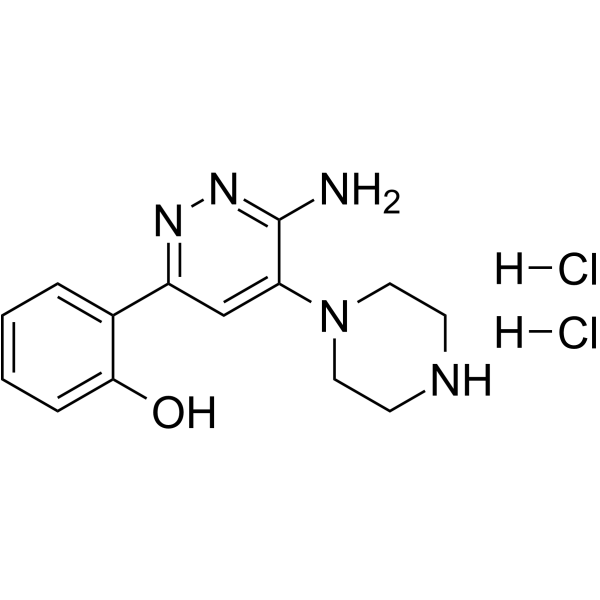
| SMILES | Cl.Cl.OC1C=CC=CC=1C1=CC(=C(N)N=N1)N1CCNCC1 |
| InChi Key | INBBYNMFULAIMY-UHFFFAOYSA-N |
| InChi Code | InChI=1S/C14H17N5O.2ClH/c15-14-12(19-7-5-16-6-8-19)9-11(17-18-14)10-3-1-2-4-13(10)20;;/h1-4,9,16,20H,5-8H2,(H2,15,18);2*1H |
| Chemical Name | 2-(6-amino-5-piperazin-1-ylpyridazin-3-yl)phenol;dihydrochloride |
| HS Tariff Code | 2934.99.9001 |
| Storage |
Powder-20°C 3 years 4°C 2 years In solvent -80°C 6 months -20°C 1 month Note: Please store this product in a sealed and protected environment, avoid exposure to moisture. |
| Shipping Condition | Room temperature (This product is stable at ambient temperature for a few days during ordinary shipping and time spent in Customs) |
Biological Activity
| Targets |
SMARCA-BD ligand 1 for Protac dihydrochloride targets SMARCA2 bromodomain (BD) (Ki = 1.8 μM) [1] SMARCA-BD ligand 1 for Protac dihydrochloride targets SMARCA4 bromodomain (BD) (Ki = 2.5 μM) [1] |
| ln Vitro |
1. Bromodomain binding activity: - SMARCA-BD ligand 1 for Protac dihydrochloride specifically binds to SMARCA2 BD and SMARCA4 BD, with no significant binding to other bromodomains (e.g., BRD4, CREBBP) [1] - The binding affinity (Ki) for SMARCA2 BD is 1.8 μM and for SMARCA4 BD is 2.5 μM, as determined by AlphaScreen assay [1] 2. PROTAC-mediated protein degradation: - When conjugated into a PROTAC (SMARCA-PROTAC), SMARCA-BD ligand 1 for Protac dihydrochloride mediates concentration-dependent degradation of SMARCA2 and SMARCA4 in HCT116 colon cancer cells [1] - Maximum degradation of SMARCA2 (≥80%) and SMARCA4 (≥70%) is achieved at 10 μM of SMARCA-PROTAC after 24 hours of treatment [1] - Western blot analysis confirms reduced SMARCA2/4 protein levels, with no significant effect on non-target proteins (e.g., BRD4, GAPDH) [1] 3. Antiproliferative activity: - SMARCA-PROTAC containing SMARCA-BD ligand 1 for Protac dihydrochloride inhibits proliferation of HCT116 cells with an IC50 value of ~3 μM (72-hour MTT assay) [1] - The antiproliferative effect is accompanied by G1 cell cycle arrest, as detected by flow cytometry [1] |
| Enzyme Assay |
1. AlphaScreen-based bromodomain binding assay: - Recombinant SMARCA2 BD or SMARCA4 BD protein is diluted in assay buffer and added to 384-well plates [1] - SMARCA-BD ligand 1 for Protac dihydrochloride is serially diluted (0.1 nM to 100 μM) and mixed with the protein solution, followed by incubation at room temperature for 1 hour [1] - Biotinylated histone H4 peptide (containing acetylated lysine) and streptavidin-conjugated donor beads, as well as anti-GST acceptor beads (for GST-tagged BD proteins), are added to the reaction mixture [1] - After incubation for 1 hour in the dark, the AlphaScreen signal is measured using a microplate reader, and Ki values are calculated by fitting the dose-response curves [1] |
| Cell Assay |
1. Cell proliferation assay (MTT): - HCT116 cells are seeded in 96-well plates at a density of 5×10^3 cells per well and cultured overnight in RPMI 1640 medium supplemented with fetal bovine serum and antibiotics [1] - SMARCA-PROTAC (incorporating SMARCA-BD ligand 1 for Protac dihydrochloride) is serially diluted (0.1 μM to 50 μM) and added to the cells, followed by incubation at 37°C with 5% CO2 for 72 hours [1] - MTT reagent is added to each well and incubated for 4 hours, then the supernatant is removed and formazan crystals are dissolved in DMSO [1] - The absorbance at 570 nm is measured using a microplate reader, and IC50 values are calculated from the dose-response curves [1] 2. Western blot for protein degradation assay: - HCT116 cells are seeded in 6-well plates at a density of 2×10^5 cells per well and cultured overnight [1] - Cells are treated with different concentrations (0.1, 1, 10, 50 μM) of SMARCA-PROTAC (containing SMARCA-BD ligand 1 for Protac dihydrochloride) for 24 hours [1] - Cells are lysed with RIPA buffer containing protease inhibitors, and protein concentrations are determined using a BCA assay [1] - Equal amounts of protein are separated by SDS-PAGE, transferred to PVDF membranes, and probed with primary antibodies against SMARCA2, SMARCA4, and GAPDH (loading control) [1] - After incubation with secondary antibodies, chemiluminescent signals are detected and quantified using imaging software [1] 3. Cell cycle analysis: - HCT116 cells are treated with 3 μM SMARCA-PROTAC (incorporating SMARCA-BD ligand 1 for Protac dihydrochloride) for 24 hours [1] - Cells are harvested, washed with PBS, fixed in 70% ethanol at -20°C overnight, and stained with propidium iodide (PI) solution containing RNase A [1] - Cell cycle distribution is analyzed by flow cytometry, and the percentage of cells in G1, S, and G2/M phases is calculated [1] |
| References |
[1]. BAF complex vulnerabilities in cancer demonstrated via structure-based PROTAC design. Nat Chem Biol. 2019 Jul;15(7):672-680. |
| Additional Infomation |
- SMARCA-BD ligand 1 for Protac dihydrochloride is a small-molecule ligand designed to specifically bind the bromodomain of SMARCA2 and SMARCA4 (members of the SWI/SNF chromatin remodeling complex) [1] - It serves as the target-binding moiety in SMARCA-PROTAC, a proteolysis-targeting chimera that induces ubiquitination and proteasomal degradation of SMARCA2/4 by recruiting the E3 ubiquitin ligase VHL [1] - The ligand is developed based on structure-based design, with a binding mode that involves hydrogen bonding and hydrophobic interactions with key residues in the SMARCA2/4 bromodomain pocket [1] - SMARCA2/4 are essential for chromatin remodeling and gene expression, and their degradation by SMARCA-PROTAC leads to inhibition of cancer cell proliferation, highlighting the ligand’s role in targeting BAF complex vulnerabilities in cancer [1] |
Solubility Data
| Solubility (In Vitro) | DMSO : ~2.7 mg/mL (~7.84 mM) |
| Solubility (In Vivo) |
Note: Listed below are some common formulations that may be used to formulate products with low water solubility (e.g. < 1 mg/mL), you may test these formulations using a minute amount of products to avoid loss of samples. Injection Formulations (e.g. IP/IV/IM/SC) Injection Formulation 1: DMSO : Tween 80: Saline = 10 : 5 : 85 (i.e. 100 μL DMSO stock solution → 50 μL Tween 80 → 850 μL Saline) *Preparation of saline: Dissolve 0.9 g of sodium chloride in 100 mL ddH ₂ O to obtain a clear solution. Injection Formulation 2: DMSO : PEG300 :Tween 80 : Saline = 10 : 40 : 5 : 45 (i.e. 100 μL DMSO → 400 μLPEG300 → 50 μL Tween 80 → 450 μL Saline) Injection Formulation 3: DMSO : Corn oil = 10 : 90 (i.e. 100 μL DMSO → 900 μL Corn oil) Example: Take the Injection Formulation 3 (DMSO : Corn oil = 10 : 90) as an example, if 1 mL of 2.5 mg/mL working solution is to be prepared, you can take 100 μL 25 mg/mL DMSO stock solution and add to 900 μL corn oil, mix well to obtain a clear or suspension solution (2.5 mg/mL, ready for use in animals). Injection Formulation 4: DMSO : 20% SBE-β-CD in saline = 10 : 90 [i.e. 100 μL DMSO → 900 μL (20% SBE-β-CD in saline)] *Preparation of 20% SBE-β-CD in Saline (4°C,1 week): Dissolve 2 g SBE-β-CD in 10 mL saline to obtain a clear solution. Injection Formulation 5: 2-Hydroxypropyl-β-cyclodextrin : Saline = 50 : 50 (i.e. 500 μL 2-Hydroxypropyl-β-cyclodextrin → 500 μL Saline) Injection Formulation 6: DMSO : PEG300 : castor oil : Saline = 5 : 10 : 20 : 65 (i.e. 50 μL DMSO → 100 μLPEG300 → 200 μL castor oil → 650 μL Saline) Injection Formulation 7: Ethanol : Cremophor : Saline = 10: 10 : 80 (i.e. 100 μL Ethanol → 100 μL Cremophor → 800 μL Saline) Injection Formulation 8: Dissolve in Cremophor/Ethanol (50 : 50), then diluted by Saline Injection Formulation 9: EtOH : Corn oil = 10 : 90 (i.e. 100 μL EtOH → 900 μL Corn oil) Injection Formulation 10: EtOH : PEG300:Tween 80 : Saline = 10 : 40 : 5 : 45 (i.e. 100 μL EtOH → 400 μLPEG300 → 50 μL Tween 80 → 450 μL Saline) Oral Formulations Oral Formulation 1: Suspend in 0.5% CMC Na (carboxymethylcellulose sodium) Oral Formulation 2: Suspend in 0.5% Carboxymethyl cellulose Example: Take the Oral Formulation 1 (Suspend in 0.5% CMC Na) as an example, if 100 mL of 2.5 mg/mL working solution is to be prepared, you can first prepare 0.5% CMC Na solution by measuring 0.5 g CMC Na and dissolve it in 100 mL ddH2O to obtain a clear solution; then add 250 mg of the product to 100 mL 0.5% CMC Na solution, to make the suspension solution (2.5 mg/mL, ready for use in animals). Oral Formulation 3: Dissolved in PEG400 Oral Formulation 4: Suspend in 0.2% Carboxymethyl cellulose Oral Formulation 5: Dissolve in 0.25% Tween 80 and 0.5% Carboxymethyl cellulose Oral Formulation 6: Mixing with food powders Note: Please be aware that the above formulations are for reference only. InvivoChem strongly recommends customers to read literature methods/protocols carefully before determining which formulation you should use for in vivo studies, as different compounds have different solubility properties and have to be formulated differently. (Please use freshly prepared in vivo formulations for optimal results.) |
| Preparing Stock Solutions | 1 mg | 5 mg | 10 mg | |
| 1 mM | 2.9050 mL | 14.5248 mL | 29.0495 mL | |
| 5 mM | 0.5810 mL | 2.9050 mL | 5.8099 mL | |
| 10 mM | 0.2905 mL | 1.4525 mL | 2.9050 mL |