SKLB4771 (also known as FLT3-IN-1) is a novel, potent and selective inhibitor of FLT3 (fms-like tyrosine kinase 3) that may be potentially used for the treatment of cutaneous inflammation and psoriasis-like symptoms of disease. In a dose-dependent manner, SKLB4771 inhibited FLT3 phosphorylation. At concentrations >0.1 μM, there was a significant inhibition of the phosphorylation of STAT5 and ERK1/2, downstream signaling proteins, which is consistent with the downregulation of FLT3 phosphorylation. A single daily dosage of 100 mg/kg of SKLB4771 for eighteen days resulted in total tumor regression in an MV4-11 xenograft mouse model with no discernible side effects.
Physicochemical Properties
| Molecular Formula | C25H27N7O3S2 | |
| Molecular Weight | 537.656981706619 | |
| Exact Mass | 537.162 | |
| Elemental Analysis | C, 55.85; H, 5.06; N, 18.24; O, 8.93; S, 11.93 | |
| CAS # | 1370256-78-2 | |
| Related CAS # |
|
|
| PubChem CID | 57412684 | |
| Appearance | Light yellow to yellow solid powder | |
| LogP | 4.769 | |
| Hydrogen Bond Donor Count | 2 | |
| Hydrogen Bond Acceptor Count | 10 | |
| Rotatable Bond Count | 9 | |
| Heavy Atom Count | 37 | |
| Complexity | 712 | |
| Defined Atom Stereocenter Count | 0 | |
| SMILES | 0 |
|
| InChi Key | LKXFSTAQMOENSC-UHFFFAOYSA-N | |
| InChi Code | InChI=1S/C25H27N7O3S2/c1-17-3-5-18(6-4-17)28-23(33)29-24-30-31-25(37-24)36-22-20-8-7-19(15-21(20)26-16-27-22)35-12-2-9-32-10-13-34-14-11-32/h3-8,15-16H,2,9-14H2,1H3,(H2,28,29,30,33) | |
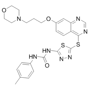
| Chemical Name | 1-(4-methylphenyl)-3-[5-[7-(3-morpholin-4-ylpropoxy)quinazolin-4-yl]sulfanyl-1,3,4-thiadiazol-2-yl]urea | |
| Synonyms |
|
|
| HS Tariff Code | 2934.99.9001 | |
| Storage |
Powder-20°C 3 years 4°C 2 years In solvent -80°C 6 months -20°C 1 month |
|
| Shipping Condition | Room temperature (This product is stable at ambient temperature for a few days during ordinary shipping and time spent in Customs) |
Biological Activity
| Targets |
Flt3 (IC50 = 10 nM); Flt4 (IC50 = 3.7 μM); Aurora A (IC50 = 1.5 μM); c-kit (IC50 = 6.8 μM); FMS (IC50 = 2.8 μM)
SKLB4771 targets human fms-like tyrosine kinase 3 (FLT3) (IC50 = 1.5 nM for wild-type FLT3; IC50 = 2.1 nM for FLT3-ITD mutant) [1] SKLB4771 exhibits >100-fold selectivity over other kinases including c-Kit (IC50 = 210 nM), VEGFR2 (IC50 = 320 nM), EGFR (IC50 = 450 nM), and PDGFRβ (IC50 = 380 nM) [1] |
| ln Vitro |
SKLB4771 merely inhibits c-Kit, FMS, 2.8 μM, 3.7 μM, and 1.5 μM of Aurora A, FMS, and FLT4, respectively. SKLB4771 demonstrates essentially no inhibitory action on the remaining 13 protein kinases that were chosen. With an IC50 value of 0.006 μM, SKLB4771 potently inhibits the growth of MV4-11 cells that express FLT3-ITD. Human T lymphoma Jurkat cells, human Burkitt's lymphoma Ramos cells, human lung cancer PC-9 and H292 cells, and human epithelial carcinoma A431 cells all show very weak inhibitory activity against SKLB4771 (IC50: 3.05 μM, 6.25 μM, 3.72 μM, 6.94 μM, and 8.91 μM, respectively).[1] In recombinant FLT3 kinase activity assay, SKLB4771 dose-dependently inhibited wild-type FLT3 and FLT3-ITD mutant kinase activity with IC50 values of 1.5 nM and 2.1 nM, respectively, by competing with ATP for the kinase domain [1] - In human acute myeloid leukemia (AML) cell lines: MV4-11 (FLT3-ITD), MOLM-13 (FLT3-ITD), and HL-60 (FLT3-WT), SKLB4771 inhibited cell proliferation with IC50 values of 3.8 nM, 5.2 nM, and 28.6 nM, respectively, after 72-hour treatment (CCK-8 assay) [1] - In MV4-11 cells, SKLB4771 (5-20 nM) dose-dependently induced apoptosis: 20 nM treatment increased apoptotic rate from 5.3% (vehicle) to 38.6% (Annexin V-FITC/PI staining), accompanied by activation of caspase-3 (3.2-fold increase) and cleavage of PARP [1] - Western blot analysis showed that SKLB4771 (10 nM) inhibited FLT3 phosphorylation (p-FLT3) by ~90% in MV4-11 cells, and reduced downstream STAT5, AKT, and ERK1/2 phosphorylation by ~85%, ~78%, and ~72%, respectively [1] - In FLT3(+) CD11c(+) dendritic cells (DCs) isolated from psoriatic lesions, SKLB4771 (10-50 nM) dose-dependently inhibited DC proliferation (IC50 = 18.5 nM) and reduced secretion of pro-inflammatory cytokines (IL-12, TNF-α, IFN-γ) by ~40-60% at 50 nM [2] - SKLB4771 (up to 100 nM) did not significantly affect the viability of normal human peripheral blood mononuclear cells (PBMCs) [1][2] |
| ln Vivo |
SKLB4771 treatment at 100 mg/kg/d causes all of the mice in this group's MV4-11 xenograft model to rapidly and completely lose their tumors. Tumor growth is dramatically slowed down by SKLB4771 treatment at doses of 20 mg/kg/d and 40 mg/kg/d; the corresponding tumor inhibition rates are 66% and 84%. Additionally, none of the SKLB4771-treated mice showed any discernible toxicity during the course of the trial, nor did any of them significantly lose weight. There are noticeably fewer Ki67 (tumor mitotic index)-positive cells in the tumor tissues from the SKLB4771-treated groups. The TUNEL data clearly demonstrate a time-dependent rise in the proportion of apoptotic cells. [1] In nude mice bearing MV4-11 (FLT3-ITD) AML xenografts, oral administration of SKLB4771 (20 mg/kg/day, 40 mg/kg/day, or 80 mg/kg/day for 21 days) dose-dependently inhibited tumor growth: high-dose treatment resulted in a tumor growth inhibition (TGI) rate of 75% and prolonged median survival from 28 days (vehicle) to 46 days [1] - SKLB4771 (40 mg/kg/day for 21 days) reduced p-FLT3 and p-STAT5 expression in tumor tissues by ~80% and ~75%, respectively, as detected by immunohistochemistry [1] - In imiquimod (IMQ)-induced psoriasis-like mouse model, intraperitoneal injection of SKLB4771 (10 mg/kg/day or 20 mg/kg/day for 7 days) dose-dependently improved skin lesions: high-dose treatment reduced epidermal thickness by ~65% and decreased inflammatory cell infiltration (CD11c(+) DCs, CD4(+) T cells) by ~55-60% [2] - SKLB4771 (20 mg/kg/day for 7 days) reduced serum levels of IL-12, TNF-α, and IFN-γ by ~50%, ~55%, and ~48%, respectively, in IMQ-induced mice [2] |
| Enzyme Assay |
In a dose-dependent manner, SKLB4771 inhibited the phosphorylation of FLT3. The phosphorylation of downstream signaling proteins STAT5 and ERK1/2 was also significantly inhibited at concentrations >0.1 μM, which is consistent with the downregulation of FLT3 phosphorylation. FLT3 kinase activity assay: Recombinant human wild-type FLT3 and FLT3-ITD mutant kinase domains were incubated with reaction buffer containing ATP (10 μM) and a fluorescent peptide substrate. Serial dilutions of SKLB4771 (0.001-1000 nM) were added, and the mixture was incubated at 37°C for 60 minutes. The reaction was terminated by adding EDTA-containing stop solution, and fluorescence intensity (excitation 485 nm, emission 535 nm) was measured to assess peptide phosphorylation. IC50 values were calculated by nonlinear regression of dose-response curves [1] - Kinase selectivity assay: A panel of 25 recombinant kinases (including c-Kit, VEGFR2, EGFR, PDGFRβ) was subjected to the same kinase assay protocol. SKLB4771 (0.001-1000 nM) was tested to determine IC50 values for these kinases, confirming selective inhibition of FLT3 [1] |
| Cell Assay |
In a 96-well plate, 1−4 × 10⁴ leukemia cells are seeded per well, and each well is filled with an equal volume of medium containing escalating inhibitor concentrations. Following the 72-hour incubation period at 37°C, 20 μL of the 5 mg/mL MTT reagent is added to each well for a 2- to 4-hour incubation period, and the cells are lysed using 50 μL of 20% acidified SDS per well. Serial dilutions of inhibitors in culture medium are used to replace the medium after the other cell lines are seeded in 96-well plates at a density of 2.5 × 10³ cells/well for 24 hours. After incubating for 72 hours, the cells are dissolved using 100% DMSO and then the MTT reagent is added and incubated for an additional 24 hours. AML cell proliferation assay: MV4-11, MOLM-13, and HL-60 cells were seeded in 96-well plates at 5×10³ cells/well. After 24-hour attachment, serial dilutions of SKLB4771 (0.1-100 nM) were added, and cells were cultured for 72 hours. CCK-8 reagent was added, and absorbance at 450 nm was measured to calculate cell viability and IC50 values [1] - Apoptosis and western blot assay: MV4-11 cells were seeded in 6-well plates (2×10⁵ cells/well) and treated with SKLB4771 (5-20 nM) for 48 hours. Apoptotic rate was determined by Annexin V-FITC/PI staining and flow cytometry. For western blot, cells were lysed in RIPA buffer, and proteins were probed with antibodies against p-FLT3, FLT3, p-STAT5, STAT5, p-AKT, AKT, p-ERK1/2, ERK1/2, caspase-3, PARP, and GAPDH [1] - Psoriatic DC function assay: FLT3(+) CD11c(+) DCs were isolated from psoriatic lesion tissues and seeded in 24-well plates (1×10⁵ cells/well). Cells were treated with SKLB4771 (10-50 nM) for 24 hours. DC proliferation was assessed by CCK-8 assay, and culture supernatants were collected to quantify IL-12, TNF-α, and IFN-γ levels by ELISA [2] - Normal PBMC viability assay: Human PBMCs were isolated and seeded in 96-well plates (1×10⁴ cells/well). Cells were treated with SKLB4771 (0.1-100 nM) for 72 hours, and viability was measured by CCK-8 assay [1][2] |
| Animal Protocol |
female NOD-SCID mouse (6−7 weeks old) 20 mg/kg, 40 mg/kg, 100 mg/kg IV MV4-11 AML xenograft model: Female BALB/c nude mice (4-6 weeks old) were subcutaneously implanted with 5×10⁶ MV4-11 cells. When tumors reached ~100 mm³, mice were randomly divided into vehicle control, SKLB4771 20 mg/kg, 40 mg/kg, and 80 mg/kg groups (n=6 per group). The drug was dissolved in 0.5% methylcellulose + 0.2% Tween 80 and administered by oral gavage once daily for 21 days. Tumor volume was measured every 3 days, and survival was recorded for 60 days. Tumor tissues were collected for immunohistochemical staining of p-FLT3 and p-STAT5 [1] - IMQ-induced psoriasis-like mouse model: Male C57BL/6 mice (6-8 weeks old) were topically treated with 5% IMQ cream on the back skin for 7 consecutive days to induce psoriasis-like lesions. From day 1 to day 7, mice were randomly divided into vehicle control and SKLB4771 10 mg/kg, 20 mg/kg groups (n=8 per group). The drug was dissolved in DMSO and diluted with saline (final DMSO ≤5%) and administered by intraperitoneal injection once daily. Skin thickness was measured on day 7, and skin tissues were collected for hematoxylin-eosin (HE) staining and inflammatory cell counting. Serum was collected to detect cytokine levels [2] |
| ADME/Pharmacokinetics |
Oral bioavailability: In mice, oral administration of SKLB4771 (40 mg/kg) resulted in an oral bioavailability of ~58% [1] - Plasma half-life (t1/2): In mice, the terminal plasma half-life of SKLB4771 was 5.2 ± 0.7 hours after oral administration (40 mg/kg) [1] - Peak plasma concentration (Cmax): In mice, oral SKLB4771 (40 mg/kg) achieved a Cmax of 426 ± 53 ng/mL at 1.2 ± 0.3 hours post-dosing [1] - AUC0-∞: In mice, AUC0-∞ of SKLB4771 was 2890 ± 320 ng·h/mL after a single oral dose of 40 mg/kg [1] - Volume of distribution (Vd/F): In mice, the apparent volume of distribution was 15.3 ± 2.1 L/kg (oral 40 mg/kg) [1] - Clearance (CL/F): Apparent oral clearance in mice was 13.8 ± 1.5 mL/min/kg (oral 40 mg/kg) [1] |
| Toxicity/Toxicokinetics |
In vitro cytotoxicity: SKLB4771 exhibited CC50 > 100 nM in human PBMCs, indicating low toxicity to normal cells [1][2] - Acute toxicity in mice: Single oral administration of SKLB4771 up to 200 mg/kg did not cause mortality or overt toxicity (lethargy, weight loss, abnormal behavior) [1] - Chronic toxicity in mice: Repeated oral administration of SKLB4771 (80 mg/kg/day for 21 days) did not induce significant changes in hematological parameters (RBC, WBC, platelets) or serum biochemical markers (ALT, AST, creatinine, BUN) [1] - Plasma protein binding: SKLB4771 exhibited plasma protein binding of 92-94% in mouse plasma and 93-95% in human plasma (equilibrium dialysis) [1] |
| References |
[1]. Discovery of the novel potent and selective FLT3 inhibitor 1-{5-[7-(3- morpholinopropoxy)quinazolin-4-ylthio]-[1,3,4]thiadiazol-2-yl}-3-p-tolylurea and its anti-acute myeloid leukemia (AML) activities in vitro and in vivo. J Med Chem. 2012 Apr 26;55(8):3852-66. [2]. Accumulation of FLT3(+) CD11c (+) dendritic cells in psoriatic lesions and the anti-psoriatic effect of a selective FLT3 inhibitor. Immunol Res. 2014 Oct;60(1):112-26. |
| Additional Infomation |
SKLB4771 is a potent, selective small-molecule inhibitor of FLT3 (wild-type and ITD mutant), developed for the treatment of acute myeloid leukemia (AML) and psoriasis [1][2] - The therapeutic mechanism of SKLB4771 involves selective inhibition of FLT3 kinase activity, blocking downstream STAT5/AKT/ERK signaling pathways, thereby suppressing proliferation and inducing apoptosis of FLT3-driven AML cells; in psoriasis, it inhibits FLT3(+) CD11c(+) DC proliferation and pro-inflammatory cytokine secretion, alleviating skin inflammation [1][2] - SKLB4771 shows high selectivity for FLT3 over other kinases, minimizing off-target effects [1] - Preclinical data demonstrate that SKLB4771 exhibits significant in vitro and in vivo efficacy against FLT3-dependent AML and psoriasis, with favorable pharmacokinetic profiles and low toxicity, supporting its potential as a targeted therapy for these diseases [1][2] |
Solubility Data
| Solubility (In Vitro) |
|
|||
| Solubility (In Vivo) |
Solubility in Formulation 1: ≥ 2.5 mg/mL (4.65 mM) (saturation unknown) in 10% DMSO + 40% PEG300 + 5% Tween80 + 45% Saline (add these co-solvents sequentially from left to right, and one by one), clear solution. For example, if 1 mL of working solution is to be prepared, you can add 100 μL of 25.0 mg/mL clear DMSO stock solution to 400 μL PEG300 and mix evenly; then add 50 μL Tween-80 to the above solution and mix evenly; then add 450 μL normal saline to adjust the volume to 1 mL. Preparation of saline: Dissolve 0.9 g of sodium chloride in 100 mL ddH₂ O to obtain a clear solution. Solubility in Formulation 2: 2.5 mg/mL (4.65 mM) in 10% DMSO + 90% (20% SBE-β-CD in Saline) (add these co-solvents sequentially from left to right, and one by one), suspension solution; with ultrasonication. For example, if 1 mL of working solution is to be prepared, you can add 100 μL of 25.0 mg/mL clear DMSO stock solution to 900 μL of 20% SBE-β-CD physiological saline solution and mix evenly. Preparation of 20% SBE-β-CD in Saline (4°C,1 week): Dissolve 2 g SBE-β-CD in 10 mL saline to obtain a clear solution. Solubility in Formulation 3: ≥ 2.5 mg/mL (4.65 mM) (saturation unknown) in 10% DMSO + 90% Corn Oil (add these co-solvents sequentially from left to right, and one by one), clear solution. For example, if 1 mL of working solution is to be prepared, you can add 100 μL of 25.0 mg/mL clear DMSO stock solution to 900 μL of corn oil and mix evenly. (Please use freshly prepared in vivo formulations for optimal results.) |
| Preparing Stock Solutions | 1 mg | 5 mg | 10 mg | |
| 1 mM | 1.8599 mL | 9.2996 mL | 18.5991 mL | |
| 5 mM | 0.3720 mL | 1.8599 mL | 3.7198 mL | |
| 10 mM | 0.1860 mL | 0.9300 mL | 1.8599 mL |