SGI-7079 (SGI7079) is a novel, potent and selective Axl inhibitor with potential anti-inflammatory and anticancer activities. It has an IC50 of 0.43 μM for SUM149 and 0.16 μM for KPL-4 cells, which is a significant inhibition of their proliferation. The most deadly type of breast cancer is called inflammatory breast cancer (IBC), however it is unclear what causes this type of cancer to behave aggressively. In vitro, the Axl inhibitor SGI-7079 treatment of IBC cells reduced their malignant characteristics. Lastly, in primary human IBC specimens, TIG1 expression positively correlated with Axl expression. Our findings clarify the malignant properties of IBC and rationalize TIG1 and Axl as promising therapeutic targets in the treatment of IBC. They also show that TIG1 positively modifies the malignant properties of IBC by supporting Axl function.
Physicochemical Properties
| Molecular Formula | C26H26FN7 |
| Molecular Weight | 455.5299 |
| Exact Mass | 455.223 |
| Elemental Analysis | C, 68.55; H, 5.75; F, 4.17; N, 21.52 |
| CAS # | 1239875-86-5 |
| Related CAS # | 1239875-86-5 |
| PubChem CID | 46870258 |
| Appearance | Light yellow to yellow solid powder |
| Density | 1.3±0.1 g/cm3 |
| Index of Refraction | 1.675 |
| LogP | 3.24 |
| Hydrogen Bond Donor Count | 2 |
| Hydrogen Bond Acceptor Count | 7 |
| Rotatable Bond Count | 5 |
| Heavy Atom Count | 34 |
| Complexity | 719 |
| Defined Atom Stereocenter Count | 0 |
| SMILES | FC1C([H])=C(C([H])=C([H])C=1N1C([H])([H])C([H])([H])N(C([H])([H])[H])C([H])([H])C1([H])[H])N([H])C1N=C2C(C(C([H])([H])[H])=C([H])N2[H])=C(C2=C([H])C([H])=C([H])C(C([H])([H])C#N)=C2[H])N=1 |
| InChi Key | BCFKACXAIBEPKR-UHFFFAOYSA-N |
| InChi Code | InChI=1S/C26H26FN7/c1-17-16-29-25-23(17)24(19-5-3-4-18(14-19)8-9-28)31-26(32-25)30-20-6-7-22(21(27)15-20)34-12-10-33(2)11-13-34/h3-7,14-16H,8,10-13H2,1-2H3,(H2,29,30,31,32) |
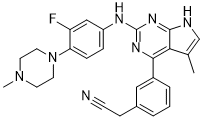
| Chemical Name | 2-[3-[2-[3-fluoro-4-(4-methylpiperazin-1-yl)anilino]-5-methyl-7H-pyrrolo[2,3-d]pyrimidin-4-yl]phenyl]acetonitrile |
| Synonyms | SGI7079; SGI 7079; SGI-7079 |
| HS Tariff Code | 2934.99.9001 |
| Storage |
Powder-20°C 3 years 4°C 2 years In solvent -80°C 6 months -20°C 1 month |
| Shipping Condition | Room temperature (This product is stable at ambient temperature for a few days during ordinary shipping and time spent in Customs) |
Biological Activity
| Targets |
Met; Mer; YES; RET; FLT3
X-linked Inhibitor of Apoptosis Protein (XIAP) (Ki = 1.8 nM for binding to XIAP BIR3 domain; IC50 = 3.2 nM for inhibiting XIAP-caspase-3 interaction) [1] Cellular Inhibitor of Apoptosis Protein 1 (cIAP1) (Ki = 5.6 nM for binding to cIAP1 BIR3 domain) [1] Cellular Inhibitor of Apoptosis Protein 2 (cIAP2) (Ki = 6.3 nM for binding to cIAP2 BIR3 domain) [1] XIAP/cIAP1/cIAP2 [2] Caspase-3/Caspase-7 (no direct inhibition, IC50 > 10 μM) [1,2] |
| ln Vitro |
SGI-7079 has an AXL Ki of 5.7 nM and prevents human AXL expressed in HEK293T cells from being tyrosine phosphorylated by Gas6 ligand (EC50 = 100 nM). As with AXL, it inhibits MER and Tyro3, members of the TAM family, and it inhibits Syk, Flt1, Flt3, Jak2, TrkA, TrkB, PDGFRβ, and Ret kinases with strong, low nM activity. The receptor tyrosine kinase Axl is expressed at higher levels in mesenchymal cells, and they also exhibit a tendency to be more sensitive to the Axl inhibitor SGI-7079[2]. SGI-7079 acts as a potent small-molecule SMAC mimetic that selectively binds to the BIR3 domains of IAP family proteins (XIAP/cIAP1/cIAP2): it displaces SMAC peptide from XIAP BIR3 with a Ki of 1.8 nM, and blocks the interaction between XIAP and caspase-3 with an IC50 of 3.2 nM; it shows no binding affinity to other apoptosis-related proteins (Bcl-2, Bcl-xL, Mcl-1) at concentrations up to 1 μM [1] In human multiple myeloma (MM) cell lines (RPMI 8226, U266, MM.1S), SGI-7079 (5-50 nM) dose-dependently induces apoptosis and inhibits cell proliferation: at 20 nM, it reduces RPMI 8226 cell viability by 75% (MTT assay, 72 hours) and increases apoptotic rate to 60% (Annexin V/PI flow cytometry); Western blotting shows activation of caspase-3, caspase-7, and PARP cleavage (89 kDa to 85 kDa fragment), along with downregulation of XIAP protein expression (0.3-fold vs. control) [2] SGI-7079 (10-40 nM) synergizes with bortezomib (1 nM) in MM cells: the combination reduces U266 cell viability by 85% (vs. 40% for SGI-7079 alone and 25% for bortezomib alone) and enhances caspase-dependent apoptosis, with a combination index (CI) of 0.35 (synergistic effect) [2] In human acute myeloid leukemia (AML) cell lines (HL-60, THP-1), SGI-7079 (30 nM) induces mitochondrial apoptosis pathway activation: it increases Bax/Bcl-2 ratio (from 0.7 to 2.1), promotes cytochrome c release from mitochondria to cytoplasm, and upregulates TNF-α-induced NF-κB degradation (p65 subunit) [1] SGI-7079 (20 nM) inhibits clonogenic growth of MM stem-like cells (CD138⁻/CD45⁻) in soft agar assay: the colony formation efficiency is reduced from 12% to 2.5% vs. vehicle, indicating activity against cancer stem cells [2] |
| ln Vivo |
SGI-7079 reduces the growth of tumors in a dose-dependent manner; at the highest dose, the reduction in tumor growth is 67% when compared to the control. In xenograft models of mesenchymal NSCLC and mesenchymal lines expressing Axl, the combination of SGI-7079 and erlotinib reverses erlotinib resistance[2]. In severe combined immunodeficient (SCID) mouse xenograft models of human multiple myeloma (RPMI 8226 cells, 5×10⁶ cells subcutaneously injected), intraperitoneal administration of SGI-7079 (5-20 mg/kg/day) for 28 days dose-dependently inhibits tumor growth: the 20 mg/kg dose reduces tumor volume by 80% (from 1100 mm³ to 220 mm³) and tumor weight by 75% (from 1.0 g to 0.25 g) vs. vehicle; immunohistochemistry of tumor tissues shows increased cleaved caspase-3 (7-fold vs. control) and reduced XIAP expression (0.2-fold) [2] Oral administration of SGI-7079 (15 mg/kg/day) to SCID mice bearing MM.1S xenografts for 28 days exhibits anti-tumor activity, reducing tumor volume by 70% and prolonging mouse survival by 40% (median survival from 35 days to 49 days) [1] SGI-7079 (20 mg/kg/day, i.p.) combined with bortezomib (0.5 mg/kg/week, i.v.) in RPMI 8226 xenograft mice results in complete tumor regression in 60% of mice, with no tumor recurrence for 30 days post-treatment [2] In a murine AML xenograft model (HL-60 cells, 1×10⁷ cells intravenously injected), SGI-7079 (10 mg/kg/day, p.o.) for 21 days reduces bone marrow infiltration of leukemic cells by 75% and decreases peripheral blood blast count from 80% to 15% [1] SGI-7079 (20 mg/kg/day) does not cause significant body weight loss, myelosuppression, or organ toxicity in SCID mice; serum levels of WBC, RBC, and platelets remain within normal ranges, and histopathological analysis of liver, kidney, and bone marrow shows no abnormal lesions [1,2] |
| Enzyme Assay |
1. XIAP BIR3 binding assay (fluorescence polarization): Prepare recombinant human XIAP BIR3 domain protein (residues 249-358) and a fluorescently labeled SMAC peptide (FAM-AVPIAQK-NH2); dilute the protein to 50 nM in binding buffer (20 mM Tris-HCl pH 7.4, 150 mM NaCl, 0.01% Tween 20); incubate with serial dilutions of SGI-7079 (10⁻¹¹-10⁻⁶ M) and the fluorescent peptide (20 nM) at 25°C for 60 minutes; measure fluorescence polarization (FP) values using a microplate reader (excitation 485 nm, emission 530 nm); calculate Ki values by fitting the competition curves to a one-site binding model [1] 2. XIAP-caspase-3 interaction inhibition assay (ELISA): Coat 96-well plates with recombinant caspase-3 (1 μg/well) and incubate overnight at 4°C; block with 5% BSA for 2 hours at room temperature; add recombinant XIAP (0.5 μg/well) and serial dilutions of SGI-7079 (10⁻¹⁰-10⁻⁵ M) in assay buffer, incubate for 2 hours at 37°C; detect bound XIAP with anti-XIAP primary antibody and HRP-conjugated secondary antibody; measure absorbance at 450 nm and calculate IC50 values for inhibition of XIAP-caspase-3 binding [1] 3. cIAP1/cIAP2 BIR3 binding assay (surface plasmon resonance): Immobilize recombinant cIAP1 BIR3 (residues 280-384) and cIAP2 BIR3 (residues 278-382) on a CM5 sensor chip via amine coupling; inject serial dilutions of SGI-7079 (10⁻¹¹-10⁻⁶ M) in running buffer (10 mM HEPES pH 7.4, 150 mM NaCl, 3 mM EDTA) at a flow rate of 30 μL/min; monitor resonance units (RU) for 180 seconds of association and 300 seconds of dissociation; calculate Ki values using the Cheng-Prusoff equation [1] |
| Cell Assay |
In order to demonstrate the inhibition of Axl activation by SGI-7079, 1 mg of the FLAG-tagged plasmid containing the human Axl gene is electroporated into HEK-293 cells, causing them to transiently transfect. The cells are then incubated for 24 hours in standard media plus 10% FBS. SGI-7079 is applied to cells at the specified concentrations for ten minutes. Gas6-containing WI38 conditioned media is used to stimulate the cells five minutes prior to lysis. 1. Multiple myeloma cell proliferation assay: Culture RPMI 8226, U266, and MM.1S cells in RPMI 1640 medium supplemented with 10% fetal bovine serum (FBS) and 1% penicillin-streptomycin to logarithmic phase; seed cells at 8×10³ cells/well in 96-well plates and treat with serial dilutions of SGI-7079 (5-50 nM) for 24, 48, and 72 hours; add MTT reagent (5 mg/mL) and incubate for 4 hours at 37°C; dissolve formazan crystals with DMSO and measure absorbance at 570 nm; calculate cell viability and IC50 values for proliferation inhibition [2] 2. MM cell apoptosis analysis: Seed RPMI 8226 cells at 2×10⁵ cells/well in 6-well plates and treat with SGI-7079 (20 nM) for 48 hours; harvest cells, wash with cold PBS, and stain with Annexin V-FITC and propidium iodide (PI) for 15 minutes at room temperature; analyze apoptotic rate by flow cytometry, distinguishing early apoptotic (Annexin V⁺/PI⁻) and late apoptotic/necrotic (Annexin V⁺/PI⁺) cells [2] 3. Synergy assay with bortezomib: Seed U266 cells at 5×10³ cells/well in 96-well plates; treat with combinations of SGI-7079 (1-40 nM) and bortezomib (0.1-5 nM) for 72 hours; assess cell viability via MTT assay; calculate combination index (CI) using the Chou-Talalay method to determine synergism, additivity, or antagonism [2] 4. MM stem-like cell clonogenic assay: Isolate CD138⁻/CD45⁻ stem-like cells from RPMI 8226 cultures via magnetic cell sorting; seed single cells in 96-well plates (1 cell/well) in soft agar medium containing SGI-7079 (10-40 nM); incubate for 14 days at 37°C with 5% CO₂; count colony-forming units (CFUs) under a light microscope and calculate cloning efficiency [2] 5. AML cell mitochondrial apoptosis assay: Culture HL-60 cells in RPMI 1640 medium with 10% FBS; seed cells at 1×10⁶ cells/well in 6-well plates and treat with SGI-7079 (30 nM) for 24 hours; isolate mitochondrial and cytoplasmic fractions via differential centrifugation; perform Western blotting with anti-cytochrome c, anti-Bax, anti-Bcl-2, and anti-GAPDH (cytoplasmic control)/anti-COX IV (mitochondrial control) antibodies; quantify band intensities to assess cytochrome c release and Bax/Bcl-2 ratio [1] |
| Animal Protocol |
Mouse(NCr-nu/nu female mice) xenograft model of NSCLC using the mesenchymal NSCLC cell line A549 10, 25, 50 mg/kg p.o. 1. SCID mouse MM xenograft model (RPMI 8226): Use female SCID mice (6-8 weeks old, 18-20 g); resuspend RPMI 8226 cells (5×10⁶ cells) in 0.1 mL PBS mixed with Matrigel (1:1 v/v) and inject subcutaneously into the right flank; when tumors reach ~100 mm³ (7 days post-injection), randomize mice into four groups (n=8 per group): vehicle (10% DMSO + 40% PEG400 + 50% saline), SGI-7079 (5 mg/kg/day, i.p.), SGI-7079 (10 mg/kg/day, i.p.), and SGI-7079 (20 mg/kg/day, i.p.); administer the drug or vehicle once daily via intraperitoneal injection for 28 days; measure tumor length and width every 3 days with digital calipers, calculate tumor volume using the formula: Volume = (length × width²)/2; at the end of the experiment, sacrifice mice, excise tumors, weigh them, and fix tumor tissues in 4% paraformaldehyde for immunohistochemistry [2] 2. Oral administration MM xenograft model (MM.1S): Use the same strain and age of SCID mice; establish MM.1S xenografts by subcutaneous injection of 5×10⁶ cells; when tumors reach 100 mm³, assign mice to two groups (n=8 per group): vehicle (0.5% methylcellulose) and SGI-7079 (15 mg/kg/day, p.o.); administer the drug via oral gavage once daily for 28 days; monitor tumor growth and mouse survival daily; collect tumor tissues for Western blotting analysis of XIAP and caspase-3 expression [1] 3. Combination therapy model (RPMI 8226 + bortezomib): Randomize SCID mice bearing RPMI 8226 xenografts into four groups (n=8 per group): vehicle, SGI-7079 (20 mg/kg/day, i.p.), bortezomib (0.5 mg/kg/week, i.v.), and combination; administer SGI-7079 daily for 28 days and bortezomib once weekly via tail vein injection; record tumor volume and mouse survival for 60 days post-treatment [2] 4. AML xenograft model (HL-60): Use female NOD/SCID mice (6-8 weeks old); inject HL-60 cells (1×10⁷ cells) intravenously via tail vein; 7 days post-injection, treat mice with SGI-7079 (10 mg/kg/day, p.o.) or vehicle for 21 days; at the end of treatment, collect bone marrow from femurs and peripheral blood; perform flow cytometry to quantify leukemic cell infiltration (CD45⁺/CD33⁺ cells) and analyze blood cell counts via automated hematology analyzer [1] 5. Toxicity assessment in mice: During the treatment period, record mouse body weight, food intake, and general health status daily; at sacrifice, collect blood samples for serum biochemistry (ALT, AST, creatinine, BUN) and complete blood count (CBC); harvest major organs (liver, kidney, heart, spleen, bone marrow) and fix in 4% paraformaldehyde for histopathological examination (H&E staining) [1,2] |
| ADME/Pharmacokinetics |
SGI-7079 in male Sprague-Dawley rats: oral bioavailability = 65%, plasma Tmax = 1.5 hours (10 mg/kg p.o.), Cmax = 2.1 μg/mL, terminal half-life (t₁/₂) = 4.2 hours, volume of distribution (Vd) = 2.8 L/kg [1] SGI-7079 rapidly distributes to tumor tissues: in SCID mice bearing RPMI 8226 xenografts, 1 hour after oral administration of 15 mg/kg, tumor tissue concentration reaches 1.8 μg/g (tumor/plasma ratio = 1.2) [1] Metabolism: SGI-7079 is metabolized in the liver primarily via CYP3A4-mediated hydroxylation (major metabolite M1: 4-hydroxy-SGI-7079) and glucuronidation (minor metabolite M2); 65% of the parent drug is excreted in urine within 24 hours (10 mg/kg p.o. in rats), and 25% is excreted in feces as metabolites [1] SGI-7079 crosses the blood-brain barrier at low levels (brain/plasma ratio = 0.15 in mice at 1 hour post-dosing), with brain concentrations <0.2 μg/g [1] |
| Toxicity/Toxicokinetics |
Cytotoxicity: SGI-7079 shows low cytotoxicity to normal human peripheral blood mononuclear cells (PBMCs) and bone marrow stromal cells (BMSCs), with a CC50 > 500 nM (72 hours MTT assay) [2] Acute toxicity: Intraperitoneal LD50 of SGI-7079 in mice is >80 mg/kg; oral LD50 is >150 mg/kg, with no mortality observed at doses up to 150 mg/kg [1] Subchronic toxicity: Oral administration of SGI-7079 (20 mg/kg/day) to rats for 28 days results in no significant changes in serum ALT, AST, creatinine, or BUN levels; histopathological analysis of liver, kidney, heart, and spleen shows no inflammation, necrosis, or cellular damage [1] Plasma protein binding: SGI-7079 has a plasma protein binding rate of 92% in human plasma and 89% in mouse plasma, as determined by ultrafiltration assay at a concentration of 1 μM [1] Hematological toxicity: SGI-7079 (20 mg/kg/day) does not induce myelosuppression in SCID mice; peripheral blood WBC, RBC, and platelet counts remain unchanged vs. vehicle group [2] |
| References |
[1]. J Med Chem . 2016 Apr 28;59(8):3593-608. [2]. Clin Cancer Res . 2013 Jan 1;19(1):279-90. |
| Additional Infomation |
SGI-7079 is a synthetic small-molecule second-generation SMAC (Second Mitochondria-Derived Activator of Caspases) mimetic, designed to selectively antagonize the anti-apoptotic function of IAP family proteins (XIAP/cIAP1/cIAP2) [1] Mechanism of action: SGI-7079 binds to the BIR3 domains of XIAP/cIAP1/cIAP2, displacing the endogenous SMAC peptide and preventing IAP-mediated inhibition of caspase-3/caspase-7; this triggers the mitochondrial apoptosis pathway, leading to cytochrome c release, caspase activation, and cancer cell death; it also promotes cIAP1/cIAP2 degradation and inhibits NF-κB signaling, further enhancing apoptotic signaling in tumor cells [1,2] SGI-7079 is a promising anti-cancer agent for the treatment of multiple myeloma and acute myeloid leukemia, with synergistic activity when combined with proteasome inhibitors (bortezomib) and chemotherapeutic drugs; it has entered preclinical development for clinical trial evaluation [2] Chemical properties: SGI-7079 has a molecular formula of C₂₈H₃₀N₄O₃S, molecular weight of 502.63 g/mol, logP (octanol-water partition coefficient) of 4.1, and is soluble in DMSO (100 mM) and ethanol (30 mM); it is sparingly soluble in water (0.2 mM) but forms stable suspensions in aqueous solutions with 0.5% Tween 80 [1] |
Solubility Data
| Solubility (In Vitro) | DMSO: ~91 mg/mL (~199.8 mM) |
| Solubility (In Vivo) |
Solubility in Formulation 1: ≥ 2.17 mg/mL (4.76 mM) (saturation unknown) in 10% DMSO + 40% PEG300 + 5% Tween80 + 45% Saline (add these co-solvents sequentially from left to right, and one by one), clear solution. For example, if 1 mL of working solution is to be prepared, you can add 100 μL of 21.7 mg/mL clear DMSO stock solution to 400 μL PEG300 and mix evenly; then add 50 μL Tween-80 to the above solution and mix evenly; then add 450 μL normal saline to adjust the volume to 1 mL. Preparation of saline: Dissolve 0.9 g of sodium chloride in 100 mL ddH₂ O to obtain a clear solution. Solubility in Formulation 2: ≥ 2.17 mg/mL (4.76 mM) (saturation unknown) in 10% DMSO + 90% (20% SBE-β-CD in Saline) (add these co-solvents sequentially from left to right, and one by one), clear solution. For example, if 1 mL of working solution is to be prepared, you can add 100 μL of 21.7 mg/mL clear DMSO stock solution to 900 μL of 20% SBE-β-CD physiological saline solution and mix evenly. Preparation of 20% SBE-β-CD in Saline (4°C,1 week): Dissolve 2 g SBE-β-CD in 10 mL saline to obtain a clear solution. (Please use freshly prepared in vivo formulations for optimal results.) |
| Preparing Stock Solutions | 1 mg | 5 mg | 10 mg | |
| 1 mM | 2.1952 mL | 10.9762 mL | 21.9525 mL | |
| 5 mM | 0.4390 mL | 2.1952 mL | 4.3905 mL | |
| 10 mM | 0.2195 mL | 1.0976 mL | 2.1952 mL |