SC75741 (SC-75741; SC 75741) is a novel, selective and potent NF-κB inhibitor with potential antiviral and anti-inflammatory activity. With an EC50 of 200 nM, it blocks NF-κB. In non-toxic concentrations, SC75741 prevents influenza viruses (IV) from replicating. Reduced expression of cytokines, chemokines, and pro-apoptotic factors is the result of SC75741's impairment of NF-κB subunit p65's DNA binding. Following this, SC75741 prevents caspase activation and obstructs the nuclear export of viral ribonucleoproteins caused by caspase. The inhibitory potential of 5 uM SC75741 on virus replication in MDCK cells is greatest later in infection. SC75741 caused an effective, dose-dependent decline in viral titers in the human alveolar type II epithelial cell line A549 that was infected with the H5N1 and H7N7 viruses.
Physicochemical Properties
| Molecular Formula | C29H23N7O2S2 | |
| Molecular Weight | 565.67 | |
| Exact Mass | 567.151 | |
| Elemental Analysis | C, 61.58; H, 4.10; N, 17.33; O, 5.66; S, 11.34 | |
| CAS # | 913822-46-5 | |
| Related CAS # |
|
|
| PubChem CID | 23661638 | |
| Appearance | Light yellow to brown solid powder | |
| LogP | 5.377 | |
| Hydrogen Bond Donor Count | 2 | |
| Hydrogen Bond Acceptor Count | 9 | |
| Rotatable Bond Count | 6 | |
| Heavy Atom Count | 40 | |
| Complexity | 915 | |
| Defined Atom Stereocenter Count | 0 | |
| SMILES | O=C(C1=CSC(C2CCN(C3C4=C(C=CS4)N=CN=3)CC2)=N1)NC1NC2C(=CC=C(C(C3C=CC=CC=3)=O)C=2)N=1 |
|
| InChi Key | QNZVBFMXWNWVKG-UHFFFAOYSA-N | |
| InChi Code | InChI=1S/C29H23N7O2S2/c37-24(17-4-2-1-3-5-17)19-6-7-20-22(14-19)34-29(33-20)35-27(38)23-15-40-28(32-23)18-8-11-36(12-9-18)26-25-21(10-13-39-25)30-16-31-26/h1-7,10,13-16,18H,8-9,11-12H2,(H2,33,34,35,38) | |
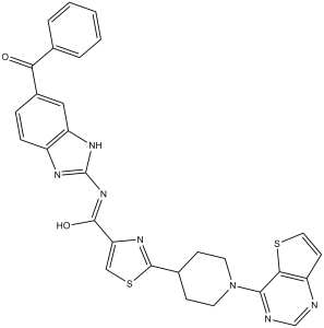
| Chemical Name | N-(6-benzoyl-1H-benzimidazol-2-yl)-2-(1-thieno[3,2-d]pyrimidin-4-ylpiperidin-4-yl)-1,3-thiazole-4-carboxamide | |
| Synonyms |
|
|
| HS Tariff Code | 2934.99.9001 | |
| Storage |
Powder-20°C 3 years 4°C 2 years In solvent -80°C 6 months -20°C 1 month |
|
| Shipping Condition | Room temperature (This product is stable at ambient temperature for a few days during ordinary shipping and time spent in Customs) |
Biological Activity
| Targets |
NF-κB (EC50 = 200 nM) Nuclear Factor-κB (NF-κB) p65 Subunit: SC75741 selectively inhibits the nuclear translocation and DNA-binding activity of the NF-κB p65 subunit. It inhibits NF-κB-driven reporter gene activity in HEK293T cells with an IC50 of 0.8 ± 0.1 μM, and suppresses influenza A virus (IAV)-induced p65 nuclear translocation in A549 cells with an EC50 of 1.1 ± 0.2 μM [2] - Influenza A Virus (IAV) Replication: SC75741 indirectly inhibits IAV replication by targeting the NF-κB pathway (not direct viral proteins), with EC50 values of 1.2 ± 0.3 μM (H5N1), 1.5 ± 0.2 μM (H1N1), and 1.4 ± 0.3 μM (H3N2) in MDCK cells (determined by viral titer measurement) [2] |
| ln Vitro |
SC75741 exhibits immunosuppressive activity by limiting the proliferation of human PBMCs with IC50 of 2.2 μM.[1] SC75741 inhibits transcriptional levels of NF-κB mediated signaling, which prevents the replication of influenza A and B viruses. Additionally, SC75741 exhibits a high barrier to the emergence of virus variants with resistance.[2] Inhibition of IAV Replication (Multiple Strains): MDCK cells (canine kidney epithelial cells) were infected with IAV strains (H5N1, H1N1, H3N2) at a multiplicity of infection (MOI) of 0.01, then treated with SC75741 (0.1–20 μM) for 48 hours. Viral titers (measured by TCID50 assay) showed concentration-dependent inhibition: at 1 μM, titers of H5N1, H1N1, and H3N2 were reduced by 68%, 62%, and 65% respectively; at 5 μM, titers were reduced by >90% for all strains. No significant inhibition of viral entry (measured by viral nucleoprotein (NP) internalization at 1 hour post-infection) or early replication (viral RNA synthesis at 6 hours post-infection) was observed, indicating SC75741 acts at the late stage of IAV replication [2] - Suppression of NF-κB Activation in IAV-Infected Cells: A549 cells (human alveolar epithelial cells) were infected with H5N1 (MOI=0.1) and treated with SC75741 (0.5–5 μM) for 24 hours. Western blot analysis showed that SC75741 dose-dependently reduced IAV-induced p65 nuclear translocation (by 45% at 1 μM, 72% at 3 μM, 90% at 5 μM) without affecting IκBα phosphorylation or degradation (upstream of p65). RT-PCR and ELISA confirmed that SC75741 also decreased IAV-induced mRNA and protein levels of pro-inflammatory cytokines: TNF-α (reduced by 70% at 5 μM), IL-6 (reduced by 65% at 5 μM), and IFN-β (reduced by 60% at 5 μM) [1,2] |
| ln Vivo |
SC75741 (15 mg/kg i.p.) reduces virus replication and cytokine expression in mice lungs after H5N1 influenza virus infection. Mice that have been exposed to SC75741 (intraperitoneal injection; 15 mg/kg; for 2 days) have 90% less H5N1 virus mRNA spread throughout their lungs. The half-life of SC74751 is approximately 40 minutes, and its plasma levels (intravenously administered at 5 mg/kg and intraperitoneally administered at 15 mg/kg; for 3.5 and 6 hours) decrease mono-exponentially after intravenous administration. SC75741 has a half-life of 55 minutes after being administered intravenously, and it appears that this is due to a slow peritoneal uptake of the drug. Protection Against Lethal H5N1 Infection in Mice: Female BALB/c mice (6–8 weeks old) were intranasally infected with a lethal dose (10×50% egg infectious dose, EID50) of H5N1 virus. SC75741 was administered via intraperitoneal injection at a dose of 20 mg/kg/day at 1 hour, 24 hours, and 48 hours post-infection (3 total doses). Compared to the vehicle control group (DMSO + saline), SC75741 treatment significantly improved survival rate (from 0% to 60% at 14 days post-infection) and reduced body weight loss (from 35% to 12% at 7 days post-infection). Lung viral titers (measured by TCID50) in the treatment group were 100-fold lower than controls at 3 days post-infection. Histopathological analysis of lung tissue showed that SC75741 attenuated H5N1-induced pulmonary inflammation: reduced alveolar edema, neutrophil infiltration, and epithelial cell damage (histology score decreased from 8.5 ± 1.2 to 3.2 ± 0.8) [1] - Reduction of Systemic Inflammation in Mice: Serum from H5N1-infected mice treated with SC75741 (20 mg/kg) showed significantly lower levels of TNF-α (from 950 ± 80 pg/mL to 280 ± 40 pg/mL) and IL-6 (from 1200 ± 100 pg/mL to 320 ± 50 pg/mL) at 3 days post-infection (measured by ELISA). Immunohistochemical staining of lung tissue revealed that SC75741 inhibited p65 nuclear accumulation in alveolar epithelial cells and macrophages (positive cell rate decreased from 65% to 20%) [1] |
| Enzyme Assay |
In accordance with the manufacturer's instructions, the A549-NF-κB-SEAP cell line is used to prepare the NF-κB reporter gene assay. In essence, pNF-κB-SEAP reporter gene plasmid-stably transfected A549 cells are plated and given the night to attach. The cells are then stimulated with 10 ng/ml TNF-α for 22 hours after being incubated with the aforementioned compounds at concentrations of 100, 30, 10, 3, 1, 0.3, 0.1, and 0 μM for 5 hours. Using a chemiluminescent SEAP reporter gene assay, the cell's supernatant is examined for SEAP activity. NF-κB Reporter Gene Assay: HEK293T cells were co-transfected with an NF-κB-luciferase reporter plasmid (pNF-κB-luc) and a Renilla luciferase internal control plasmid (pRL-TK) using a transfection reagent. Twenty-four hours post-transfection, cells were treated with SC75741 (0.1–10 μM) for 2 hours, then stimulated with TNF-α (10 ng/mL) for 6 hours. Cells were lysed with passive lysis buffer, and luciferase activity was measured using a dual-luciferase assay system. The relative luciferase activity (firefly/Renilla) was calculated, and the IC50 was determined as the concentration of SC75741 that inhibited 50% of TNF-α-induced reporter activity [2] - NF-κB p65 DNA-Binding Assay (EMSA): Nuclear extracts were prepared from H5N1-infected A549 cells (MOI=0.1, 24 hours post-infection) treated with SC75741 (0.5–5 μM). Extracts (10 μg protein) were incubated with a 32P-labeled double-stranded oligonucleotide containing the NF-κB consensus binding site (5′-AGTTGAGGGGACTTTCCCAGGC-3′) in binding buffer (20 mM Tris-HCl pH7.5, 50 mM NaCl, 1 mM DTT, 10% glycerol, 0.5 μg poly(dI-dC)) for 20 minutes at room temperature. Samples were separated by 6% non-denaturing polyacrylamide gel electrophoresis, dried, and exposed to X-ray film. Band intensity of the p65-DNA complex was quantified using ImageJ, and inhibition rate was calculated relative to infected, untreated controls [2] |
| Cell Assay |
Cell viability assay is prepared using a CellTiter-BluTM Cell Viability Assay. Four replicates are measured for each compound concentration. IAV Viral Titer Measurement (TCID50 Assay): MDCK cells were seeded in 96-well plates at 2×10⁴ cells/well and cultured overnight. Cells were infected with IAV (H5N1, H1N1, H3N2) at MOI=0.01 for 1 hour at 37°C, then the inoculum was removed and replaced with medium containing SC75741 (0.1–20 μM). After 48 hours of incubation, culture supernatants were collected and serially diluted (10⁻¹ to 10⁻⁸) in fresh medium. Diluted supernatants were added to new MDCK cell monolayers and incubated for 72 hours. Cytopathic effect (CPE) was observed under a microscope, and the 50% tissue culture infectious dose (TCID50) was calculated using the Reed-Muench method [2] - Western Blot for NF-κB Activation Markers: A549 cells were infected with H5N1 (MOI=0.1) and treated with SC75741 (0.5–5 μM) for 24 hours. Cytoplasmic and nuclear extracts were prepared using a nuclear extraction kit. Equal amounts of protein (30 μg) were separated by 10% SDS-PAGE, transferred to PVDF membranes, and blocked with 5% non-fat milk for 1 hour. Membranes were incubated overnight at 4°C with primary antibodies against p65 (nuclear/cytoplasmic), IκBα, phospho-IκBα (Ser32), or β-actin (cytoplasmic control)/histone H3 (nuclear control). After washing, membranes were incubated with HRP-conjugated secondary antibodies for 1 hour, and bands were visualized with ECL reagent. Band intensity was quantified using ImageJ [1,2] - RT-PCR for Pro-Inflammatory Cytokines: A549 cells were infected with H5N1 (MOI=0.1) and treated with SC75741 (0.5–5 μM) for 24 hours. Total RNA was extracted using TRIzol reagent, and 1 μg RNA was reverse-transcribed to cDNA. RT-PCR was performed with specific primers for TNF-α, IL-6, IFN-β, and GAPDH (internal control). Reaction conditions: 95°C for 5 minutes, 35 cycles of 95°C (30s), 58°C (30s), 72°C (30s), and final extension at 72°C for 10 minutes. PCR products were separated by 1.5% agarose gel electrophoresis, stained with ethidium bromide, and quantified using ImageJ [1] |
| Animal Protocol |
Inbred female C57BL/6 mice at the age of 6-8 weeks[2] 15 mg/kg Intraperitoneal injection; for 2 days Lethal H5N1 Mouse Infection Model: Female BALB/c mice (6–8 weeks old, n=10 per group) were anesthetized with isoflurane and intranasally infected with 10×EID50 of H5N1 influenza virus (0.05 mL volume). SC75741 was dissolved in DMSO (10% final concentration) and diluted with sterile physiological saline to a concentration of 2 mg/mL. Mice in the treatment group received intraperitoneal injections of SC75741 at 20 mg/kg/day at 1 hour, 24 hours, and 48 hours post-infection. The vehicle control group received an equal volume of 10% DMSO in saline. Mice were monitored daily for survival rate and body weight for 14 days. At 3 days post-infection, 3 mice per group were euthanized by cervical dislocation: lung tissues were collected for viral titer (TCID50) and histopathological analysis, and serum was collected for cytokine (TNF-α, IL-6) measurement by ELISA [1] |
| Toxicity/Toxicokinetics |
In Vitro Cytotoxicity: MTT assay was performed in MDCK, A549, and HEK293T cells treated with SC75741 (0.1–50 μM) for 48 hours. Cell viability remained >90% at concentrations ≤20 μM; at 50 μM, viability decreased by ~15% (MDCK), ~12% (A549), and ~10% (HEK293T) compared to vehicle controls [1,2] - In Vivo Safety (Mice): BALB/c mice treated with SC75741 (20 mg/kg/day, 3 doses) showed no significant changes in body weight (excluding infection-related loss) or organ weights (liver, kidney, lung) compared to vehicle controls. Serum biochemical analysis (ALT, AST, BUN, creatinine) at 3 days post-infection revealed no abnormal values, indicating no obvious hepatic or renal toxicity. Histopathological examination of liver and kidney tissues showed no signs of damage [1] |
| References |
[1]. The NF-kappaB inhibitor SC75741 protects mice against highly pathogenic avian influenza A virus. Antiviral Res. 2013 Sep;99(3):336-44. [2]. The NF-κB inhibitor SC75741 efficiently blocks influenza virus propagation and confers a high barrier for development of viral resistance. Cell Microbiol. 2013 Jul;15(7):1198-211. |
| Additional Infomation |
Mechanism of Antiviral Action: SC75741 does not directly target viral proteins (e.g., hemagglutinin, neuraminidase, RNA polymerase) but inhibits IAV replication by blocking NF-κB p65 nuclear translocation. NF-κB activation is required for late-stage IAV replication (viral assembly/release) and excessive pro-inflammatory responses; thus, SC75741 exerts both antiviral and anti-inflammatory effects [1,2] - Broad Antiviral Spectrum: SC75741 inhibits replication of multiple IAV subtypes (H5N1, H1N1, H3N2) in vitro, including highly pathogenic avian influenza (HPAI) H5N1 and seasonal influenza strains, suggesting potential utility against diverse influenza outbreaks [2] - High Barrier to Viral Resistance: In a resistance selection assay, MDCK cells were infected with H1N1 virus and cultured with sub-optimal concentrations of SC75741 (0.5×EC50) for 15 consecutive passages. No significant increase in EC50 (≤1.2-fold change) was observed, indicating that IAV has a low tendency to develop resistance to SC75741—likely due to the drug targeting a host pathway (NF-κB) rather than viral proteins [2] |
Solubility Data
| Solubility (In Vitro) |
|
|||
| Solubility (In Vivo) |
Solubility in Formulation 1: ≥ 2.08 mg/mL (3.68 mM) (saturation unknown) in 10% DMSO + 40% PEG300 + 5% Tween80 + 45% Saline (add these co-solvents sequentially from left to right, and one by one), clear solution. For example, if 1 mL of working solution is to be prepared, you can add 100 μL of 20.8 mg/mL clear DMSO stock solution to 400 μL PEG300 and mix evenly; then add 50 μL Tween-80 to the above solution and mix evenly; then add 450 μL normal saline to adjust the volume to 1 mL. Preparation of saline: Dissolve 0.9 g of sodium chloride in 100 mL ddH₂ O to obtain a clear solution. Solubility in Formulation 2: 2.08 mg/mL (3.68 mM) in 10% DMSO + 90% (20% SBE-β-CD in Saline) (add these co-solvents sequentially from left to right, and one by one), suspension solution; with ultrasonication. For example, if 1 mL of working solution is to be prepared, you can add 100 μL of 20.8 mg/mL clear DMSO stock solution to 900 μL of 20% SBE-β-CD physiological saline solution and mix evenly. Preparation of 20% SBE-β-CD in Saline (4°C,1 week): Dissolve 2 g SBE-β-CD in 10 mL saline to obtain a clear solution. Solubility in Formulation 3: ≥ 2.08 mg/mL (3.68 mM) (saturation unknown) in 10% DMSO + 90% Corn Oil (add these co-solvents sequentially from left to right, and one by one), clear solution. For example, if 1 mL of working solution is to be prepared, you can add 100 μL of 20.8 mg/mL clear DMSO stock solution to 900 μL of corn oil and mix evenly. (Please use freshly prepared in vivo formulations for optimal results.) |
| Preparing Stock Solutions | 1 mg | 5 mg | 10 mg | |
| 1 mM | 1.7678 mL | 8.8391 mL | 17.6782 mL | |
| 5 mM | 0.3536 mL | 1.7678 mL | 3.5356 mL | |
| 10 mM | 0.1768 mL | 0.8839 mL | 1.7678 mL |