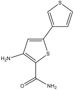
SC-514 (SC 514; SC514) is a novel, potent, orally bioactive, selective and reversible, and ATP-competitive IKK-2 inhibitor with potential anti-inflammatory activity. With an IC50 of 3–12 μM, it only inhibits IKK2 and does not affect other IKK isoforms. The treatment of osteoclastogenesis may be possible with SC-514.
Physicochemical Properties
| Molecular Formula | C9H8N2OS2 | |
| Molecular Weight | 224.3 | |
| Exact Mass | 224.007 | |
| Elemental Analysis | C, 48.19; H, 3.60; N, 12.49; O, 7.13; S, 28.59 | |
| CAS # | 354812-17-2 | |
| Related CAS # |
|
|
| PubChem CID | 2807869 | |
| Appearance | Light yellow to yellow solid powder | |
| Density | 1.5±0.1 g/cm3 | |
| Boiling Point | 399.2±42.0 °C at 760 mmHg | |
| Melting Point | 209-211°C | |
| Flash Point | 195.2±27.9 °C | |
| Vapour Pressure | 0.0±0.9 mmHg at 25°C | |
| Index of Refraction | 1.715 | |
| LogP | 1.52 | |
| Hydrogen Bond Donor Count | 2 | |
| Hydrogen Bond Acceptor Count | 4 | |
| Rotatable Bond Count | 2 | |
| Heavy Atom Count | 14 | |
| Complexity | 237 | |
| Defined Atom Stereocenter Count | 0 | |
| SMILES | S1C(C(N([H])[H])=O)=C(C([H])=C1C1=C([H])SC([H])=C1[H])N([H])[H] |
|
| InChi Key | BMUACLADCKCNKZ-UHFFFAOYSA-N | |
| InChi Code | InChI=1S/C9H8N2OS2/c10-6-3-7(5-1-2-13-4-5)14-8(6)9(11)12/h1-4H,10H2,(H2,11,12) | |
| Chemical Name | 3-amino-5-thiophen-3-ylthiophene-2-carboxamide | |
| Synonyms |
|
|
| HS Tariff Code | 2934.99.9001 | |
| Storage |
Powder-20°C 3 years 4°C 2 years In solvent -80°C 6 months -20°C 1 month |
|
| Shipping Condition | Room temperature (This product is stable at ambient temperature for a few days during ordinary shipping and time spent in Customs) |
Biological Activity
| Targets |
IKK-2 (IC50 = 11.2 μM); CDK2/A (IC50 = 61 μM); AUR2 (IC50 = 71 μM); PRAK (IC50 = 75 μM); MSK (IC50 = 123 μM) IκB Kinase 2 (IKK2/IKKβ): SC-514 is a selective inhibitor of IKK2. It inhibits recombinant human IKK2 kinase activity with an IC50 of 15 ± 2 μM (measured by phosphorylating GST-IκBα substrate) [1] - NF-κB Signaling Pathway: In IL-1β-stimulated synovial fibroblasts, SC-514 inhibits NF-κB activation with an EC50 of 12 ± 1.5 μM (determined by IL-6 mRNA expression inhibition) [1] - IKK2-Mediated Oxidative Stress Regulation: In melanoma cells, SC-514 modulates oxidative stress via IKK2 inhibition, with an EC50 of 8 ± 1.5 μM for enhancing dacarbazine (DTIC)-induced apoptosis [2] |
| ln Vitro |
SC-514 inhibits the native IKK complex or recombinant human IKK-1/IKK-2 heterodimer and IKK-2 homodimer similarly. In IL-1β-induced synovial fibroblasts (RASFs) derived from rheumatoid arthritis, SC-514 inhibits transcription of NF-κB-dependent IL-6, IL-8, and COX-2 genes with IC50 values of 20, 20, and 8 μM. 100 μM In RASFs treated with IL-1, SC-514 decreases the level of p65 translocation into the nucleus, inhibits the phosphorylation and degradation of IκBα , and blocks the phosphorylation and degradation of IκBα. Activation and phosphorylation of the IKK complex are not inhibited by SC-514. IB phosphorylation and degradation are partially blocked by SC-514, but not entirely. P65 import into the nucleus in SC-514 treatment cells is slightly slowed and reduced, and p65 export from the nucleus is accelerated. Either I-B or p65 are equally inhibited by SC-514's ability to phosphorylate them.[1] Inhibition of NF-κB Target Genes in Synovial Fibroblasts: Human rheumatoid arthritis synovial fibroblasts (RASFs) were pre-treated with SC-514 (5–30 μM) for 1 hour, then stimulated with IL-1β (10 ng/mL) for 6 hours. RT-PCR showed concentration-dependent reduction in IL-6 mRNA (by 40% at 10 μM, 75% at 20 μM, 90% at 30 μM) and IL-8 mRNA (by 35% at 10 μM, 70% at 20 μM, 88% at 30 μM). Western blot revealed decreased nuclear translocation of NF-κB p65 (by 65% at 20 μM) and reduced phosphorylation of IκBα (by 70% at 20 μM) [1] - Sensitization of Melanoma Cells to Alkylating Agents: Human melanoma A375 cells were pre-treated with SC-514 (2.5–20 μM) for 2 hours, then co-incubated with dacarbazine (DTIC, 100 μM) for 48 hours. MTT assay showed: SC-514 alone (20 μM) inhibited proliferation by 18%; DTIC alone inhibited by 30%; combination inhibited by 65% (synergistic index = 0.7). Annexin V-FITC/PI staining showed apoptosis rate increased from 12% (DTIC alone) to 45% (combination of 10 μM SC-514 + DTIC). DCFH-DA assay revealed SC-514 (10 μM) increased DTIC-induced ROS production by 2.3-fold; GSH assay showed reduced intracellular GSH levels by 40% [2] - Suppression of IKK2 Downstream Signaling in Melanoma Cells: Western blot of A375 cells treated with SC-514 (5–20 μM) showed dose-dependent reduction in phosphorylated IKKβ (p-IKKβ, by 50% at 10 μM, 80% at 20 μM) and phosphorylated p65 (p-p65, by 45% at 10 μM, 75% at 20 μM). Combination with DTIC further decreased p-p65 (by 90%) and increased cleaved caspase-3 (3.5-fold) and cleaved PARP (2.8-fold) [2] |
| ln Vivo |
In an acute model of inflammation that involves LPS-induced serum TNF-α production, SC-514 is effective. In vivo, SC-514 (50 mg/kg, i.p.) reduces TTNF-α-production by 70%. [1] Enhanced Antitumor Efficacy in Melanoma Xenografts: Nude mice (BALB/c nu/nu, 6–8 weeks old) were subcutaneously injected with 5×10⁶ A375 cells. When tumors reached ~100 mm³, mice were randomized into 4 groups (n=6 per group): Vehicle (0.1% DMSO + saline), SC-514 (20 mg/kg/day), DTIC (50 mg/kg/week), SC-514 + DTIC. SC-514 was administered intraperitoneally daily; DTIC was administered intraperitoneally once weekly, for 3 weeks. Compared to vehicle: - SC-514 alone: Tumor volume reduced by 22%, weight reduced by 18%; - DTIC alone: Tumor volume reduced by 45%, weight reduced by 40%; - Combination: Tumor volume reduced by 78%, weight reduced by 72%. Immunohistochemistry of tumors showed: Combination group had reduced Ki-67 (proliferation marker, 65% reduction) and increased cleaved caspase-3 (4.2-fold increase) vs. DTIC alone [2] |
| Enzyme Assay |
Using a NEMO antibody (3-10 μg) and protein A-agarose beads, IKK complexes are immunoprecipitated from IL-1β-treated RASF cell lysates (0.5-2 mg). Antibody complexes are centrifuged and then washed twice in kinase buffer (25 mM HEPES, pH 7.6, 2 mM MgCl2, 2 mM MnCl2, 10 mM NaF, 5 mM DTT, and 1 mM phenylmethylsulfonyl fluoride) before being washed three times in cold whole-cell lysis buffer. In a reaction with 10 mM biotinylated IB peptide as substrate and 1 mM [γ-33P]ATP (2500 Ci/mmol), 100–200 g of immunoprecipitated IKK are used to measure the kinase activity. The kinase activity of 100–200 μg of immunoprecipitated IKK is assessed in a reaction with 10 μM biotinylated IB peptide as substrate and 1 μM [γ-33P]ATP (2500 Ci/mmol). A SAM 96 biotin capture plate is then filled with 25 μL of the reaction mixture following a 30-minute incubation at room temperature. Following each wash step, the plate was allowed to air dry before each well received 25 μL of scintillation fluid. A Top-Count NXT[1] is used to measure [γ-33P]ATP incorporation. Recombinant IKK2 Kinase Activity Assay: The reaction mixture contained 50 mM Tris-HCl (pH 7.5), 10 mM MgCl₂, 2 mM ATP, 1 μg recombinant human IKK2, 2 μg GST-IκBα (substrate), and SC-514 (5–50 μM). The mixture was incubated at 30°C for 30 minutes, then stopped by adding 5×SDS sample buffer. Samples were separated by 10% SDS-PAGE, transferred to PVDF membranes, and probed with anti-phospho-IκBα (Ser32) antibody. Band intensity was quantified using ImageJ. The IC50 was calculated as the concentration of SC-514 that inhibited 50% of IKK2-mediated GST-IκBα phosphorylation [1] |
| Cell Assay |
Melanoma cell lines (1×10⁴) are plated in 60 mm dishes and either left untreated or pretreated with SC-514 (50 µM) and/or Fotemustine for the crystal violet staining assay. Following formalin fixation and crystal violet staining, cells are prepared. The optical density at 595 nm (OD595) of solubilized crystal violet from formalin-fixed cells is used to calculate the number of cells. The MTT reduction assay is another method for determining cytotoxicity. RASF NF-κB Target Gene Expression Assay: Human RASFs were seeded in 6-well plates (5×10⁴ cells/well) and cultured overnight. Cells were pre-treated with SC-514 (5–30 μM) for 1 hour, then stimulated with IL-1β (10 ng/mL) for 6 hours (mRNA) or 15 minutes (protein). For mRNA: Total RNA was extracted, reverse-transcribed to cDNA, and RT-PCR was performed with primers for IL-6, IL-8, and GAPDH. For protein: Cells were lysed (nuclear/cytoplasmic extraction kit), and western blot was performed with anti-NF-κB p65 (nuclear/cytoplasmic), anti-phospho-IκBα (Ser32), and anti-β-actin (cytoplasmic)/histone H3 (nuclear) [1] - A375 Melanoma Cell Proliferation and Apoptosis Assay: A375 cells were seeded in 96-well plates (5×10³ cells/well) for MTT or 6-well plates (2×10⁵ cells/well) for apoptosis/ROS/GSH assays. MTT: Cells were treated with SC-514 (2.5–20 μM) ± DTIC (100 μM) for 48 hours, incubated with MTT (5 mg/mL) for 4 hours, lysed with DMSO, and absorbance measured at 570 nm. Apoptosis: Cells were stained with Annexin V-FITC/PI and analyzed by flow cytometry. ROS: Cells were loaded with DCFH-DA (10 μM) for 30 minutes, and fluorescence intensity (488 nm excitation) was measured. GSH: Intracellular GSH was detected using a GSH assay kit (colorimetric method) at 405 nm [2] |
| Animal Protocol |
Rats: Adult male Wistar rats are given either SC-514 or vehicle (2% Me2SO in saline) by intraperitoneal injection (10 and 50 mg/kg) or oral gavage (50 mg/kg) after an overnight fast. One mg/kg of LPS (Escherichia coli) in saline is given intraperitoneally two hours after compound treatment. Ninety minutes later, the animals are bled, and serum TNF levels are measured using a rat-specific TNFα ELISA. Mice: Six-week-old male nu/nu BALB/c mice are kept in separate ventilated cages. Reconstituted A375 or G361 (5×10⁶) cells are inoculated subcutaneously into the backs of naked mice, where they are allowed to grow for 7 days. After that, mice are divided into 4 groups at random (n=6) and given intraperitoneal injections of a 200 µL solution containing 30% PEG/5% Tween-80 as the vehicle control, 25 mg/kg SC-514, or 25 mg/kg Fotemustine, once daily for 13–15 days. Every three days, the tumor volume and body weight are measured. Using a caliper, tumor volumes are measured and calculated. Mice are killed at the conclusion of the experiment, and tumor xenografts are gathered. For a Western blot analysis, tumor tissues are kept at -80°C. A375 Melanoma Xenograft Model: Male BALB/c nu/nu mice (6–8 weeks old) were housed under specific pathogen-free conditions. 5×10⁶ A375 cells (in 0.2 mL PBS) were injected subcutaneously into the right flank. When tumors reached ~100 mm³ (day 0), mice were divided into 4 groups (n=6): 1. Vehicle: 0.1% DMSO in sterile saline, intraperitoneal injection (i.p.) daily; 2. SC-514: Dissolved in 0.1% DMSO + saline to 2 mg/mL, i.p. 20 mg/kg/day; 3. DTIC: Dissolved in saline to 5 mg/mL, i.p. 50 mg/kg once weekly; 4. SC-514 + DTIC: Combined administration as above. Tumor volume (length × width² / 2) was measured every 2 days. After 3 weeks, mice were euthanized by cervical dislocation. Tumors were weighed, fixed in 10% neutral buffered formalin for immunohistochemistry (anti-Ki-67, anti-cleaved caspase-3), or frozen at -80°C for protein extraction [2] |
| Toxicity/Toxicokinetics |
In Vitro Cytotoxicity: MTT assay in normal cells (human foreskin fibroblasts, NHDF) and RASFs showed SC-514 (≤30 μM) had no significant cytotoxicity (cell viability >90%). At 50 μM, viability decreased by ~15% (NHDF) and ~12% (RASFs) [1,2] - In Vivo Safety: In the A375 xenograft model, SC-514 (20 mg/kg/day, i.p., 3 weeks) alone or in combination with DTIC did not cause significant changes in body weight (no difference vs. vehicle), organ weights (liver, kidney, spleen), or serum biochemical parameters (ALT, AST, BUN, creatinine). Histopathological examination of liver and kidney tissues revealed no inflammation or necrosis [2] |
| References |
[1]. A selective IKK-2 inhibitor blocks NF-kappa B-dependent gene expression in interleukin-1 beta-stimulated synovial fibroblasts. J Biol Chem. 2003 Aug 29;278(35):32861-71. [2]. Sensitization of melanoma cells to alkylating agent-induced DNA damage and cell death via orchestrating oxidative stress and IKKβ inhibition. Redox Biol. 2017 Apr;11:562-576. |
| Additional Infomation |
3-amino-5-(3-thiophenyl)-2-thiophenecarboxamide is a ring assembly and a member of thiophenes. Mechanism of Action: - Anti-Inflammation (Literature 1): SC-514 binds to the ATP-binding pocket of IKK2, inhibiting its kinase activity, thereby reducing IκBα phosphorylation and NF-κB p65 nuclear translocation, and suppressing transcription of pro-inflammatory cytokines (IL-6, IL-8) [1] - Antitumor Synergy (Literature 2): SC-514 enhances alkylating agent (DTIC) efficacy by dual mechanisms: 1) Inhibiting IKK2-NF-κB signaling to reduce cancer cell survival; 2) Increasing intracellular ROS and decreasing GSH to amplify DTIC-induced oxidative stress and DNA damage [2] - Therapeutic Potential: - Inflammatory Diseases: SC-514 may be useful for rheumatoid arthritis by targeting synovial fibroblast-mediated inflammation [1] - Cancer: SC-514 has potential as a chemo-sensitizer for melanoma treatment, improving the efficacy of alkylating agents while minimizing systemic toxicity [2] |
Solubility Data
| Solubility (In Vitro) |
|
|||
| Solubility (In Vivo) |
Solubility in Formulation 1: ≥ 2.5 mg/mL (11.15 mM) (saturation unknown) in 10% DMSO + 40% PEG300 + 5% Tween80 + 45% Saline (add these co-solvents sequentially from left to right, and one by one), clear solution. For example, if 1 mL of working solution is to be prepared, you can add 100 μL of 25.0 mg/mL clear DMSO stock solution to 400 μL PEG300 and mix evenly; then add 50 μL Tween-80 to the above solution and mix evenly; then add 450 μL normal saline to adjust the volume to 1 mL. Preparation of saline: Dissolve 0.9 g of sodium chloride in 100 mL ddH₂ O to obtain a clear solution. Solubility in Formulation 2: ≥ 2.5 mg/mL (11.15 mM) (saturation unknown) in 10% DMSO + 90% (20% SBE-β-CD in Saline) (add these co-solvents sequentially from left to right, and one by one), clear solution. For example, if 1 mL of working solution is to be prepared, you can add 100 μL of 25.0 mg/mL clear DMSO stock solution to 900 μL of 20% SBE-β-CD physiological saline solution and mix evenly. Preparation of 20% SBE-β-CD in Saline (4°C,1 week): Dissolve 2 g SBE-β-CD in 10 mL saline to obtain a clear solution. Solubility in Formulation 3: ≥ 2.5 mg/mL (11.15 mM) (saturation unknown) in 10% DMSO + 90% Corn Oil (add these co-solvents sequentially from left to right, and one by one), clear solution. For example, if 1 mL of working solution is to be prepared, you can add 100 μL of 25.0 mg/mL clear DMSO stock solution to 900 μL of corn oil and mix evenly. Solubility in Formulation 4: 2% DMSO+30% PEG 300+2% Tween 80+ddH2O: 4mg/mL (Please use freshly prepared in vivo formulations for optimal results.) |
| Preparing Stock Solutions | 1 mg | 5 mg | 10 mg | |
| 1 mM | 4.4583 mL | 22.2916 mL | 44.5831 mL | |
| 5 mM | 0.8917 mL | 4.4583 mL | 8.9166 mL | |
| 10 mM | 0.4458 mL | 2.2292 mL | 4.4583 mL |