Physicochemical Properties
| Molecular Formula | C20H15CLFN3O |
| Molecular Weight | 367.80 |
| Exact Mass | 367.088 |
| Elemental Analysis | C, 65.31; H, 4.11; Cl, 9.64; F, 5.17; N, 11.42; O, 4.35 |
| CAS # | 350228-36-3 |
| Related CAS # | SB 202190;152121-30-7 |
| PubChem CID | 135451580 |
| Appearance | Light yellow to yellow solid powder |
| LogP | 5.452 |
| Hydrogen Bond Donor Count | 3 |
| Hydrogen Bond Acceptor Count | 4 |
| Rotatable Bond Count | 3 |
| Heavy Atom Count | 26 |
| Complexity | 415 |
| Defined Atom Stereocenter Count | 0 |
| InChi Key | XOQSUTIMRLPFPB-UHFFFAOYSA-N |
| InChi Code | InChI=1S/C20H14FN3O.ClH/c21-16-5-1-13(2-6-16)18-19(14-9-11-22-12-10-14)24-20(23-18)15-3-7-17(25)8-4-15;/h1-12,25H,(H,23,24);1H |
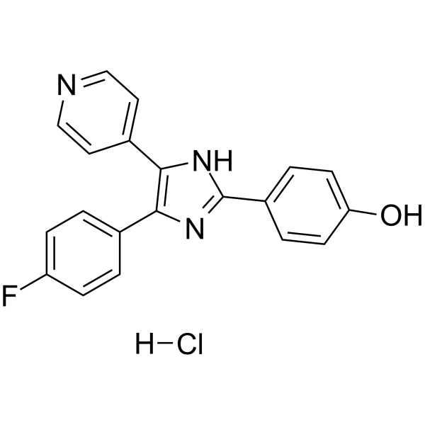
| Chemical Name | 4-[4-(4-fluorophenyl)-5-pyridin-4-yl-1H-imidazol-2-yl]phenol;hydrochloride |
| Synonyms | 350228-36-3; SB 202190 Hydrochloride; SB 202190 (hydrochloride); 4-(4-(4-FLUOROPHENYL)-5-(PYRIDIN-4-YL)-1H-IMIDAZOL-2-YL)PHENOL HYDROCHLORIDE; SB202190 hydrochloride; FHPI HCL; SB202190 HCl; 4-[4-(4-fluorophenyl)-5-pyridin-4-yl-1,3-dihydroimidazol-2-ylidene]cyclohexa-2,5-dien-1-one;hydrochloride; |
| HS Tariff Code | 2934.99.9001 |
| Storage |
Powder-20°C 3 years 4°C 2 years In solvent -80°C 6 months -20°C 1 month Note: Please store this product in a sealed and protected environment, avoid exposure to moisture. |
| Shipping Condition | Room temperature (This product is stable at ambient temperature for a few days during ordinary shipping and time spent in Customs) |
Biological Activity
| Targets | p38α 50 nM (IC50) p38β2 100 nM (IC50) |
| ln Vitro | In a dose- and time-dependent way, SB 202190 (0-10 μM; 0-72 hours) hydrochloride inhibits the proliferation of certain CRC cell lines, including RKO, CACO2, and SW480. Within this similar subset of CRC lines (RKO, CACO2, and SW480), SB 202190 hydrochloride significantly reduced colony formation and anchorage-independent growth (10 μM for 7–10 days) and increased apoptotic cell death (10 μM for 72 hours)[2]. p38i selectively inhibits mTORC1 signaling in RKO, CACO2, and SW480 cells by hydrolyzing SB 202190 (10 μM; 2 hours) to phosphorylate S6K1 (T389) and S6(S235/236) but not AKT (S473)[2]. |
| ln Vivo |
SB 202190 (5 mg/kg; once day for 10–12 days; intraperitoneal injection) hydrochloride exhibits proliferation and survival of malignant cells inhibition[2]. In the passive transfer mouse model, administration of SB202190, which inhibits p38, prevents PV IgG-induced blister formation.[5] Treatment with SB202190 results in a statistically significant survival advantage over control in the endotoxin model of sepsis.[8] SB202190 and SN-50 resulted in significant survival benefit in the lipopolysaccharide model (P = 0.0006) but not bacterial or CLP models (P = 0.9 and 0.3, respectively). SB-202190 and SN-50, in combination with antibiotic, resulted in a significant survival benefit in the CLP model (P = 0.0001 and 0.006, respectively). Circulating levels of both tumor necrosis factor-alpha and interleukin-6 were significantly reduced at 2 h (P = 0.047 and 0.036, respectively) and Western blot demonstrated down-regulation of p38 kinase 2 h after CLP in animals treated with p38MAPK and SN-50 inhibitors in combination with antibiotics. Conclusions: We have demonstrated that p-38 and NF-kappaB inhibition improve survival in endotoxin shock, whereas the survival benefit in polymicrobial sepsis requires coexistent antibiotic treatment. Vascular dementia (VaD) is a common age-related neurodegenerative disease resulting from chronic hypoxia. In the present study, we examined the protective effects of p38 MAPK inhibitor SB202190 against hippocampal apoptosis and spatial learning and memory deficits in a chronic hypoperfusion rat model of VaD established by permanent bilateral carotid occlusion (2-VO). Sixty rats were randomly divided into sham-operated, VaD model, and VaD plus SB202190 groups (n = 20/group). After sham/2-VO surgery, rats were administered 0.1% DMSO (sham-operated and VaD groups) or SB202190 by intracerebroventricular injection. One week after inhibitor/vehicle treatment, hippocampal p38 MAPK phosphorylation was higher in the model group than in the SB202190 group (P < 0.01). Compared to the model group, the SB202190 group exhibited significantly shorter escape latencies in the Morris water maze hidden platform trials (P < 0.01) and longer times in the original platform quadrant during probe trials (P < 0.01). The SB202190 group also showed significantly reduced neuronal apoptosis in the hippocampus compared to VaD model rats (P < 0.01) as well as higher (antiapoptotic) Bcl-2 expression and lower (proapoptotic) caspase-3 expression (P < 0.01 for both). In conclusion, blockade of the p38 MAPK signaling pathway by SB202190 following permanent 2-OV reduced apoptosis of hippocampal neurons and rescued spatial learning and memory deficits[12]. |
| Enzyme Assay |
The p38α and p38β are measured in 25 mM Tris-HCl, pH 7.5, with 0.1 mM EGTA and 0.33 mg/mL of myelin basic protein as the substrate. When using [γ-33P]ATP, assays can be run manually for 10 minutes at 30 °C in 50 L incubations or automatically with a Biomek 2000 Laboratory Automation Workstation in a 96-well format for 40 minutes at room temperature in 25 L incubations. Magnesium acetate and ATP have concentrations of 10 and 0.1 mM, respectively. MgATP is used to start every assay. To end a manual assay, aliquots of the incubation are spotted on phosphocellulose paper and then submerged in 50 mM phosphoric acid. Robotic assays are ended by adding 5 μL of 0.5 M phosphoric acid, followed by the spotting of aliquots onto P30 filter mats. All papers are then cleaned four times in 50 mM phosphoric acid to remove ATP, once in acetone (for manual incubations) or methanol (for robotic incubations), dried, and radioactivity is counted.
Protein Kinase Assays[2] MAPKAPK 2 assays were performed as described previously. Briefly, Jurkat cells were serum-starved for 24 h and then incubated with or without the specific p38 inhibitor SB202190 for 30 min prior to treatment with anti-Fas mAb (100 ng/ml) for 2 h or left alone as indicated in the figure legends. The cells were harvested in lysis buffer and clarified by centrifugation. Endogenous MAPKAPK 2 was immunoprecipitated with anti-MAPKAPK 2 polyclonal antibody for 3 h at 4 °C. The activity of the immune complex was assayed at 30 °C for 30 min in 30 μl of kinase buffer in the presence of 1 μm ATP/10 μCi [γ-32P]ATP (10 Ci/mmol) with GST-hsp27 as a substrate. The reactions were terminated with Laemmli sample buffer. The proteins were resolved by 13% SDS-polyacrylamide gel electrophoresis followed by autoradiography. The phosphorylated proteins were quantitated by a PhosphorImager. Caspase Activity Assays[2] Jurkat/neo or Jurkat/bcl-2 cells (106 cells) were treated with or without SB202190 (50 μm), PD098059 (50 μm) in the presence or absence of caspase inhibitor benzyloxycarbonyl-Val-Ala-Asp (zVAD)-fluoromethylketone for 24 h. The cells were then harvested in lysis buffer (25 mm Hepes, pH 7.4, 0.25% Nonidet P-40, 10 μg/ml leupeptin, 10 μg/ml aprotinin, 5 mm EDTA, 2 mm dithiothreitol, and 10 mm digitonin). The lysates were clarified by centrifugation, and the supernatants were used for caspase assays. The caspase activity was measured in a reaction mixture containing 20 μg of cell extracts, 20 μm fluoregenic peptide acetyl-Asp-Glu-Val-Asp-aminomethylcoumarin (DEVD-AMC) as described. Fluorescent AMC product formation was measured at excitation 360 nm, emission 460 nm using a Cytofluor II fluorescent plate reader. |
| Cell Assay |
Serum-starved cells are treated for 24 hours with various concentrations of SB 202190. Flow cytometry analysis is done after either a trypan blue exclusion or a propidium iodide exclusion is used to determine the viability of the cells. H33258 staining is used to see the apoptotic nuclei. The roles of p38 MAP kinases and ERK in UVB induced cox-2 gene expression were studied in a human keratinocyte cell line, HaCaT. UVB significantly increased cox-2 gene expression at both protein and mRNA levels. As we reported previously, p38 and ERK were significantly activated after UVB irradiation in HaCaT cells. In addition, treating the cells with p38 inhibitor SB202190 or MEK inhibitor PD98059 specifically inhibited UVB induced p38 or ERK activation, respectively. In this study, we further examined the roles of p38 and ERK in UVB induced cox-2 gene expression in HaCaT cells. We found that SB202190 strongly inhibited UVB induced COX-2 protein expression at different time points and various UVB doses. Furthermore, SB202190 markedly inhibited UVB induced cox-2 mRNA. Our data indicated that ERK did not play a role in UVB induced cox-2 gene expression in human keratinocytes since suppression of ERK did not significantly alter UVB induced increase of COX-2 protein and mRNA. These results suggested, for the first time, that activation of p38 is required for UVB induced cox-2 gene expression in human keratinocytes. Since cox-2 expression plays an important role in UV carcinogenesis, p38 could be a potential molecular target for chemoprevention of skin cancer.[2] During renal injury, activation of p38 mitogen-activated protein kinase (MAPK) in proximal tubular cells plays an important role in the inflammatory events that eventually lead to renal fibrosis. We hypothesized that local inhibition of p38 within these cells may be an interesting approach for the treatment of renal fibrosis. To effectuate this, we developed a renal-specific conjugate of the p38 inhibitor SB202190 [4-(4-fluorophenyl)-2-(4-hydroxyphenyl)-5-(4-pyridyl)1H-imidazole] and the carrier lysozyme. First, we demonstrated that SB202190 inhibited the expression of albumin-induced proinflammatory (monocyte chemoattractant protein-1) and transforming growth factor (TGF)-beta1-induced profibrotic (procollagen-Ialpha1) genes over 50% in renal tubular cells (normal rat kidney-52E). Next, we conjugated SB202190 via a carbamate linkage to lysozyme. However, this conjugate rapidly released the drug upon incubation in serum. Therefore, we applied a new platinum(II)-based linker approach, the so-called universal linkage system (ULS), which forms a coordinative bond with SB202190. The SB202190-ULS-lysozyme remained stable in serum but released the drug in kidney homogenates. SB202190-ULS-lysozyme accumulated efficiently in renal tubular cells and provided a local drug reservoir during a period of 3 days after a single intravenous injection. Treatment with SB202190-ULS-lysozyme inhibited TGF-beta1-induced gene expression for procollagen-Ialpha1 by 64% in HK-2 cells. Lastly, we evaluated the efficacy of a single dose of the conjugate in the unilateral renal ischemia-reperfusion rat model. A reduction of intrarenal p38 phosphorylation and alpha-smooth muscle actin protein expression was observed 4 days after the ischemia-reperfusion injury. In conclusion, we have developed a novel strategy for local delivery of the p38 MAPK inhibitor SB202190, which may be of use in the treatment of renal fibrosis[3]. |
| Animal Protocol |
C57BL/6J mice injected i.d. with a sterile solution of either control IgG or PV IgG 12.5 μg Administered via i.d. The 60 Wistar rats were randomly assigned to the sham-operated group, the VaD model group, and the SB202190 group (20 animals each) using a random number table. The VaD rat model (n = 40) was established by separating and ligating the bilateral carotid artery via two-vessel occlusion (2-VO). For animals of the sham-operated group (n = 20), the bilateral carotid artery was separated using the same methods but without ligation. After recovery, animals of the SB202190 group received intracerebroventricular (ICV) injection of SB202190 (dissolved in 100% DMSO and then diluted in normal saline (NS) for a final concentration of DMSO of 0.1%) and both the VaD model and sham-operated groups received ICV injection of equal volume 0.1% DMSO. In each group, eight rats were examined in the Morris water maze to assess spatial learning and memory, six rats were sacrificed and brain sections were prepared for TUNEL staining and Bcl-2/caspase-3 immunohistochemistry, and six rats were sacrificed and tissue homogenates were prepared for Western blot assay of phospho-p38 MAPK expression.[12] All animals were administered 0.1% DMSO or SB202190 immediately after sham or 2-VO surgery by ICV injection. Briefly, after anesthesia by IP injection of a 10% chloral hydrate (0.35 mL/100 g body weight), the animal was secured within a Jiangwan I-C rat stereotaxic frame. The site for ICV injection was 0.8 mm caudal to bregma and 1.5 mm to the right of the midline. A hole was drilled horizontally with an electric drill and 5 μL of 0.1% DMSO or 10 μmol/L SB202190 solution was injected using a microinjector. The injection was completed in 4 min and the needle kept in position for an additional 2 min. One week after ICV injection, animals were examined in the Morris water maze or sacrificed and brain tissues were prepared for TUNEL staining, immunofluorescence, and Western blot.[2] Intraperitoneal lipopolysaccharide (30 mg/kg), tail vein injection of bacteria (Staphylococcus aureus + Salmonella Typhimurium, 5 x 10(7) colony forming units/kg) and cecal ligation and puncture (CLP) with or without antibiotics (Augmentin, 100 mg/kg) were the septic models used. Animals received control, SB-202190 (a p38 inhibitor), or SN-50 (an NF-kappaB inhibitor), and mortality was assessed by log-rank analysis. Blood was collected at different time points for cytokine analysis, and splenic tissue was used for cytoplasmic protein extraction to assess kinase activation.[8] |
| References |
[1]. Biochem J. 2000 Oct 1;351(Pt 1):95-105. [2]. J Biol Chem. 1998 Jun 26;273(26):16415-20. [3]. Oncogene. 2001 Jun 28;20(29):3921-6. [4]. J Pharmacol Exp Ther. 2006 Oct;319(1):8-19. [5]. Proc Natl Acad Sci U S A. 2006 Aug 22;103(34):12855-60. [6]. Cell Signal. 2008 Apr;20(4):675-83. [7]. Leuk Res. 2009 May;33(5):693-9. [8]. J Surg Res. 2009 Mar;152(1):46-53. [9]. Cancer Res. 2011 Feb 1;71(3):1041-9. [10]. Mol Cancer Ther. 2012 Mar;11(3):561-71. [11]. EMBO Rep. 2017 Nov;18(11):2067-2078. [12]. Biomed Res Int. 2013:2013:215798. |
| Additional Infomation |
SB-202190 is a member of the class of imidazoles that is 1H-imidazole in which the hydrogens at positions 2, 4, and 5 are replaced by 4-hydroxyphenyl, pyridin-4-yl, and 4-fluorophenyl groups, respectively. It is a widely used inhibitor of mitogen-activated protein kinase (MAPK) alpha and beta. It has a role as an EC 2.7.11.24 (mitogen-activated protein kinase) inhibitor and an apoptosis inducer. It is a member of imidazoles, a member of phenols, a member of pyridines and an organofluorine compound. 4-(4-Fluorophenyl)-2-(4-hydroxyphenyl)-5-(4-pyridyl)-1H-imidazole has been reported in Mammea siamensis with data available. p38, a subfamily of the mitogen-activated protein kinase, regulates gene expression in response to various extracellular stimuli. The pyridinyl imidazoles like SB202190 are specific inhibitors of p38alpha and p38beta and have been widely used in investigation of the biological functions of p38. Here we show that SB202190 by itself was sufficient to induce cell death, with typical apoptotic features such as nucleus condensation and intranucleosomal DNA fragmentation. SB202190 stimulated the activity of CPP32-like caspases, and its apoptotic effect was completely blocked by the protease inhibitor benzyloxycarbonyl-Val-Ala-Asp-fluoromethyl ketone and expression of bcl-2. In addition, SB202190 was able to potentiate apoptosis induced by Fas(APO-1) ligation or UV irradiation. Expression of p38beta attenuated the apoptotic effect of SB202190 and the cell death induced by Fas ligation and UV irradiation. In contrast, expression of p38alpha induced cell death mildly. These results indicate that SB202190 induces apoptosis through activation of CPP32-like caspases and suggest that distinct members of the p38 subfamily of mitogen-activated protein kinase have different functions in apoptosis.[1] |
Solubility Data
| Solubility (In Vitro) | DMSO : 83.33 mg/mL (226.56 mM) |
| Solubility (In Vivo) |
Note: Listed below are some common formulations that may be used to formulate products with low water solubility (e.g. < 1 mg/mL), you may test these formulations using a minute amount of products to avoid loss of samples. Injection Formulations (e.g. IP/IV/IM/SC) Injection Formulation 1: DMSO : Tween 80: Saline = 10 : 5 : 85 (i.e. 100 μL DMSO stock solution → 50 μL Tween 80 → 850 μL Saline) *Preparation of saline: Dissolve 0.9 g of sodium chloride in 100 mL ddH ₂ O to obtain a clear solution. Injection Formulation 2: DMSO : PEG300 :Tween 80 : Saline = 10 : 40 : 5 : 45 (i.e. 100 μL DMSO → 400 μLPEG300 → 50 μL Tween 80 → 450 μL Saline) Injection Formulation 3: DMSO : Corn oil = 10 : 90 (i.e. 100 μL DMSO → 900 μL Corn oil) Example: Take the Injection Formulation 3 (DMSO : Corn oil = 10 : 90) as an example, if 1 mL of 2.5 mg/mL working solution is to be prepared, you can take 100 μL 25 mg/mL DMSO stock solution and add to 900 μL corn oil, mix well to obtain a clear or suspension solution (2.5 mg/mL, ready for use in animals). Injection Formulation 4: DMSO : 20% SBE-β-CD in saline = 10 : 90 [i.e. 100 μL DMSO → 900 μL (20% SBE-β-CD in saline)] *Preparation of 20% SBE-β-CD in Saline (4°C,1 week): Dissolve 2 g SBE-β-CD in 10 mL saline to obtain a clear solution. Injection Formulation 5: 2-Hydroxypropyl-β-cyclodextrin : Saline = 50 : 50 (i.e. 500 μL 2-Hydroxypropyl-β-cyclodextrin → 500 μL Saline) Injection Formulation 6: DMSO : PEG300 : castor oil : Saline = 5 : 10 : 20 : 65 (i.e. 50 μL DMSO → 100 μLPEG300 → 200 μL castor oil → 650 μL Saline) Injection Formulation 7: Ethanol : Cremophor : Saline = 10: 10 : 80 (i.e. 100 μL Ethanol → 100 μL Cremophor → 800 μL Saline) Injection Formulation 8: Dissolve in Cremophor/Ethanol (50 : 50), then diluted by Saline Injection Formulation 9: EtOH : Corn oil = 10 : 90 (i.e. 100 μL EtOH → 900 μL Corn oil) Injection Formulation 10: EtOH : PEG300:Tween 80 : Saline = 10 : 40 : 5 : 45 (i.e. 100 μL EtOH → 400 μLPEG300 → 50 μL Tween 80 → 450 μL Saline) Oral Formulations Oral Formulation 1: Suspend in 0.5% CMC Na (carboxymethylcellulose sodium) Oral Formulation 2: Suspend in 0.5% Carboxymethyl cellulose Example: Take the Oral Formulation 1 (Suspend in 0.5% CMC Na) as an example, if 100 mL of 2.5 mg/mL working solution is to be prepared, you can first prepare 0.5% CMC Na solution by measuring 0.5 g CMC Na and dissolve it in 100 mL ddH2O to obtain a clear solution; then add 250 mg of the product to 100 mL 0.5% CMC Na solution, to make the suspension solution (2.5 mg/mL, ready for use in animals). Oral Formulation 3: Dissolved in PEG400 Oral Formulation 4: Suspend in 0.2% Carboxymethyl cellulose Oral Formulation 5: Dissolve in 0.25% Tween 80 and 0.5% Carboxymethyl cellulose Oral Formulation 6: Mixing with food powders Note: Please be aware that the above formulations are for reference only. InvivoChem strongly recommends customers to read literature methods/protocols carefully before determining which formulation you should use for in vivo studies, as different compounds have different solubility properties and have to be formulated differently. (Please use freshly prepared in vivo formulations for optimal results.) |
| Preparing Stock Solutions | 1 mg | 5 mg | 10 mg | |
| 1 mM | 2.7189 mL | 13.5943 mL | 27.1887 mL | |
| 5 mM | 0.5438 mL | 2.7189 mL | 5.4377 mL | |
| 10 mM | 0.2719 mL | 1.3594 mL | 2.7189 mL |