Physicochemical Properties
| Molecular Formula | C10H18O3 |
| Molecular Weight | 186.2481 |
| Exact Mass | 186.125 |
| CAS # | 14113-05-4 |
| PubChem CID | 5312738 |
| Appearance | White to off-white solid powder |
| Density | 1.0±0.1 g/cm3 |
| Boiling Point | 339.2±15.0 °C at 760 mmHg |
| Flash Point | 173.1±16.9 °C |
| Vapour Pressure | 0.0±1.7 mmHg at 25°C |
| Index of Refraction | 1.486 |
| LogP | 1.99 |
| Hydrogen Bond Donor Count | 2 |
| Hydrogen Bond Acceptor Count | 3 |
| Rotatable Bond Count | 8 |
| Heavy Atom Count | 13 |
| Complexity | 152 |
| Defined Atom Stereocenter Count | 0 |
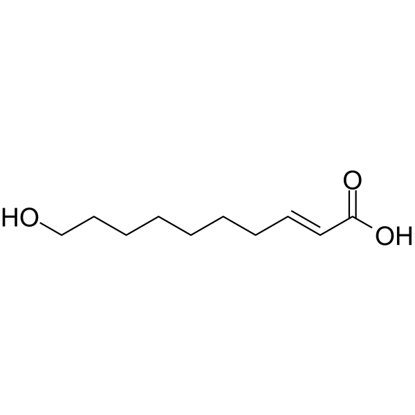
| SMILES | C(CCC/C=C/C(=O)O)CCCO |
| InChi Key | QHBZHVUGQROELI-SOFGYWHQSA-N |
| InChi Code | InChI=1S/C10H18O3/c11-9-7-5-3-1-2-4-6-8-10(12)13/h6,8,11H,1-5,7,9H2,(H,12,13)/b8-6+ |
| Chemical Name | (E)-10-hydroxydec-2-enoic acid |
| HS Tariff Code | 2934.99.9001 |
| Storage |
Powder-20°C 3 years 4°C 2 years In solvent -80°C 6 months -20°C 1 month |
| Shipping Condition | Room temperature (This product is stable at ambient temperature for a few days during ordinary shipping and time spent in Customs) |
Biological Activity
| Targets |
Queen Bee Acid (QBA) is described as a modulator of estrogen receptor (ER) function. It binds to an allosteric site on the ER without competing with estradiol, acting as an agonist in the absence of estradiol. It also inhibits histone deacetylases (HDACs), which is linked to caste determination in bees. The downstream neurotrophic and neuroprotective effects are suggested to involve the activation of Extracellular signal-regulated kinase (Erk) signaling pathways. [1]. |
| ln Vitro |
Queen Bee Acid (QBA) concentration-dependently increased the growth (neurite extension) of primary rat hippocampal neurons, with a half-maximal effective concentration (EC50) of 75.9 ± 29.1 nM. Maximal growth promotion occurred between 0.3–30 μM. Concentrations above 30 μM resulted in a reduction from the maximal effect [1]. Queen Bee Acid (QBA) protected primary rat hippocampal neurons from hypoxia-induced cell death (<0.3% O2 for 48 h), with a half-maximal inhibitory concentration (IC50) of 64.6 ± 17.9 nM. It also improved mitochondrial health in these neurons with an EC50 of 156.9 ± 58.4 nM [1]. Queen Bee Acid (QBA) protected primary rat hippocampal neurons from glutamate-induced cell death (25 μM for 24 h), with an IC50 of 56.0 ± 27.3 nM. It also improved mitochondrial health under glutamate challenge with an EC50 of 317.5 ± 200.3 nM [1]. The literature cites previous work indicating that Queen Bee Acid (QBA) (1–100 μM) can stimulate neuronal differentiation while inhibiting astrocyte differentiation from neural stem/progenitor cells [1]. |
| ln Vivo |
Long-term dietary administration of Queen Bee Acid (QBA) (12 or 24 mg/kg/day for approximately 5 months) to aged male Sprague-Dawley rats significantly reduced anxiety-like behavior. This was evidenced by increased time spent in the open arms and an increased number of head dips in the elevated plus maze test [1]. The same long-term QBA administration (12 or 24 mg/kg/day) to aged male rats significantly increased body weight gain and helped maintain weight during periods of behavioral testing stress, with no difference in food consumption compared to controls [1]. Dietary administration of Queen Bee Acid (QBA) at doses equivalent to the rat doses (0, 30, or 60 mg/kg/day for 4 months) to adult BALB/c mice induced sex-dependent changes in body composition. In male mice, QBA increased overall body weight, muscle mass, and adiposity. In female mice, QBA did not affect overall weight but significantly decreased adiposity and increased trabecular bone density [1]. No significant effects of Queen Bee Acid (QBA) were observed on locomotor activity (open field test), spatial learning and memory (Barnes maze), or depressive-like behavior (forced swim test) in the aged male rat study [1]. |
| Cell Assay |
Neurite Extension Assay: Primary hippocampal neurons were isolated from embryonic day 17–18 rat pups and cultured in laminin/poly-D-lysine coated plates. Cells were incubated with QBA (dissolved in methanol, final concentration 0–30 μM) for 7 days, with half of the culture medium being replaced with fresh medium containing the corresponding QBA concentration every other day. After 7 days, cultures were fixed and immunostained for the neuron-specific marker MAP2a, and nuclei were counterstained with DAPI. Fluorescent images were acquired using confocal microscopy. Quantitative analysis of neurite growth was performed by calculating the percentage area covered by MAP2a staining normalized to the number of neuronal nuclei (DAPI-positive) in the field of view [1]. Hypoxia Challenge Assay: Primary hippocampal neurons were cultured for 7 days, then pretreated with QBA (0–30 μM) for 48 hours. Subsequently, cultures were exposed to a hypoxic atmosphere (<0.3% O2) for 48 hours. Following the challenge, cells were stained with fluorescent probes: calcein AM for live cells, ethidium homodimer for dead cells, and tetramethylrhodamine ethyl ester (TMRE) for mitochondrial membrane potential (health). Fluorescence intensity for each marker was quantified using a fluorimeter to assess cell viability, death, and mitochondrial health [1]. Glutamate Challenge Assay: The procedure was similar to the hypoxia challenge. Neurons cultured for 7 days were pretreated with QBA (0–30 μM) for 48 hours and then exposed to a toxic concentration of glutamate (25 μM) for 24 hours. Cell viability, death, and mitochondrial health were assessed using the same triple-staining method (calcein AM, ethidium homodimer, TMRE) and fluorimetric quantification [1]. |
| Animal Protocol |
Rat Behavioral and Weight Maintenance Study: Aged (12 months old at study start) male Sprague-Dawley rats were pair-housed. They were fed custom-prepared diets based on the AIN-93G formulation, which were specifically designed to deliver 0, 12, or 24 mg/kg/day of Queen Bee Acid (QBA). Diets were balanced for all fatty acids except for QBA content. The dietary intervention began at least 3.5 months prior to behavioral testing and continued throughout the study. Body weight was measured weekly, and food consumption per cage was monitored. After prolonged exposure (starting at ~15 months of age), rats underwent a behavioral test battery: Open Field Test (for general activity), Barnes Maze (for spatial learning and memory over 4 training days and a probe trial), Elevated Plus Maze (for anxiety-like behavior), and Forced Swim Test (for depressive-like behavior) [1]. Mouse Body Composition Study: Adult (2 months old at start) male and female BALB/c mice were group-housed. They were fed custom diets formulated to deliver QBA at doses of 0, 30, or 60 mg/kg/day. These doses were calculated to be equivalent (by body surface area) to the doses given to rats. The feeding period lasted for 4 months. Food intake was monitored. Mice were weighed weekly. Body composition (fat mass, lean mass, free fluid) was assessed non-invasively every 7 days using a time-domain nuclear magnetic resonance (TD-NMR) analyzer. No anesthesia was required for this brief procedure. After 4 months, final measurements were taken, mice were euthanized, and their hind limbs were collected for bone density analysis via peripheral quantitative computed tomography (pQCT) to measure trabecular and cortical bone density [1]. |
| References |
[1]. Long-Term Administration of Queen Bee Acid (QBA) to Rodents Reduces Anxiety-Like Behavior, Promotes Neuronal Health and Improves Body Composition. Nutrients. 2017 Dec 23;10(1). |
| Additional Infomation |
(E)-10-hydroxydec-2-enoic acid is an omega-hydroxy amino acid that is 2-decenoic acid in which one of the hydrogens attached to the terminal carbon is replaced by a hydroxy group and in which the C=C double bond has E configuration. It is a component of royal jelly. It has a role as an animal metabolite and a geroprotector. It is an alpha,beta-unsaturated monocarboxylic acid, a straight-chain fatty acid, a hydroxy monounsaturated fatty acid and an omega-hydroxy-medium-chain fatty acid. Queen Bee Acid (QBA) is identified as the predominant fatty acid in royal jelly, constituting approximately 2-6% of its wet weight and about 40% of its total fatty acid content [1]. The neuroprotective, anxiolytic, and body composition-modifying effects observed in this study are proposed to be mediated, at least in part, through QBA's interaction with estrogen receptors and the subsequent modulation of downstream signaling pathways, including Erk activation [1]. The sex-dependent effects of QBA on body composition (increasing adiposity and muscle in males, decreasing adiposity and increasing bone density in females) strongly suggest an interaction with endogenous sex hormone pathways [1]. The research was fully funded and conducted by authors employed by a nutritional products company. It is noted that this company does not commercially sell or manufacture royal jelly or purified QBA [1]. |
Solubility Data
| Solubility (In Vitro) | DMSO : ~100 mg/mL (~536.91 mM) |
| Solubility (In Vivo) |
Solubility in Formulation 1: ≥ 2.5 mg/mL (13.42 mM) (saturation unknown) in 10% DMSO + 40% PEG300 + 5% Tween80 + 45% Saline (add these co-solvents sequentially from left to right, and one by one), clear solution. For example, if 1 mL of working solution is to be prepared, you can add 100 μL of 25.0 mg/mL clear DMSO stock solution to 400 μL PEG300 and mix evenly; then add 50 μL Tween-80 to the above solution and mix evenly; then add 450 μL normal saline to adjust the volume to 1 mL. Preparation of saline: Dissolve 0.9 g of sodium chloride in 100 mL ddH₂ O to obtain a clear solution. Solubility in Formulation 2: ≥ 2.5 mg/mL (13.42 mM) (saturation unknown) in 10% DMSO + 90% (20% SBE-β-CD in Saline) (add these co-solvents sequentially from left to right, and one by one), clear solution. For example, if 1 mL of working solution is to be prepared, you can add 100 μL of 25.0 mg/mL clear DMSO stock solution to 900 μL of 20% SBE-β-CD physiological saline solution and mix evenly. Preparation of 20% SBE-β-CD in Saline (4°C,1 week): Dissolve 2 g SBE-β-CD in 10 mL saline to obtain a clear solution. Solubility in Formulation 3: ≥ 2.5 mg/mL (13.42 mM) (saturation unknown) in 10% DMSO + 90% Corn Oil (add these co-solvents sequentially from left to right, and one by one), clear solution. For example, if 1 mL of working solution is to be prepared, you can add 100 μL of 25.0 mg/mL clear DMSO stock solution to 900 μL of corn oil and mix evenly. Solubility in Formulation 4: 10 mg/mL (53.69 mM) in 0.5% CMC-Na/saline water (add these co-solvents sequentially from left to right, and one by one), suspension solution; with ultrasonication. Preparation of saline: Dissolve 0.9 g of sodium chloride in 100 mL ddH₂ O to obtain a clear solution. (Please use freshly prepared in vivo formulations for optimal results.) |
| Preparing Stock Solutions | 1 mg | 5 mg | 10 mg | |
| 1 mM | 5.3691 mL | 26.8456 mL | 53.6913 mL | |
| 5 mM | 1.0738 mL | 5.3691 mL | 10.7383 mL | |
| 10 mM | 0.5369 mL | 2.6846 mL | 5.3691 mL |