Physicochemical Properties
| Molecular Formula | C30H48O5 |
| Molecular Weight | 488.6991 |
| Exact Mass | 488.35 |
| CAS # | 20137-37-5 |
| PubChem CID | 12315075 |
| Appearance | White to off-white solid powder |
| Density | 1.2±0.1 g/cm3 |
| Boiling Point | 622.8±55.0 °C at 760 mmHg |
| Melting Point | 272-274℃ |
| Flash Point | 344.5±28.0 °C |
| Vapour Pressure | 0.0±4.1 mmHg at 25°C |
| Index of Refraction | 1.581 |
| LogP | 5.75 |
| Hydrogen Bond Donor Count | 4 |
| Hydrogen Bond Acceptor Count | 5 |
| Rotatable Bond Count | 2 |
| Heavy Atom Count | 35 |
| Complexity | 945 |
| Defined Atom Stereocenter Count | 11 |
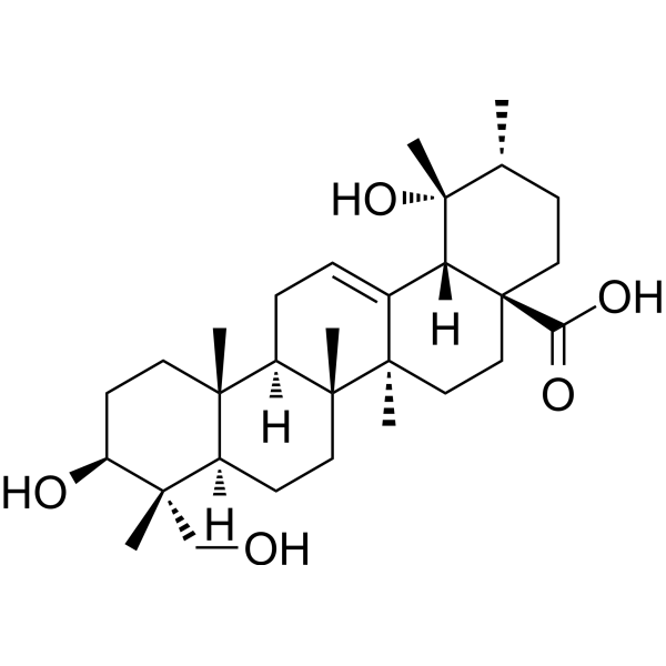
| SMILES | C[C@@H]1CC[C@@]2(CC[C@@]3(C(=CC[C@H]4[C@]3(CC[C@@H]5[C@@]4(CC[C@@H]([C@@]5(C)CO)O)C)C)[C@@H]2[C@]1(C)O)C)C(=O)O |
| InChi Key | YLHQFGOOMKJFLP-LTFXOGOQSA-N |
| InChi Code | InChI=1S/C30H48O5/c1-18-9-14-30(24(33)34)16-15-27(4)19(23(30)29(18,6)35)7-8-21-25(2)12-11-22(32)26(3,17-31)20(25)10-13-28(21,27)5/h7,18,20-23,31-32,35H,8-17H2,1-6H3,(H,33,34)/t18-,20-,21-,22+,23-,25+,26+,27-,28-,29-,30+/m1/s1 |
| Chemical Name | (1R,2R,4aS,6aR,6aS,6bR,8aR,9R,10S,12aR,14bS)-1,10-dihydroxy-9-(hydroxymethyl)-1,2,6a,6b,9,12a-hexamethyl-2,3,4,5,6,6a,7,8,8a,10,11,12,13,14b-tetradecahydropicene-4a-carboxylic acid |
| HS Tariff Code | 2934.99.9001 |
| Storage |
Powder-20°C 3 years 4°C 2 years In solvent -80°C 6 months -20°C 1 month Note: This product requires protection from light (avoid light exposure) during transportation and storage. |
| Shipping Condition | Room temperature (This product is stable at ambient temperature for a few days during ordinary shipping and time spent in Customs) |
Biological Activity
| Targets |
AKT/mTOR signaling pathway (HepG2 cell IC₅₀ = 25.3 μM; SMMC-7721 cell IC₅₀ = 21.7 μM) [1] MAPK signaling pathway (ERK1/2, JNK, p38) [1] DNA damage response (γ-H2AX) [1] |
| ln Vitro |
Rotundic acid exhibits potent antiproliferative activity against human hepatocellular carcinoma (HCC) cells, with IC₅₀ values of 25.3 μM (HepG2) and 21.7 μM (SMMC-7721) after 48 hours of treatment (MTT assay). [1] It induces significant DNA damage in HCC cells: Comet assay shows a dose-dependent increase in tail moment (1.8-fold and 2.5-fold higher than control at 20 μM and 40 μM, respectively), and Western blot confirms upregulated expression of γ-H2AX (a marker of DNA double-strand breaks). [1] Rotundic acid promotes HCC cell apoptosis in a dose-dependent manner: Annexin V-FITC/PI staining reveals that 40 μM treatment increases the apoptotic rate to 38.6% (HepG2) and 42.3% (SMMC-7721) after 48 hours; it also activates caspase-3 and caspase-9, and induces cleavage of PARP, as evidenced by Western blot. [1] It inhibits the AKT/mTOR pathway: Treatment with Rotundic acid (10-40 μM) dose-dependently reduces the phosphorylation levels of AKT (p-AKT) and mTOR (p-mTOR) without affecting total AKT/mTOR protein expression. [1] It modulates the MAPK pathway: Rotundic acid decreases phosphorylated ERK1/2 (p-ERK1/2) levels while increasing phosphorylated JNK (p-JNK) and phosphorylated p38 (p-p38) levels in a dose-dependent manner. [1] Rotundic acid suppresses the clonogenic potential of HCC cells: Colony formation assay shows that 20 μM and 40 μM treatments reduce the number of colonies by 56.2% and 78.5% (HepG2), and 61.3% and 82.1% (SMMC-7721), respectively, compared to the control group. [1] It has no significant cytotoxicity against normal human hepatocytes (LO2) at concentrations up to 40 μM (cell viability > 85%). [1] |
| ln Vivo |
In a HepG2 xenograft nude mouse model, intraperitoneal administration of Rotundic acid (20 mg/kg and 40 mg/kg, 3 times/week for 4 weeks) significantly inhibits tumor growth: Tumor volume is reduced by 45.8% (20 mg/kg) and 68.3% (40 mg/kg), and tumor weight is decreased by 42.5% (20 mg/kg) and 65.7% (40 mg/kg) compared to the vehicle control group (P < 0.05). [1] Tumor tissue analysis confirms that Rotundic acid induces DNA damage (upregulated γ-H2AX expression) and apoptosis (increased TUNEL-positive cells) in vivo. [1] It modulates signaling pathways in tumor tissues: Western blot and immunohistochemistry show reduced p-AKT/p-mTOR levels and increased p-JNK/p-p38 levels in tumor tissues from treated mice. [1] |
| Cell Assay |
MTT cell viability assay: HepG2, SMMC-7721, and LO2 cells are seeded in 96-well plates (5×10³ cells/well) and cultured overnight. Rotundic acid is added at concentrations of 0, 5, 10, 20, 40, 80 μM, and cells are incubated for 48 hours. MTT reagent is added, and after 4 hours of incubation, the absorbance at 570 nm is measured to calculate cell viability and IC₅₀ values. [1] Colony formation assay: HCC cells are seeded in 6-well plates (1×10³ cells/well) and cultured for 24 hours. Rotundic acid (0, 20, 40 μM) is added, and cells are cultured for 14 days. Colonies are fixed, stained, and counted to evaluate clonogenic potential. [1] Comet assay for DNA damage: HepG2 cells are treated with Rotundic acid (0, 20, 40 μM) for 24 hours. Cells are embedded in agarose gel, subjected to electrophoresis, stained with a DNA-binding dye, and observed under a fluorescence microscope. Tail moment is quantified to assess DNA damage. [1] Apoptosis detection: HCC cells are treated with Rotundic acid (0, 20, 40 μM) for 48 hours, stained with Annexin V-FITC and PI, and analyzed by flow cytometry to determine the apoptotic rate. [1] Western blot assay: HCC cells are treated with Rotundic acid (0, 10, 20, 40 μM) for 24 hours. Total proteins are extracted, separated by SDS-PAGE, transferred to membranes, and probed with antibodies against γ-H2AX, AKT, p-AKT, mTOR, p-mTOR, ERK1/2, p-ERK1/2, JNK, p-JNK, p38, p-p38, caspase-3, caspase-9, PARP, and cleaved PARP. GAPDH is used as an internal reference. [1] Immunofluorescence staining for γ-H2AX: HepG2 cells are seeded on coverslips, treated with Rotundic acid (0, 20, 40 μM) for 24 hours, fixed, permeabilized, and incubated with anti-γ-H2AX antibody and fluorescent secondary antibody. Nuclei are stained with DAPI, and γ-H2AX foci are observed under a confocal microscope. [1] |
| Animal Protocol |
HepG2 xenograft nude mouse model: Male BALB/c nude mice (6-8 weeks old) are subcutaneously injected with 5×10⁶ HepG2 cells into the right axilla. When tumors reach a volume of ~100 mm³, mice are randomly divided into 3 groups (n=6/group): vehicle control group (0.1% DMSO in normal saline), low-dose Rotundic acid group (20 mg/kg), and high-dose Rotundic acid group (40 mg/kg). The drug is administered via intraperitoneal injection 3 times a week for 4 weeks. Tumor volume (measured every 3 days) and body weight (measured weekly) are recorded. At the end of the experiment, mice are euthanized, tumors are excised, weighed, and fixed in 4% paraformaldehyde for histological and immunohistochemical analysis. [1] Tumor tissue analysis: Paraffin-embedded tumor sections are subjected to HE staining (to observe histological changes) and TUNEL staining (to detect apoptotic cells). Immunohistochemistry is performed to detect the expression of p-AKT, p-mTOR, p-JNK, p-p38, and γ-H2AX in tumor tissues. [1] |
| Toxicity/Toxicokinetics |
In the in vivo study, Rotundic acid (20-40 mg/kg, intraperitoneal injection) does not cause significant changes in mouse body weight, food intake, or general behavior during the 4-week treatment period. [1] Serum biochemical analysis shows no significant abnormalities in liver function (ALT, AST) or kidney function (BUN, CREA) in treated mice compared to the control group. [1] Histopathological examination of major organs (liver, kidney, heart, lung, spleen) reveals no obvious toxic lesions in Rotundic acid-treated groups. [1] |
| References |
[1]. Rotundic Acid Induces DNA Damage and Cell Death in Hepatocellular Carcinoma Through AKT/mTOR and MAPK Pathways. Front Oncol. 2019 Jun 26;9:545. |
| Additional Infomation |
Rotundic acid is a triterpenoid. It has a role as a metabolite. Rotundic acid has been reported in Enkianthus campanulatus, Mussaenda macrophylla, and other organisms with data available. Rotundic acid is a natural triterpenoid compound isolated from medicinal plants (e.g., Clematis chinensis Osbeck). [1] It exerts antitumor effects on hepatocellular carcinoma through a dual mechanism: inhibiting the AKT/mTOR pathway (which promotes cell survival and proliferation) and modulating the MAPK pathway (which mediates stress response and apoptosis), ultimately inducing DNA damage and caspase-dependent cell death. [1] The selective cytotoxicity of Rotundic acid against HCC cells (with minimal toxicity to normal hepatocytes) and favorable in vivo safety profile make it a potential lead compound for the development of HCC therapeutic agents. [1] |
Solubility Data
| Solubility (In Vitro) | DMSO : ~100 mg/mL (~204.62 mM) |
| Solubility (In Vivo) |
Solubility in Formulation 1: 2.5 mg/mL (5.12 mM) in 10% DMSO + 40% PEG300 + 5% Tween80 + 45% Saline (add these co-solvents sequentially from left to right, and one by one), suspension solution; with sonication. For example, if 1 mL of working solution is to be prepared, you can add 100 μL of 25.0 mg/mL clear DMSO stock solution to 400 μL PEG300 and mix evenly; then add 50 μL Tween-80 to the above solution and mix evenly; then add 450 μL normal saline to adjust the volume to 1 mL. Preparation of saline: Dissolve 0.9 g of sodium chloride in 100 mL ddH₂ O to obtain a clear solution. Solubility in Formulation 2: 2.5 mg/mL (5.12 mM) in 10% DMSO + 90% (20% SBE-β-CD in Saline) (add these co-solvents sequentially from left to right, and one by one), suspension solution; with ultrasonication. For example, if 1 mL of working solution is to be prepared, you can add 100 μL of 25.0 mg/mL clear DMSO stock solution to 900 μL of 20% SBE-β-CD physiological saline solution and mix evenly. Preparation of 20% SBE-β-CD in Saline (4°C,1 week): Dissolve 2 g SBE-β-CD in 10 mL saline to obtain a clear solution. Solubility in Formulation 3: ≥ 2.5 mg/mL (5.12 mM) (saturation unknown) in 10% DMSO + 90% Corn Oil (add these co-solvents sequentially from left to right, and one by one), clear solution. For example, if 1 mL of working solution is to be prepared, you can add 100 μL of 25.0 mg/mL clear DMSO stock solution to 900 μL of corn oil and mix evenly. (Please use freshly prepared in vivo formulations for optimal results.) |
| Preparing Stock Solutions | 1 mg | 5 mg | 10 mg | |
| 1 mM | 2.0462 mL | 10.2312 mL | 20.4625 mL | |
| 5 mM | 0.4092 mL | 2.0462 mL | 4.0925 mL | |
| 10 mM | 0.2046 mL | 1.0231 mL | 2.0462 mL |