Physicochemical Properties
| Molecular Formula | C29H31NO8 |
| Molecular Weight | 521.558348894119 |
| Exact Mass | 521.204 |
| CAS # | 1139253-73-8 |
| PubChem CID | 49793307 |
| Appearance | White to off-white solid powder |
| LogP | 2.9 |
| Hydrogen Bond Donor Count | 2 |
| Hydrogen Bond Acceptor Count | 8 |
| Rotatable Bond Count | 7 |
| Heavy Atom Count | 38 |
| Complexity | 829 |
| Defined Atom Stereocenter Count | 5 |
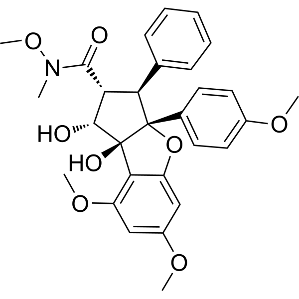
| SMILES | O1C2=CC(OC)=CC(OC)=C2[C@]2(O)[C@H](O)[C@H](C(N(OC)C)=O)[C@@H](C3=CC=CC=C3)[C@]12C1=CC=C(OC)C=C1 |
| InChi Key | TZDAVNWDKGYBCW-IDAMAFBJSA-N |
| InChi Code | InChI=1S/C29H31NO8/c1-30(37-5)27(32)23-24(17-9-7-6-8-10-17)29(18-11-13-19(34-2)14-12-18)28(33,26(23)31)25-21(36-4)15-20(35-3)16-22(25)38-29/h6-16,23-24,26,31,33H,1-5H3/t23-,24-,26-,28+,29+/m1/s1 |
| Chemical Name | (1R,2R,3S,3aR,8bS)-1,8b-dihydroxy-N,6,8-trimethoxy-3a-(4-methoxyphenyl)-N-methyl-3-phenyl-2,3-dihydro-1H-cyclopenta[b][1]benzofuran-2-carboxamide |
| HS Tariff Code | 2934.99.9001 |
| Storage |
Powder-20°C 3 years 4°C 2 years In solvent -80°C 6 months -20°C 1 month |
| Shipping Condition | Room temperature (This product is stable at ambient temperature for a few days during ordinary shipping and time spent in Customs) |
Biological Activity
| ln Vitro | For AML cell lines and FLT3-ITD-positive AML cell lines, rohinitib (6.25–50 nM; 72 h) causes cell apoptosis[1]. Normal bone marrow (BM) is less sensitive to Rohinitib (25 nM; 72 h) than primary AML cells, and FLT3-ITD-positive cells are more sensitive than FLT3 wild-type AML cells[1]. |
| ln Vivo | In vivo, rohinitib (0.75 and 1.0 mg/kg; sc once daily for 5 days until mice get moribund) exhibits anti-AML effects[1]. |
| Cell Assay |
Apoptosis Analysis[1] Cell Types: AML cell lines Tested Concentrations: 6.25, 12.5, 25 and 50 nM Incubation Duration: 72 h Experimental Results: Dose-dependently induced apoptosis of MOLM-13, MOLM-14, MV4;11, OCI-AML3, THP-1, HL-60, Kasumi-1 and NB4 cell lines. And Dramatically induced cell apoptosis of FLT3-ITD, FLT3-ITD-expressing murine Ba/F3 and human OCI-AML3 cells. |
| Animal Protocol |
Animal/Disease Models: Female NSG mice with AML xenografts generated by intravenous (iv)injections of MOLM-13 cells[1] Doses: 0.75 and 1.0 mg/kg Route of Administration: subcutaneous (sc)injection; 0.75 and 1.0 mg/kg one time/day 5 days a week until mice get moribund Experimental Results: Dramatically diminished the leukemia burden, circulating and BM leukemic human CD45+ cells. Dose-dependently prolonged the survival rate of mice. |
| References |
[1]. J. Inhibition of translation initiation factor eIF4a inactivates heat shock factor 1 (HSF1) and exerts anti-leukemia activity in AML. Leukemia. 2021 Sep;35(9):2469-2481. |
Solubility Data
| Solubility (In Vitro) | DMSO : 200 mg/mL (383.46 mM) |
| Solubility (In Vivo) |
Solubility in Formulation 1: ≥ 5 mg/mL (9.59 mM) (saturation unknown) in 10% DMSO + 90% (20% SBE-β-CD in Saline) (add these co-solvents sequentially from left to right, and one by one), clear solution. For example, if 1 mL of working solution is to be prepared, you can add 100 μL of 50.0 mg/mL clear DMSO stock solution to 900 μL of 20% SBE-β-CD physiological saline solution and mix evenly. Preparation of 20% SBE-β-CD in Saline (4°C,1 week): Dissolve 2 g SBE-β-CD in 10 mL saline to obtain a clear solution. Solubility in Formulation 2: ≥ 5 mg/mL (9.59 mM) (saturation unknown) in 10% DMSO + 90% Corn Oil (add these co-solvents sequentially from left to right, and one by one), clear solution. For example, if 1 mL of working solution is to be prepared, you can add 100 μL of 50.0 mg/mL clear DMSO stock solution to 900 μL of corn oil and mix evenly. (Please use freshly prepared in vivo formulations for optimal results.) |
| Preparing Stock Solutions | 1 mg | 5 mg | 10 mg | |
| 1 mM | 1.9173 mL | 9.5866 mL | 19.1732 mL | |
| 5 mM | 0.3835 mL | 1.9173 mL | 3.8346 mL | |
| 10 mM | 0.1917 mL | 0.9587 mL | 1.9173 mL |