Physicochemical Properties
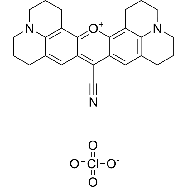
| Molecular Formula | C26H26CLN3O5 |
| Molecular Weight | 495.9547 |
| Exact Mass | 495.156 |
| CAS # | 137993-41-0 |
| PubChem CID | 127750 |
| Appearance | Dark purple to black solid powder |
| Hydrogen Bond Donor Count | 0 |
| Hydrogen Bond Acceptor Count | 7 |
| Rotatable Bond Count | 0 |
| Heavy Atom Count | 35 |
| Complexity | 1020 |
| Defined Atom Stereocenter Count | 0 |
| InChi Key | HACOCUMLBPNDIN-UHFFFAOYSA-M |
| InChi Code | InChI=1S/C26H26N3O.ClHO4/c27-15-22-20-13-16-5-1-9-28-11-3-7-18(23(16)28)25(20)30-26-19-8-4-12-29-10-2-6-17(24(19)29)14-21(22)26;2-1(3,4)5/h13-14H,1-12H2;(H,2,3,4,5)/q+1;/p-1 |
| Chemical Name | 3-oxa-23-aza-9-azoniaheptacyclo[17.7.1.15,9.02,17.04,15.023,27.013,28]octacosa-1(27),2(17),4,9(28),13,15,18-heptaene-16-carbonitrile;perchlorate |
| HS Tariff Code | 2934.99.9001 |
| Storage |
Powder-20°C 3 years 4°C 2 years In solvent -80°C 6 months -20°C 1 month Note: Please store this product in a sealed and protected environment (e.g. under nitrogen), avoid exposure to moisture and light. |
| Shipping Condition | Room temperature (This product is stable at ambient temperature for a few days during ordinary shipping and time spent in Customs) |
Biological Activity
| Targets | The literature describes Rhodamine 800 as a potential near-infrared (NIR)-active contrast agent responsive to cellular energy states, specifically the mitochondrial membrane potential.Its “target” is the physicochemical environment (e.g., membrane potential, solvent polarity) within energized mitochondria.[1] |
| ln Vitro |
1. Parts of the working solution for rhodamine 800 1.1 Prepare the stock solution. Remove the 5 mM stock solution and mix 1 mg of Rhodamine 800 with 525 μL of anhydrous DMSO. 1.2 Components of working solution To prepare a 1-20 μM Rhodamine 800 working solution, use pre-prepared cell-free bone marrow or PBS storage solution. Note: Please prepare Rhodamine 800 working solution for use by adjusting its concentration to suit the current circumstances. 2. Suspended cell staining (2.1): Centrifuge cells, add PBS, then wash twice for five minutes each time. The density of cells is 1×10⁶/mL. 2.2 Add 1 mL of the working solution Rhodamine 800, then centrifuge for 30 to 60 minutes. 2.3 After centrifuging for three to four minutes at 400 g, remove the supernatant. 2.4 Wash the cells twice with PBS, giving them five minutes each time. 2.5 Re-suspend the cells in 1 mL of PBS or serum-free cells, and use a flow cytometer or fluorescence microscope to observe. 3. 3.1 Culture the adherent cells 3.2 Take off the coverslip from the cells and absorb all of the iodine in the process of staining cells (adhesion). 3.3 Add 100 μL dye working solution, cover the cells with a gentle shake, and let them remain sterile for five to thirty minutes. 3.4 Aspirate the dye working solution, rinse with culture medium two or three times for five minutes each, and use a flow cytometer or fluorescence microscope to observe. In isolated rat liver mitochondria, the absorption spectrum of Rhodamine 800 showed a red shift upon energization (state 4), with peaks at 706 nm and 648 nm. Addition of the uncoupler CCCP caused a blue shift to 690 nm and 628 nm, resembling its spectrum in aqueous buffer.[1] The fluorescence intensity of Rhodamine 800 was strongly quenched in energized mitochondria (state 4), decreasing to 22% of its intensity in aqueous buffer, and recovered to about 70% upon uncoupling.[1] Changes in absorbance (measured at 730-685 nm or 730-800 nm) and fluorescence intensity (measured at 692 nm) of Rhodamine 800 varied linearly with the valinomycin-induced potassium diffusion potential (ΔψK+) across the mitochondrial membrane. The sensitivity of fluorescence quenching to membrane potential was approximately 2.3% per 10 mV.[1] In isolated rat hepatocytes under normoxia, Rhodamine 800 exhibited spectral characteristics similar to those in energized mitochondria. Upon induction of anoxia (complete reduction of cytochrome oxidase), these characteristics remained unchanged for about 5 minutes, indicating the cells were still energized. Subsequent addition of CCCP caused a rapid shift in absorbance and fluorescence to resemble the de-energized/uncoupled state.[1] The uptake of Rhodamine 800 into energized mitochondria was concentration-dependent. At concentrations below 5 µM, the uptake-to-free dye ratio was around 5-6, corresponding to an intra-mitochondrial concentration approximately 8000 times higher than in the medium. The ratio reached a maximum (~20) at about 15 µM added dye. In uncoupled mitochondria, the uptake ratio remained constant at about 1.[1] Adenine nucleotides (ADP and ATP) statically quenched the fluorescence of Rhodamine 800 in solution, but this effect was minor compared to the quenching induced by changes in membrane potential.[1] At concentrations below 5 µM, Rhodamine 800 had no significant effect on the respiratory control ratio (RCR) or oxygen consumption rates (in state 3 or state 4) of isolated mitochondria.[1] |
| Cell Assay |
For optical measurements in isolated hepatocytes, hepatocytes were isolated from rat liver via collagenase perfusion. Cell viability was assessed using trypan blue exclusion, and preparations with >90% intact cells were used.[1] Hepatocytes were incubated with Rhodamine 800 (e.g., 3.3 µM) in a physiological buffer at 37°C. The cell suspension was placed in a quartz cuvette within the spectrophotometer/fluorometer.[1] Simultaneous measurements of oxygen concentration (using a Clark-type electrode), redox state of cytochrome oxidase (via absorbance at 620-605 nm), and Rhodamine 800 signals (absorbance at 730-685 nm or fluorescence) were performed during the transition from aerobic to anaerobic conditions. Uncoupler (CCCP, 2 µM) was added after anoxia to collapse the membrane potential.[1] Fluorescence emission spectra of Rhodamine 800 in hepatocytes were recorded with excitation at 660 nm, using a cutoff filter to block excitation light. Spectra were compared before and after CCCP addition.[1] |
| Toxicity/Toxicokinetics |
At concentrations up to 5 µM, Rhodamine 800 did not affect mitochondrial respiration control ratio or rates, suggesting low toxicity to mitochondrial function at the concentrations used for probing.[1] The literature does not provide other toxicity data (e.g., LD50, organ toxicity, protein binding) for Rhodamine 800.[1] |
| References |
[1]. Rhodamine 800 as a probe of energization of cells and tissues in the near-infrared region: a study with isolated rat liver mitochondria and hepatocytes. J Biochem. 1997 Jan;121(1):29-37. [2]. Near-infrared fluorescence detection of acetylcholine in aqueous solution using a complex ofrhodamine 800 and p-sulfonatocalix[8]arene. Sensors (Basel). 2010;10(3):2438-49. |
| Additional Infomation |
Rhodamine 800 is a cationic dye whose optical properties (absorption and fluorescence spectra, intensity) are sensitive to the solvent environment. Its absorption and fluorescence peaks shift to longer wavelengths in more polar solvents, and fluorescence intensity varies significantly with solvent type.[1] The primary mechanism of its response in biological systems is potential-dependent accumulation into the mitochondrial matrix. In energized mitochondria, the dye accumulates, leading to a red shift in absorption and quenching of fluorescence. Upon de-energization (uncoupling), the dye is released, reversing these spectral changes.[1] The study proposes Rhodamine 800 as a promising contrast agent for non-invasive monitoring of tissue energy states using near-infrared spectrophotometry (NIRS), due to its NIR-active wavelengths and responsiveness to mitochondrial membrane potential.[1] The authors note that for in vivo application in blood-circulating tissues, chemical modification (e.g., adding carboxyl ester groups) of Rhodamine 800 might be necessary to improve tissue incorporation.[1] |
Solubility Data
| Solubility (In Vitro) | DMSO : ~83.33 mg/mL (~168.02 mM) |
| Solubility (In Vivo) |
Note: Listed below are some common formulations that may be used to formulate products with low water solubility (e.g. < 1 mg/mL), you may test these formulations using a minute amount of products to avoid loss of samples. Injection Formulations (e.g. IP/IV/IM/SC) Injection Formulation 1: DMSO : Tween 80: Saline = 10 : 5 : 85 (i.e. 100 μL DMSO stock solution → 50 μL Tween 80 → 850 μL Saline) *Preparation of saline: Dissolve 0.9 g of sodium chloride in 100 mL ddH ₂ O to obtain a clear solution. Injection Formulation 2: DMSO : PEG300 :Tween 80 : Saline = 10 : 40 : 5 : 45 (i.e. 100 μL DMSO → 400 μLPEG300 → 50 μL Tween 80 → 450 μL Saline) Injection Formulation 3: DMSO : Corn oil = 10 : 90 (i.e. 100 μL DMSO → 900 μL Corn oil) Example: Take the Injection Formulation 3 (DMSO : Corn oil = 10 : 90) as an example, if 1 mL of 2.5 mg/mL working solution is to be prepared, you can take 100 μL 25 mg/mL DMSO stock solution and add to 900 μL corn oil, mix well to obtain a clear or suspension solution (2.5 mg/mL, ready for use in animals). Injection Formulation 4: DMSO : 20% SBE-β-CD in saline = 10 : 90 [i.e. 100 μL DMSO → 900 μL (20% SBE-β-CD in saline)] *Preparation of 20% SBE-β-CD in Saline (4°C,1 week): Dissolve 2 g SBE-β-CD in 10 mL saline to obtain a clear solution. Injection Formulation 5: 2-Hydroxypropyl-β-cyclodextrin : Saline = 50 : 50 (i.e. 500 μL 2-Hydroxypropyl-β-cyclodextrin → 500 μL Saline) Injection Formulation 6: DMSO : PEG300 : castor oil : Saline = 5 : 10 : 20 : 65 (i.e. 50 μL DMSO → 100 μLPEG300 → 200 μL castor oil → 650 μL Saline) Injection Formulation 7: Ethanol : Cremophor : Saline = 10: 10 : 80 (i.e. 100 μL Ethanol → 100 μL Cremophor → 800 μL Saline) Injection Formulation 8: Dissolve in Cremophor/Ethanol (50 : 50), then diluted by Saline Injection Formulation 9: EtOH : Corn oil = 10 : 90 (i.e. 100 μL EtOH → 900 μL Corn oil) Injection Formulation 10: EtOH : PEG300:Tween 80 : Saline = 10 : 40 : 5 : 45 (i.e. 100 μL EtOH → 400 μLPEG300 → 50 μL Tween 80 → 450 μL Saline) Oral Formulations Oral Formulation 1: Suspend in 0.5% CMC Na (carboxymethylcellulose sodium) Oral Formulation 2: Suspend in 0.5% Carboxymethyl cellulose Example: Take the Oral Formulation 1 (Suspend in 0.5% CMC Na) as an example, if 100 mL of 2.5 mg/mL working solution is to be prepared, you can first prepare 0.5% CMC Na solution by measuring 0.5 g CMC Na and dissolve it in 100 mL ddH2O to obtain a clear solution; then add 250 mg of the product to 100 mL 0.5% CMC Na solution, to make the suspension solution (2.5 mg/mL, ready for use in animals). Oral Formulation 3: Dissolved in PEG400 Oral Formulation 4: Suspend in 0.2% Carboxymethyl cellulose Oral Formulation 5: Dissolve in 0.25% Tween 80 and 0.5% Carboxymethyl cellulose Oral Formulation 6: Mixing with food powders Note: Please be aware that the above formulations are for reference only. InvivoChem strongly recommends customers to read literature methods/protocols carefully before determining which formulation you should use for in vivo studies, as different compounds have different solubility properties and have to be formulated differently. (Please use freshly prepared in vivo formulations for optimal results.) |
| Preparing Stock Solutions | 1 mg | 5 mg | 10 mg | |
| 1 mM | 2.0163 mL | 10.0817 mL | 20.1633 mL | |
| 5 mM | 0.4033 mL | 2.0163 mL | 4.0327 mL | |
| 10 mM | 0.2016 mL | 1.0082 mL | 2.0163 mL |