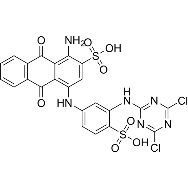
Physicochemical Properties
| Molecular Formula | C23H14CL2N6O8S2 |
| Molecular Weight | 637.4287 |
| Exact Mass | 635.969 |
| CAS # | 13324-20-4 |
| Related CAS # | 4499-01-8 (di-hydrochloride salt) |
| PubChem CID | 25863 |
| Appearance | Blue to dark blue solid powder |
| Density | 1.86g/cm3 |
| Index of Refraction | 1.774 |
| LogP | 6.405 |
| Hydrogen Bond Donor Count | 5 |
| Hydrogen Bond Acceptor Count | 14 |
| Rotatable Bond Count | 6 |
| Heavy Atom Count | 41 |
| Complexity | 1220 |
| Defined Atom Stereocenter Count | 0 |
| HS Tariff Code | 2934.99.9001 |
| Storage |
Powder-20°C 3 years 4°C 2 years In solvent -80°C 6 months -20°C 1 month Note: This product requires protection from light (avoid light exposure) during transportation and storage. |
| Shipping Condition | Room temperature (This product is stable at ambient temperature for a few days during ordinary shipping and time spent in Customs) |
Biological Activity
| ln Vitro |
Reactive Blue 4 was effectively decolorized and biotransformed by aerobic bacterial granules (ABGs) under in vitro conditions. The ABGs decolorized Reactive Blue 4 at a wide range of pH (4.0 to 11.0) and temperature (20 to 55 °C). The granules tolerated and degraded high dye concentrations up to 1000 mg L⁻¹, with a maximum decolorization rate (Vmax) of 6.16 ± 0.82 mg L⁻¹ h⁻¹ and a Michaelis constant (Km) of 227 ± 41 mg L⁻¹. The decolorization rate was higher under static conditions (5.01 mg L⁻¹ h⁻¹) compared to shaking conditions (3.71 mg L⁻¹ h⁻¹). During continuous exposure cycles, the decolorization rate increased from 1.27 mg L⁻¹ h⁻¹ (61% decolorization) in the 1st cycle to 1.79 mg L⁻¹ h⁻¹ (86% decolorization) by the 9th cycle, then decreased to 1.15 mg L⁻¹ h⁻¹ (55% decolorization) by the 16th cycle, indicating retained tolerance. Biotransformation was confirmed by HPLC, FT-IR, and GC-HRMS analysis, identifying metabolites such as 4-amino-9,10-dihydro-9,10-dioxoanthracene-2-sulfonic acid, 2-(4,6-dichloro-1,3,5-triazin-2-ylamino)-4-aminophenol, and 1-aminoanthracene-9,10-dione. Significant reduction in organic load was observed: Chemical Oxygen Demand (COD), Biological Oxygen Demand (BOD), and Total Dissolved Solids (TDS) were reduced by 90%, 91%, and 77%, respectively, after 72 h under static conditions.[2] |
| Cell Assay |
Cytotoxicity Assay: The cytotoxicity of Reactive Blue 4 and its biotransformation metabolites was evaluated on HaCat (human keratinocyte) and FHM (fish epithelial) cell lines using the MTT assay. Cells were seeded in 96-well plates (HaCat: 10⁵ cells mL⁻¹; FHM: 2×10⁵ cells mL⁻¹) in their respective media (DMEM/F12 for HaCat, MEM for FHM). The dye or its metabolites (final concentration 0.01 mg mL⁻¹), obtained at different biotransformation time points, were added to the wells. Plates were incubated for 48 h in a CO₂ incubator (5% CO₂, 37°C). After incubation, media was removed, wells were washed with PBS, and MTT solution (40 µg per well) was added followed by a 4 h incubation. The resulting formazan crystals were dissolved in dimethyl sulphoxide, and absorbance was measured at 570 nm (with background subtraction at 690 nm). Cell viability was calculated as a percentage relative to untreated control cells.[2] Genotoxicity Assay (Comet Assay): The genotoxicity of Reactive Blue 4 and its metabolites was assessed on human blood lymphocytes, HaCat, and FHM cell lines using the alkaline comet assay. For lymphocytes, cells (10⁴ cells mL⁻¹) were incubated with the dye (0.25 mg mL⁻¹) or its metabolites in RPMI medium for 1 h at 37°C. H₂O₂ (100 µM, 5 min, 4°C) served as the positive control. For HaCat and FHM cells, cells (4×10⁵ cells mL⁻¹) were treated with the dye or metabolites (0.01 mg L⁻¹) in 96-well plates for 48 h, then detached using trypsin-EDTA. Treated cells were washed, suspended in a small volume of medium, mixed with low-melting-point agarose, and layered onto microscope slides pre-coated with normal-melting-point agarose. Cells were lysed (in a solution containing NaCl, EDTA, Tris, DMSO, and Triton X-100, pH 10), followed by alkaline unwinding (in electrophoresis buffer: NaOH, EDTA, pH >13) and electrophoresis. Slides were neutralized, stained with ethidium bromide (20 µg mL⁻¹), and visualized under a fluorescence microscope. DNA damage was quantified for ~300 cells per sample using image analysis software, with percent tail DNA as the parameter.[2] |
| Toxicity/Toxicokinetics |
Phytotoxicity: Reactive Blue 4 exhibited phytotoxicity towards Triticum aestivum (wheat). At 100 mg L⁻¹, it reduced seed germination by 27%, root length by 48%, shoot length by 29%, chlorophyll-a content by 76%, and chlorophyll-b content by 53%. The biotransformed metabolites (especially after 48 h static treatment) showed no significant phytotoxic effects, with near-normal germination, growth, and chlorophyll content.[2] Cytotoxicity: Reactive Blue 4 (0.01 mg mL⁻¹) significantly reduced cell viability to 53.33% in HaCat cells and 44.97% in FHM cells. The cytotoxicity of its metabolites decreased over time. Metabolites from 48 h static treatment showed 95.83% (HaCat) and 100.80% (FHM) cell survival, indicating effective detoxification.[2] Genotoxicity: Reactive Blue 4 induced significant DNA damage. It increased % tail DNA by 3.26-fold in human lymphocytes, 2.02-fold in HaCat cells, and 11.5-fold in FHM cells compared to controls. The biotransformed metabolites (from 24 h static, 48 h static, and 24 h static + 24 h shaking treatments) showed a significant reduction in DNA damage, indicating loss of genotoxicity.[2] |
| References |
[1]. Reactive Blue 4 as a Single Colorimetric Chemosensor for Sequential Determination of Multiple Analytes with Different Optical Responses in Aqueous Media: Cu2+-Cysteine Using a Metal Ion Displacement and Cu2+-Arginine Through the Host-Guest Interaction. [2]. Effective biotransformation and detoxification of anthraquinone dye reactive blue 4 by using aerobic bacterial granules. Water Res. 2017 Oct 1;122:603-613. |
| Additional Infomation |
Reactive Blue 4 (RB4) is a water-soluble anthraquinone dye used as a colorimetric chemosensor. The sensor operates sequentially: free RB4 detects Cu²⁺ ions via a binding site-signaling subunit approach, forming a 1:1 RB4-Cu²⁺ complex with an association constant of (4.46 ± 0.12) × 10⁵ L mol⁻¹. This complex then differentially detects L-arginine (Arg) and L-cysteine (Cys). Arg is detected through a host-guest interaction involving its amino and carboxylate groups binding to the complex (binding constant 1.09 × 10⁴ L mol⁻¹). Cys is detected via a metal displacement mechanism due to its higher affinity for Cu²⁺, which releases free RB4. The limits of detection (LOD) in aqueous HEPES buffer (pH 7.0) are 1.96 μmol L⁻¹ for Cu²⁺, 1.06 μmol L⁻¹ for Arg, and 1.33 μmol L⁻¹ for Cys. The sensor works effectively in a pH range of 6.0 to 9.0, with optimal response at pH 7.0. Practical applications were demonstrated by detecting Cu²⁺ in spiked water (tap, river) and serum samples, Arg in commercial dietary supplements, and Cys in human serum samples with good recovery rates. The system was also fabricated into paper test strips for the naked-eye visual detection of Arg and Cys. Furthermore, the absorbance changes at 607 nm were used to mimic molecular logic gates: a NAND gate with inputs Cu²⁺ and Arg, and an IMPLICATION gate with inputs Cu²⁺ and Cys.[1] Reactive Blue 4 is an anthraquinone dye used in the textile industry and is a recalcitrant water pollutant. This study demonstrates that aerobic bacterial granules (ABGs) are an effective system for its bioremediation and detoxification. Microbial community analysis (16S rRNA metagenomics) revealed shifts in granule composition during dye exposure, with increased abundance of phyla like Firmicutes and Bacteroidetes, and families like Porphyromonadaceae and Gracilibacteraceae, implicating their role in degradation. PICRUSt imputed metagenomic analysis predicted the up-regulation of xenobiotic degradation pathways (e.g., naphthalene, benzoate degradation) and environmental information processing systems during dye decolorization. The proposed biotransformation pathway involves reductive cleavage and subsequent breakdown into simpler, less toxic aromatic compounds. The study highlights the potential of ABGs for treating textile effluents containing high loads of anthraquinone dyes under variable environmental conditions.[2] |
Solubility Data
| Solubility (In Vitro) | DMSO : ~83.33 mg/mL (~130.73 mM) |
| Solubility (In Vivo) |
Solubility in Formulation 1: ≥ 4.17 mg/mL (6.54 mM) (saturation unknown) in 10% DMSO + 40% PEG300 + 5% Tween80 + 45% Saline (add these co-solvents sequentially from left to right, and one by one), clear solution. For example, if 1 mL of working solution is to be prepared, you can add 100 μL of 41.7 mg/mL clear DMSO stock solution to 400 μL PEG300 and mix evenly; then add 50 μL Tween-80 to the above solution and mix evenly; then add 450 μL normal saline to adjust the volume to 1 mL. Preparation of saline: Dissolve 0.9 g of sodium chloride in 100 mL ddH₂ O to obtain a clear solution. Solubility in Formulation 2: ≥ 4.17 mg/mL (6.54 mM) (saturation unknown) in 10% DMSO + 90% (20% SBE-β-CD in Saline) (add these co-solvents sequentially from left to right, and one by one), clear solution. For example, if 1 mL of working solution is to be prepared, you can add 100 μL of 41.7 mg/mL clear DMSO stock solution to 900 μL of 20% SBE-β-CD physiological saline solution and mix evenly. Preparation of 20% SBE-β-CD in Saline (4°C,1 week): Dissolve 2 g SBE-β-CD in 10 mL saline to obtain a clear solution. Solubility in Formulation 3: ≥ 4.17 mg/mL (6.54 mM) (saturation unknown) in 10% DMSO + 90% Corn Oil (add these co-solvents sequentially from left to right, and one by one), clear solution. For example, if 1 mL of working solution is to be prepared, you can add 100 μL of 41.7 mg/mL clear DMSO stock solution to 900 μL of corn oil and mix evenly. Solubility in Formulation 4: ≥ 2.08 mg/mL (3.26 mM) (saturation unknown) in 10% DMSO + 40% PEG300 + 5% Tween80 + 45% Saline (add these co-solvents sequentially from left to right, and one by one), clear solution. For example, if 1 mL of working solution is to be prepared, you can add 100 μL of 20.8 mg/mL clear DMSO stock solution to 400 μL of PEG300 and mix evenly; then add 50 μL of Tween-80 to the above solution and mix evenly; then add 450 μL of normal saline to adjust the volume to 1 mL. Preparation of saline: Dissolve 0.9 g of sodium chloride in 100 mL ddH₂ O to obtain a clear solution. Solubility in Formulation 5: ≥ 2.08 mg/mL (3.26 mM) (saturation unknown) in 10% DMSO + 90% (20% SBE-β-CD in Saline) (add these co-solvents sequentially from left to right, and one by one), clear solution. For example, if 1 mL of working solution is to be prepared, you can add 100 μL of 20.8 mg/mL clear DMSO stock solution to 900 μL of 20% SBE-β-CD physiological saline solution and mix evenly. Preparation of 20% SBE-β-CD in Saline (4°C,1 week): Dissolve 2 g SBE-β-CD in 10 mL saline to obtain a clear solution. (Please use freshly prepared in vivo formulations for optimal results.) |
| Preparing Stock Solutions | 1 mg | 5 mg | 10 mg | |
| 1 mM | 1.5688 mL | 7.8440 mL | 15.6880 mL | |
| 5 mM | 0.3138 mL | 1.5688 mL | 3.1376 mL | |
| 10 mM | 0.1569 mL | 0.7844 mL | 1.5688 mL |