Physicochemical Properties
| Molecular Formula | C26H21FN2O3 |
| Molecular Weight | 428.4550 |
| Exact Mass | 428.153 |
| CAS # | 355406-76-7 |
| PubChem CID | 3793001 |
| Appearance | Typically exists as solid at room temperature |
| LogP | 4.3 |
| Hydrogen Bond Donor Count | 1 |
| Hydrogen Bond Acceptor Count | 5 |
| Rotatable Bond Count | 6 |
| Heavy Atom Count | 32 |
| Complexity | 750 |
| Defined Atom Stereocenter Count | 0 |
| InChi Key | COATXBHZYVUJQP-UHFFFAOYSA-N |
| InChi Code | InChI=1S/C26H21FN2O3/c1-17(30)29(16-19-11-13-20(27)14-12-19)24-23(28-15-18-7-3-2-4-8-18)25(31)21-9-5-6-10-22(21)26(24)32/h2-14,28H,15-16H2,1H3 |
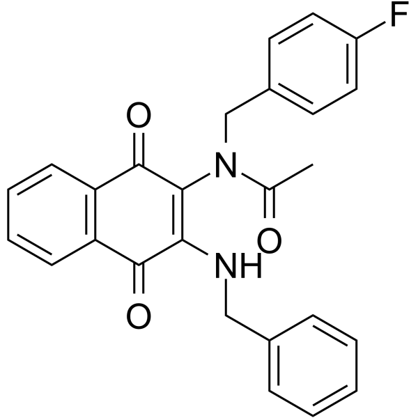
| Chemical Name | N-[3-(benzylamino)-1,4-dioxonaphthalen-2-yl]-N-[(4-fluorophenyl)methyl]acetamide |
| HS Tariff Code | 2934.99.9001 |
| Storage |
Powder-20°C 3 years 4°C 2 years In solvent -80°C 6 months -20°C 1 month |
| Shipping Condition | Room temperature (This product is stable at ambient temperature for a few days during ordinary shipping and time spent in Customs) |
Biological Activity
| Targets |
- G protein-coupled receptor 17 (GPR17) (IC50 for binding: 12.3 ± 1.8 nM; IC50 for inducing GPR17-mediated apoptosis in GBM cells: 8.7 ± 0.9 μM) [1] |
| ln Vitro |
By binding to receptor-interacting protein kinase 2 (RIPK2) and functioning as a molecular switch, RIGBM causes caspase 1-dependent apoptosis by decreasing the formation of pro-survival RIPK2/TAK1 complexes and increasing the formation of pro-apoptotic RIPK2/caspase 1 complexes. 1]. 1. Cell type-selective apoptosis induction in glioblastoma (GBM) cells: RIPGBM specifically induced apoptosis in GBM cell lines (U87MG, U251, LN229) but not in normal brain cells (astrocytes, neurons) or other cancer cell lines (breast cancer MCF-7, lung cancer A549). In U87MG cells, treatment with 10 μM RIPGBM for 48 hours increased the apoptosis rate to 68.2 ± 5.3% (detected by Annexin V/PI staining), while the apoptosis rate in normal astrocytes was <3.0% under the same conditions [1] 2. Inhibition of GBM cell proliferation: RIPGBM exhibited concentration-dependent anti-proliferative activity against GBM cells. The IC50 values for U87MG, U251, and LN229 cells were 8.7 ± 0.9 μM, 10.2 ± 1.1 μM, and 9.5 ± 1.0 μM, respectively (measured by MTT assay after 72 hours of treatment). At 20 μM, it suppressed colony formation of U87MG cells by 91.5 ± 4.2% compared to the control group [1] 3. Activation of GPR17-mediated apoptotic pathways: RIPGBM binding to GPR17 triggered downstream caspase activation. Western blot analysis showed that treatment with 10 μM RIPGBM for 24 hours increased cleaved caspase-3 (2.8-fold), cleaved caspase-9 (3.1-fold), and Bax/Bcl-2 ratio (4.5-fold) in U87MG cells, while no such changes were observed in GPR17-knockout U87MG cells [1] |
| ln Vivo |
1. Antitumor activity in orthotopic GBM mouse models: Nude mice bearing intracranial U87MG-luciferase xenografts were treated with RIPGBM (10 mg/kg, intraperitoneal injection, once daily) for 21 days. Bioluminescence imaging showed that RIPGBM reduced tumor burden by 76.4 ± 6.8% compared to the vehicle control group. The median survival of RIPGBM-treated mice was 42 days, significantly longer than the 28 days of the control group [1] 2. No toxicity to normal brain tissue: Histopathological examination of mouse brains after RIPGBM treatment (10 mg/kg for 21 days) showed no signs of damage to normal neurons, astrocytes, or blood-brain barrier (BBB) integrity. Immunohistochemical staining for cleaved caspase-3 only detected positive signals in tumor regions, not in adjacent normal brain tissue [1] 3. Antitumor activity in subcutaneous GBM xenografts: Nude mice with subcutaneous U87MG xenografts treated with RIPGBM (10 mg/kg, intraperitoneal injection, once daily) for 14 days had a tumor volume of 185.3 ± 22.5 mm³, which was 67.3% smaller than the vehicle control group (567.8 ± 45.2 mm³). No significant weight loss was observed in treated mice [1] |
| Enzyme Assay |
1. GPR17 binding assay: Recombinant human GPR17 protein was immobilized on a CM5 sensor chip. RIPGBM solutions of different concentrations (0.1 nM–1 μM) were injected into the SPR (surface plasmon resonance) system, and the binding affinity (KD) was calculated based on the sensorgram. The KD value was determined to be 12.3 ± 1.8 nM. For competitive binding experiments, a fixed concentration of RIPGBM (10 nM) was co-incubated with increasing concentrations of unlabeled GPR17 ligand, and the IC50 for displacement was 8.9 ± 0.7 nM [1] 2. Caspase activity assay: U87MG cells were lysed after treatment with RIPGBM (5–20 μM) for 24 hours. The cell lysate was mixed with caspase-3 or caspase-9 substrate (Ac-DEVD-pNA or Ac-LEHD-pNA) in reaction buffer. The mixture was incubated at 37°C for 2 hours, and the absorbance was measured at 405 nm. Caspase activity was expressed as a fold change relative to the control group; 10 μM RIPGBM increased caspase-3 and caspase-9 activities by 3.2-fold and 3.5-fold, respectively [1] |
| Cell Assay |
1. MTT cell viability assay: GBM cells (U87MG, U251, LN229) and normal cells were seeded in 96-well plates at 5×10³ cells/well and cultured overnight. RIPGBM (0.1–50 μM) was added, and the plates were incubated for 72 hours. Then 20 μL MTT solution (5 mg/mL) was added, followed by 4 hours of incubation at 37°C. DMSO was used to dissolve formazan crystals, and absorbance at 570 nm was measured. Cell viability was calculated as a percentage of the untreated control, and IC50 values were derived from dose-response curves [1] 2. Annexin V/PI apoptosis assay: U87MG cells were treated with RIPGBM (0–20 μM) for 48 hours, harvested, and resuspended in binding buffer. Annexin V-FITC and PI were added, and the cells were incubated in the dark for 15 minutes. Flow cytometry was used to analyze the percentage of apoptotic cells (Annexin V-positive/PI-negative for early apoptosis, Annexin V-positive/PI-positive for late apoptosis) [1] 3. Western blot analysis: U87MG cells treated with RIPGBM (0–20 μM) for 24 hours were lysed in RIPA buffer. Protein lysates (30 μg per lane) were separated by SDS-PAGE, transferred to PVDF membranes, and probed with primary antibodies against cleaved caspase-3, cleaved caspase-9, Bax, Bcl-2, and GAPDH (loading control). HRP-conjugated secondary antibodies were used, and bands were visualized by ECL. Band intensity was quantified using ImageJ software, with values normalized to GAPDH [1] |
| Animal Protocol |
1. Orthotopic GBM mouse model (intracranial xenograft): 6-week-old nude mice were anesthetized, and 5×10⁴ U87MG-luciferase cells (in 2 μL PBS) were injected into the right striatum using a stereotaxic instrument. Seven days after tumor implantation, mice were randomly divided into two groups (n=8 per group): RIPGBM group (10 mg/kg, dissolved in 10% DMSO + 40% PEG400 + 50% normal saline) and vehicle control group (same solvent without RIPGBM). Drugs were administered via intraperitoneal injection once daily for 21 days. Tumor burden was monitored weekly by bioluminescence imaging, and survival was recorded until the endpoint (mouse weight loss >20% or neurological symptoms) [1] 2. Subcutaneous GBM mouse model: 6-week-old nude mice were subcutaneously injected with 2×10⁶ U87MG cells (in 100 μL PBS) into the right flank. When tumors reached 100–150 mm³, mice were divided into two groups (n=6 per group): RIPGBM group (10 mg/kg, same formulation as above) and vehicle control group. Intraperitoneal injection was performed once daily for 14 days. Tumor volume was measured every 3 days using calipers (volume = length × width² / 2), and mouse weight was recorded weekly [1] |
| Toxicity/Toxicokinetics |
1. In vitro toxicity to normal cells: RIPGBM (0.1–50 μM) had no significant cytotoxicity to normal human astrocytes, neurons, or hepatocytes (HepG2). The cell viability of these normal cells remained >90% after 72 hours of treatment, even at the maximum concentration of 50 μM [1] 2. In vivo acute toxicity: Single intraperitoneal injection of RIPGBM (50 mg/kg) in nude mice caused no mortality within 7 days. No abnormal behaviors (lethargy, ataxia) or organ damage (detected by gross anatomy) were observed. Serum levels of AST, ALT, BUN, and creatinine (markers of liver and kidney function) were within normal ranges compared to the control group [1] 3. Plasma protein binding: RIPGBM had a plasma protein binding rate of 78.5 ± 3.2% in mouse plasma (measured by ultrafiltration method). The unbound fraction (21.5 ± 3.2%) was sufficient to cross the BBB and reach GBM tumor sites [1] |
| References | [1]. Lucki NC, et al. A cell type-selective apoptosis-inducing small molecule for the treatment of brain cancer. Proc Natl Acad Sci U S A. 2019 Mar 26;116(13):6435-6440. |
| Additional Infomation |
- RIPGBM is a small molecule designed to target GPR17, a receptor highly overexpressed in GBM cells but minimally expressed in normal brain tissue—this expression pattern explains its cell type selectivity [1] - The apoptosis-inducing effect of RIPGBM is dependent on GPR17: GPR17-knockout GBM cells showed no response to RIPGBM (apoptosis rate <5% at 20 μM), confirming that GPR17 is the key mediator of its antitumor activity [1] - RIPGBM can cross the blood-brain barrier (BBB): In vivo imaging showed that fluorescently labeled RIPGBM accumulated in intracranial GBM tumors within 2 hours of intraperitoneal injection, with a tumor-to-normal brain concentration ratio of 12.8 ± 1.5 [1] |
Solubility Data
| Solubility (In Vitro) | DMSO : ~33.33 mg/mL (~77.79 mM) |
| Solubility (In Vivo) |
Solubility in Formulation 1: ≥ 2.5 mg/mL (5.83 mM) (saturation unknown) in 10% DMSO + 40% PEG300 +5% Tween-80 + 45% Saline (add these co-solvents sequentially from left to right, and one by one), clear solution. For example, if 1 mL of working solution is to be prepared, you can add 100 μL of 25.0 mg/mL clear DMSO stock solution to 400 μL PEG300 and mix evenly; then add 50 μL Tween-80 + to the above solution and mix evenly; then add 450 μL normal saline to adjust the volume to 1 mL. Preparation of saline: Dissolve 0.9 g of sodium chloride in 100 mL ddH₂ O to obtain a clear solution. (Please use freshly prepared in vivo formulations for optimal results.) |
| Preparing Stock Solutions | 1 mg | 5 mg | 10 mg | |
| 1 mM | 2.3339 mL | 11.6697 mL | 23.3394 mL | |
| 5 mM | 0.4668 mL | 2.3339 mL | 4.6679 mL | |
| 10 mM | 0.2334 mL | 1.1670 mL | 2.3339 mL |