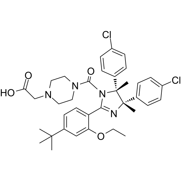
Physicochemical Properties
| Molecular Formula | C36H42CL2N4O4 |
| Molecular Weight | 665.65 |
| CAS # | 2360561-90-4 |
| Appearance | Typically exists as solid at room temperature |
| HS Tariff Code | 2934.99.9001 |
| Storage |
Powder-20°C 3 years 4°C 2 years In solvent -80°C 6 months -20°C 1 month |
| Shipping Condition | Room temperature (This product is stable at ambient temperature for a few days during ordinary shipping and time spent in Customs) |
Biological Activity
| Targets | MDM2 |
| ln Vitro |
MDM2-PROTAC activates wild-type p53 and is more effective than MDM2 inhibitors at inducing apoptosis of wild-type p53-containing breast cancer cells[1] To characterize the biological effects of our MDM2-PROTAC, we first tested it head-to-head with MDM2 inhibitors that prevent MDM2-p53 binding in p53 wild-type breast cancer cells with reported sensitivity to the MDM2 inhibitor Nutlin (25–27). Treatment of p53 wild-type MCF7 and DU4475 cells with MDM2 inhibitors (Nutlin, RG7112, RG7112D) or our MDM2-PROTAC resulted in the expected stabilization of MDM2 protein with the inhibitors and loss of MDM2 with the PROTAC (Fig. 2A). All compounds increased p53 protein levels (Fig. 2A) and significantly reduced cell survival in a concentration-dependent manner (Fig. 2B; Supplementary Fig. S2A). Of note, the IC50 of our MDM2-PROTAC in MCF7 and DU4475 cells was lower than Nutlin (2.8μM and 2.3μM versus 11μM and 4μM, respectively) and lower than or equal to RG7112D and RG7112. Similarly, increased Caspase-3 activity was detected in MDM2-PROTAC-treated cells compared to those that received the MDM2 inhibitors at the same concentrations (Fig. 2C; Supplementary Fig. S2B). Over time, there was more cleaved PARP (Fig. 2D) and subG1 apoptotic DNA (Fig. 2E) with MDM2-PROTAC treatment compared to the MDM2 inhibitors. Furthermore, similar to Nutlin and its derivatives, MDM2-PROTAC treatment of MCF7 cells resulted in increased mRNA and protein levels of pro-apoptotic p53 target genes BAX, NOXA, PUMA (Fig. 2F and 2G, respectively).[1] |
| ln Vivo |
MDM2-PROTAC is stable and effectively kills TNBC cells in vivo[1] Prior to testing our MDM2-PROTAC on tumors in vivo, we performed mouse liver microsome and pharmacokinetic (PK) studies to determine its stability in vivo. Our MDM2-PROTAC was moderately metabolically stable with a 25.9-minute half-life (Fig. 5A) and had excellent in vivo stability with plasma levels stable over 6 hours in mice after a single 10mg/kg intraperitoneal dose (Fig. 5B). Due to its stability in vivo, we then evaluated the effectiveness of our MDM2-PROTAC at killing TNBC tumors in mice. Xenografts of MDA-MB-231 and MDA-MB-436 TNBC cells grew to ~80mm3, and then tumor size-matched mice were treated with our MDM2-PROTAC, RG7112D control compound, or vehicle control. Fourteen days of MDM2-PROTAC treatment significantly extended mouse survival (Fig. 5C) and decreased tumor volume (Fig. 5D; Supplementary Fig. S4A), compared to control mice for both xenograft models. To confirm our MDM2-PROTAC was hitting its target (MDM2) in the TNBC tumors, we harvested tumors from a cohort of mice after 72 hours of MDM2-PROTAC treatment. Mice that received MDM2-PROTAC showed loss of MDM2 protein (Fig. 5E) and increased cleaved PARP (Fig. 5E), Annexin-V positivity (Fig. 5F), Caspase-3 activity (Fig. 5G), subG1 apoptotic DNA (Fig. 5H), and non-viable cells (Fig. 5I). Notably, no signs of overt toxicity in immune-competent C57Bl/6 mice (Supplementary Fig. S4B, S4C) or immune-deficient mice from the xenograft experiments (Supplementary Fig. S4D–F) were observed following MDM2-PROTAC treatment. Specifically, mouse weight was maintained and complete blood counts and the histology and cellular content of the spleen, bone marrow, and intestine were normal (Supplementary Fig. S4). Therefore, our MDM2-PROTAC showed clear in vivo efficacy against p53-inactivated TNBC tumors and no obvious toxicity to normal tissues. |
| Animal Protocol |
Tumor xenografts[1] Mouse experiments were approved by the Thomas Jefferson University Institutional Animal Care and Use Committee and followed all state and federal rules and regulations. For xenografts, 10×106 MDA-MB-231 or MDA-MB-436 cells were injected (subcutaneous) into one flank of 6–8 week-old female athymic nude mice (Envigo; RRID:RGD_5508395). Mice were randomized (tumor size-matched) into treatment groups once tumor volumes reached 80mm3, and daily 50mg/kg intraperitoneal injections of MDM2-PROTAC, RG7112D, and vehicle control began and continued for 14 consecutive days for the survival studies or 3 days for the tumor response to treatment studies. Compounds were dissolved in 10% DMSO, 10% solutol, and 80% PBS. Tumors were measured using digital calipers and volumes calculated using the ellipsoid volume formula. A blood sample was collected and complete blood counts were determined (GENESIS Veterinary Hematology Analyzer, Oxford Science) 13 days after treatment began. Mice were euthanized once tumors reached 2000mm3. Tissues harvested were formalin-fixed, paraffin-embedded, sectioned, H&E stained, and histologically evaluated. For the tumor response to treatment studies, after 72hr of treatment, apoptotic analyses, as described above, of single-cell suspensions of tumors harvested from euthanized mice were performed. Patient samples and patient derived explants[1] De-identified, fresh surgically-resected TNBC tumor and normal breast tissue from adjuvant-treated patients (Supplementary Table S1) were obtained with patient written informed consent (IRB protocol #20D.826) from the Thomas Jefferson University biorepository, which is a CAP certified lab that follows International Ethical Guidelines for Biomedical Research Involving Human Subjects. Patient-derived explants were established as previously reported (33,34). Following 48hr of treatment with the MDM2-PROTAC, control compound RG7112D, or DMSO vehicle control, explants were harvested and stained with H&E for histopathological review. A board-certified pathologist (Dr. Juan Palazzo) reviewed each blinded case for viable tumor and/or normal benign breast tissue. Immunohistochemistry for cleaved Caspase-3 (Supplementary Table S2) was performed on 4μm sections using the Vectorlab ABC-HRP detection Kit, HIER (pH 6.0) with Biocare Medical intelliPATH Autostainer. Blinded samples were scored for percent cleaved Caspase-3 positive cells by Dr. Palazzo and representative images taken. Mammosphere cultures from patient tumor samples were established, as described above. Following 72hr of treatment with the MDM2-PROTAC, RG7112, RG7112D, or DMSO vehicle control, mammospheres were evaluated for Caspase-3/7 activity and survival/ATP-production, as described above. Sequencing of TP53 cDNA was completed during or after experimentation, as published (86), except RNA was isolated using TRIzol and cDNA was generated, as described above. Primer sequences are in Supplementary Table S3. |
| References |
[1]. Targeted MDM2 Degradation Reveals a New Vulnerability for p53-Inactivated Triple-Negative Breast Cancer. Cancer Discov. 2023 May 4;13(5):1210-1229. |
| Additional Infomation | Triple-negative breast cancers (TNBC) frequently inactivate p53, increasing their aggressiveness and therapy resistance. We identified an unexpected protein vulnerability in p53-inactivated TNBC and designed a new PROteolysis TArgeting Chimera (PROTAC) to target it. Our PROTAC selectively targets MDM2 for proteasome-mediated degradation with high-affinity binding and VHL recruitment. MDM2 loss in p53 mutant/deleted TNBC cells in two-dimensional/three-dimensional culture and TNBC patient explants, including relapsed tumors, causes apoptosis while sparing normal cells. Our MDM2-PROTAC is stable in vivo, and treatment of TNBC xenograft-bearing mice demonstrates tumor on-target efficacy with no toxicity to normal cells, significantly extending survival. Transcriptomic analyses revealed upregulation of p53 family target genes. Investigations showed activation and a required role for TAp73 to mediate MDM2-PROTAC-induced apoptosis. Our data, challenging the current MDM2/p53 paradigm, show MDM2 is required for p53-inactivated TNBC cell survival, and PROTAC-targeted MDM2 degradation is an innovative potential therapeutic strategy for TNBC and superior to existing MDM2 inhibitors. Significance: p53-inactivated TNBC is an aggressive, therapy-resistant, and lethal breast cancer subtype. We designed a new compound targeting an unexpected vulnerability we identified in TNBC. Our MDM2-targeted degrader kills p53-inactivated TNBC cells, highlighting the requirement for MDM2 in TNBC cell survival and as a new therapeutic target for this disease. See related commentary by Peuget and Selivanova, p. 1043. This article is highlighted in the In This Issue feature, p. 1027.[1] |
Solubility Data
| Solubility (In Vitro) | May dissolve in DMSO (in most cases), if not, try other solvents such as H2O, Ethanol, or DMF with a minute amount of products to avoid loss of samples |
| Solubility (In Vivo) |
Note: Listed below are some common formulations that may be used to formulate products with low water solubility (e.g. < 1 mg/mL), you may test these formulations using a minute amount of products to avoid loss of samples. Injection Formulations (e.g. IP/IV/IM/SC) Injection Formulation 1: DMSO : Tween 80: Saline = 10 : 5 : 85 (i.e. 100 μL DMSO stock solution → 50 μL Tween 80 → 850 μL Saline) *Preparation of saline: Dissolve 0.9 g of sodium chloride in 100 mL ddH ₂ O to obtain a clear solution. Injection Formulation 2: DMSO : PEG300 :Tween 80 : Saline = 10 : 40 : 5 : 45 (i.e. 100 μL DMSO → 400 μLPEG300 → 50 μL Tween 80 → 450 μL Saline) Injection Formulation 3: DMSO : Corn oil = 10 : 90 (i.e. 100 μL DMSO → 900 μL Corn oil) Example: Take the Injection Formulation 3 (DMSO : Corn oil = 10 : 90) as an example, if 1 mL of 2.5 mg/mL working solution is to be prepared, you can take 100 μL 25 mg/mL DMSO stock solution and add to 900 μL corn oil, mix well to obtain a clear or suspension solution (2.5 mg/mL, ready for use in animals). Injection Formulation 4: DMSO : 20% SBE-β-CD in saline = 10 : 90 [i.e. 100 μL DMSO → 900 μL (20% SBE-β-CD in saline)] *Preparation of 20% SBE-β-CD in Saline (4°C,1 week): Dissolve 2 g SBE-β-CD in 10 mL saline to obtain a clear solution. Injection Formulation 5: 2-Hydroxypropyl-β-cyclodextrin : Saline = 50 : 50 (i.e. 500 μL 2-Hydroxypropyl-β-cyclodextrin → 500 μL Saline) Injection Formulation 6: DMSO : PEG300 : castor oil : Saline = 5 : 10 : 20 : 65 (i.e. 50 μL DMSO → 100 μLPEG300 → 200 μL castor oil → 650 μL Saline) Injection Formulation 7: Ethanol : Cremophor : Saline = 10: 10 : 80 (i.e. 100 μL Ethanol → 100 μL Cremophor → 800 μL Saline) Injection Formulation 8: Dissolve in Cremophor/Ethanol (50 : 50), then diluted by Saline Injection Formulation 9: EtOH : Corn oil = 10 : 90 (i.e. 100 μL EtOH → 900 μL Corn oil) Injection Formulation 10: EtOH : PEG300:Tween 80 : Saline = 10 : 40 : 5 : 45 (i.e. 100 μL EtOH → 400 μLPEG300 → 50 μL Tween 80 → 450 μL Saline) Oral Formulations Oral Formulation 1: Suspend in 0.5% CMC Na (carboxymethylcellulose sodium) Oral Formulation 2: Suspend in 0.5% Carboxymethyl cellulose Example: Take the Oral Formulation 1 (Suspend in 0.5% CMC Na) as an example, if 100 mL of 2.5 mg/mL working solution is to be prepared, you can first prepare 0.5% CMC Na solution by measuring 0.5 g CMC Na and dissolve it in 100 mL ddH2O to obtain a clear solution; then add 250 mg of the product to 100 mL 0.5% CMC Na solution, to make the suspension solution (2.5 mg/mL, ready for use in animals). Oral Formulation 3: Dissolved in PEG400 Oral Formulation 4: Suspend in 0.2% Carboxymethyl cellulose Oral Formulation 5: Dissolve in 0.25% Tween 80 and 0.5% Carboxymethyl cellulose Oral Formulation 6: Mixing with food powders Note: Please be aware that the above formulations are for reference only. InvivoChem strongly recommends customers to read literature methods/protocols carefully before determining which formulation you should use for in vivo studies, as different compounds have different solubility properties and have to be formulated differently. (Please use freshly prepared in vivo formulations for optimal results.) |
| Preparing Stock Solutions | 1 mg | 5 mg | 10 mg | |
| 1 mM | 1.5023 mL | 7.5115 mL | 15.0229 mL | |
| 5 mM | 0.3005 mL | 1.5023 mL | 3.0046 mL | |
| 10 mM | 0.1502 mL | 0.7511 mL | 1.5023 mL |
