Physicochemical Properties
| Molecular Formula | C33H34N6O7 |
| Molecular Weight | 626.66 |
| Exact Mass | 626.248 |
| Elemental Analysis | C, 63.25; H, 5.47; N, 13.41; O, 17.87 |
| CAS # | 2267290-96-8 |
| PubChem CID | 139593563 |
| Appearance | Light yellow to yellow solid powder |
| LogP | 2 |
| Hydrogen Bond Donor Count | 4 |
| Hydrogen Bond Acceptor Count | 9 |
| Rotatable Bond Count | 14 |
| Heavy Atom Count | 46 |
| Complexity | 1140 |
| Defined Atom Stereocenter Count | 0 |
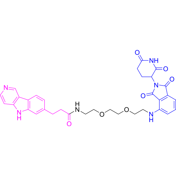
| SMILES | O=C1C(CCC(N1)=O)N1C(C2C=CC=C(C=2C1=O)NCCOCCOCCNC(CCC1C=CC2C3C=NC=CC=3NC=2C=1)=O)=O |
| InChi Key | NWTCZTAQSRLSCP-UHFFFAOYSA-N |
| InChi Code | InChI=1S/C33H34N6O7/c40-28(8-5-20-4-6-21-23-19-34-11-10-24(23)37-26(21)18-20)36-13-15-46-17-16-45-14-12-35-25-3-1-2-22-30(25)33(44)39(32(22)43)27-7-9-29(41)38-31(27)42/h1-4,6,10-11,18-19,27,35,37H,5,7-9,12-17H2,(H,36,40)(H,38,41,42) |
| Chemical Name | N-[2-[2-[2-[[2-(2,6-dioxopiperidin-3-yl)-1,3-dioxoisoindol-4-yl]amino]ethoxy]ethoxy]ethyl]-3-(5H-pyrido[4,3-b]indol-7-yl)propanamide |
| Synonyms | QC-01 C175; 2267290-96-8; QC-01-175; CHEMBL4465176; |
| HS Tariff Code | 2934.99.9001 |
| Storage |
Powder-20°C 3 years 4°C 2 years In solvent -80°C 6 months -20°C 1 month |
| Shipping Condition | Room temperature (This product is stable at ambient temperature for a few days during ordinary shipping and time spent in Customs) |
Biological Activity
| Targets | CRBN; A152T and P301L mutant tau protein |
| ln Vitro |
Tauopathies are neurodegenerative diseases characterized by aberrant forms of tau protein accumulation leading to neuronal death in focal brain areas. Positron emission tomography (PET) tracers that bind to pathological tau are used in diagnosis, but there are no current therapies to eliminate these tau species. We employed targeted protein degradation technology to convert a tau PET-probe into a functional degrader of pathogenic tau. The hetero-bifunctional molecule QC-01-175 was designed to engage both tau and Cereblon (CRBN), a substrate-receptor for the E3-ubiquitin ligase CRL4CRBN, to trigger tau ubiquitination and proteasomal degradation. QC-01-175 effected clearance of tau in frontotemporal dementia (FTD) patient-derived neuronal cell models, with minimal effect on tau from neurons of healthy controls, indicating specificity for disease-relevant forms. QC-01-175 also rescued stress vulnerability in FTD neurons, phenocopying CRISPR-mediated MAPT-knockout. This work demonstrates that aberrant tau in FTD patient-derived neurons is amenable to targeted degradation, representing an important advance for therapeutics. [1] Tau Degradation Mitigates ALS SNs-Induced Alterations in Mitochondrial Morphology [2] To determine whether reducing tau may rescue mitochondrial morphology, a novel bifunctional tau degrader, QC-01-175, which is capable of recruiting tau to the E3 ubiquitin ligase CRL4CRBN resulting in its degradation by the proteasome [36], was used to reduce pTau-S396 levels in SH-SY5Y cells following treatment with control- or ALS-derived SNs. To identify the effective dose and treatment time for QC-01-175 which would be able to reduce pTau-S396 levels, SH-SY5Y cells were first treated with 1 or 10 μM of QC-01-175 for 2, 4, and 24h following treatment with ALS SNs (10 ng/mL/24h). While 1 μM QC-01-175 had no effect on pTau-S396 levels, 10 μM of the degrader reduced pTau-S396 in ALS SNs-treated cells compared to vehicle-treated cells following 4h and this effect was absent following 24h at the same concentration (Fig. 6a, b). Therefore, SH-SY5Y cells were treated with ALS SNs (10 ng/mL/24h) followed by QC-01-175 (10 μM) for 4h before assessing pTau-S396 levels by western blots. The results revealed a significant decrease in pTau-S396 levels in ALS SNs+QC-01-175(10 μM) compared to ALS SNs-treated cells (Fig. 6c). For mitochondrial morphological assessments, SH-SY5Y cells were treated with either recombinant tau or SNs derived from control and ALS mCTX (10 ng/mL/24h) followed by treatment with QC-01-175 (10 μM/4h) before assessing mitochondrial morphological parameters (Fig. 7a). As in previous experiments, recombinant tau and ALS SNs treatment significantly shortened mitochondrial length and volume compared to vehicle- and control SNs-treated cells. Importantly, degrading tau with QC-01-175 reversed the effects of recombinant tau and ALS SNs- on mitochondria morphology. Specifically, there were fewer smaller mitochondria and more larger mitochondria in ALS SNs+QC-01-175 compared to ALS SNs-treated cells (Fig. 7b, c). Furthermore, selective degradation of tau using QC-01-175 prevented recombinant tau- and ALS SNs-induced mitochondrial length and volume alterations, as treatment with tau or ALS SNs resulted in fewer smaller mitochondria (< 2 μm in length and < 2 μm3 in volume) following QC-01-175 treatment (Suppl. Figure 7a-b). Lastly, there were more larger mitochondria (> 8 μm3 in volume) in ALS SNs+QC-01-175 compared to ALS SNs-treated cells (Suppl. Figure 7b). Mitochondrial network analyses revealed no alterations in the number of total networks/cell, similar to previous experiments (Fig. 7d). While there was an ALS SNs-induced decrease in the number of large networks/cell or mitochondrial branch length, this decrease was not observed in ALS SNs+QC-01-175 (Fig. 7e, f). Although QC-01-175 treatment more than doubled the number of large networks compared to ALS SNs treatment, this protection did not rise to statistical significance. Tau Degradation Prevents the Increase in Reactive Oxygen Species Induced by ALS SNs [2] Given that treatment with ALS SNs increases oxidative stress, and QC-01-175 ameliorates mitochondrial morphology, we next sought to determine whether QC-01-175 treatment was also able to mitigate oxidative stress induced by ALS SNs. SH-SY5Y cells were treated with either recombinant tau or SNs derived from control and ALS mCTX (10 ng/mL/24 h) followed by treatment with QC-01-175 (10 μM/4h) before assessing ROS levels using CellROX, a fluorogenic probe for measuring oxidative stress in live cells (Fig. 8a). Antimycin A (25 μM/30min) was used as positive control (Suppl. Figure 8). Similar to previous results, the treatment with both recombinant tau and ALS SNs induced an increase in ROS levels compared to both vehicle- and control SNs-treated cells (Fig. 8b). Importantly, ROS levels were significantly decreased in both recombinant tau+QC-01-175- and ALS SNs+QC-01-175-treated cells compared to recombinant tau- and ALS SNs-treated cells (Fig. 8b), indicating a rescue of oxidative stress by tau-specific clearance. |
| Enzyme Assay |
QC-01-175 Treatment [2] QC-01-175 was designed using a targeted protein degradation technology to recognize both tau and Cereblon (CRBN), a substrate receptor for the E3-ubiquitin ligase in order to specifically induce pathogenic tau ubiquitination and degradation. SH-SY5Y cells were treated with 1 or 10 μM QC-01-175 for 2, 4, and 24 h following treatment with control or ALS SNs (10 ng/mL/24 h). Western blot experiments were performed to verify the efficiency of QC-01-175 by assessing pTau-S396 levels. Mitochondrial morphological parameters were measured as previously described in SH-SY5Y cells treated with either recombinant tau protein (10 ng/mL) or SNs derived from control (n = 3) and ALS mCTX (n = 3) for 24 h followed by exposure to QC-01-175 (10 μM) for 4 h. Reactive oxygen species were measured in SH-SY5Y cells treated with either tau, control (n = 4) or ALS SNs (n = 9) (10 ng/mL/24 h) followed by treatment with QC-01-175 (10 μM/4 h). |
| Cell Assay |
Neuronal stress and viability assay [1] Stress vulnerability assays were performed as previously described (Silva et al., 2016) (Figure 7B). NPCs were plated and differentiated in 96-well plate format, for eight weeks. Either QC-01-175, QC-03–075 or vehicle (DMSO) were added directly into the media (100 μL) to a final concentration of 5 μM, and incubated for 8 hr at 37°C. Then, each well was treated with either 10 μM of amyloid-beta(1-42, or vehicle alone, for an additional 16 hr incubation. At 24 hr, viability was measured with the Alamar Blue Cell viability reagent, according to manufacturer instructions. Readings were done in the EnVision Multilabel Plate Reader. Sample preparation TMT LC-MS3 mass spectrometry [1] A152T neurons at 6 weeks of differentiation were treated with DMSO vehicle, 1 µM of degrader QC-01-175 or 1 µM negative control QC-03–075 in biological triplicates for 4 hr, or pre-treated for 30 min with 10 µM MLN4924 followed by 1 µM QC-01-175 addition for 3.5 hr, in biological duplicates. Neuronal cells were washed in PBS and collected at 3000 g centrifugation. Lysis buffer (8 M Urea, 50 mM NaCl, 50 mM 4-(2hydroxyethyl)−1-piperazineethanesulfonic acid (EPPS) pH 8.5, protease and phosphatase inhibitors were added to the cell pellets and homogenized by 20 passes through a 21 gauge (1.25 in. long) needle to achieve a cell lysate with a protein concentration between 0.25–2 mg/mL. |
| References |
[1]. Targeted degradation of aberrant tau in frontotemporal dementia patient-derived neuronal cell models. Elife. 2019 Mar 25;8:e45457. [2]. Targeting Tau Mitigates Mitochondrial Fragmentation and Oxidative Stress in Amyotrophic Lateral Sclerosis. Mol Neurobiol. 2022 Jan;59(1):683-702. |
| Additional Infomation |
Potential off-target binding for the bifunctional molecule QC-01-175 could be mediated by either the tau-binding moiety, derived from T807, or by the CRBN ligand, in this case derived from pomalidomide (Figure 1C I). To address the first hypothesis, we tested the well-known off-target activity against monoamine oxidase-B (MAO-B) and monoamine oxidase-A (MAO-A) (Lemoine et al., 2018, Vermeiren et al., 2018) by implementing an in vitro MAO inhibition assay (relocated Figure 1—figure supplement 2), which revealed that QC-01-175 showed significantly reduced inhibition of MAO relative to T807. To address pomalidomide off-target engagement, as well as overall QC-01-175 effect on the global proteome, we employed multiplexed MS-based proteomics. Upon 4 h treatment with QC-01-175, the only significant changes observed comprise the validated immune-modulatory drug (IMiD) targets ZFP91, ZNF653 and ZNF827 (Donovan et al., 2018), while no QC-01-175 specific off-targets were observed (see Figure 6, and the Results subsection “Evaluation of degrader specificity in neurons”). We have also done this analysis upon 24 h treatment (not included) and the results were very similar with the same three IMiD targets showing the largest and only off-target significant effect (after tau). Future work to avoid IMiD target effect is now discussed in the third paragraph of the Discussion section.
Also, one cannot exclude other interactions by QC-01-175 that do not alter protein levels but modulate function and enzymatic activity. This is a very relevant and complex point that is brought up again in the Discussion section, where potential challenges for in vivo and clinical applications are discussed. [1] Collectively, our results in a large cohort of human post-mortem mCTX suggest that hyperphosphorylated tau at S396 may underlie mitochondrial fragmentation in ALS by interacting with the pro-fission GTPase DRP1. Lastly, our data provide the groundwork to assess QC-01-175 as a novel potential therapeutic strategy to improve mitochondrial morphology and function, and, in turn, motor neuron survival in ALS. [2] |
Solubility Data
| Solubility (In Vitro) | May dissolve in DMSO (in most cases), if not, try other solvents such as H2O, Ethanol, or DMF with a minute amount of products to avoid loss of samples |
| Solubility (In Vivo) |
Note: Listed below are some common formulations that may be used to formulate products with low water solubility (e.g. < 1 mg/mL), you may test these formulations using a minute amount of products to avoid loss of samples. Injection Formulations (e.g. IP/IV/IM/SC) Injection Formulation 1: DMSO : Tween 80: Saline = 10 : 5 : 85 (i.e. 100 μL DMSO stock solution → 50 μL Tween 80 → 850 μL Saline) *Preparation of saline: Dissolve 0.9 g of sodium chloride in 100 mL ddH ₂ O to obtain a clear solution. Injection Formulation 2: DMSO : PEG300 :Tween 80 : Saline = 10 : 40 : 5 : 45 (i.e. 100 μL DMSO → 400 μLPEG300 → 50 μL Tween 80 → 450 μL Saline) Injection Formulation 3: DMSO : Corn oil = 10 : 90 (i.e. 100 μL DMSO → 900 μL Corn oil) Example: Take the Injection Formulation 3 (DMSO : Corn oil = 10 : 90) as an example, if 1 mL of 2.5 mg/mL working solution is to be prepared, you can take 100 μL 25 mg/mL DMSO stock solution and add to 900 μL corn oil, mix well to obtain a clear or suspension solution (2.5 mg/mL, ready for use in animals). Injection Formulation 4: DMSO : 20% SBE-β-CD in saline = 10 : 90 [i.e. 100 μL DMSO → 900 μL (20% SBE-β-CD in saline)] *Preparation of 20% SBE-β-CD in Saline (4°C,1 week): Dissolve 2 g SBE-β-CD in 10 mL saline to obtain a clear solution. Injection Formulation 5: 2-Hydroxypropyl-β-cyclodextrin : Saline = 50 : 50 (i.e. 500 μL 2-Hydroxypropyl-β-cyclodextrin → 500 μL Saline) Injection Formulation 6: DMSO : PEG300 : castor oil : Saline = 5 : 10 : 20 : 65 (i.e. 50 μL DMSO → 100 μLPEG300 → 200 μL castor oil → 650 μL Saline) Injection Formulation 7: Ethanol : Cremophor : Saline = 10: 10 : 80 (i.e. 100 μL Ethanol → 100 μL Cremophor → 800 μL Saline) Injection Formulation 8: Dissolve in Cremophor/Ethanol (50 : 50), then diluted by Saline Injection Formulation 9: EtOH : Corn oil = 10 : 90 (i.e. 100 μL EtOH → 900 μL Corn oil) Injection Formulation 10: EtOH : PEG300:Tween 80 : Saline = 10 : 40 : 5 : 45 (i.e. 100 μL EtOH → 400 μLPEG300 → 50 μL Tween 80 → 450 μL Saline) Oral Formulations Oral Formulation 1: Suspend in 0.5% CMC Na (carboxymethylcellulose sodium) Oral Formulation 2: Suspend in 0.5% Carboxymethyl cellulose Example: Take the Oral Formulation 1 (Suspend in 0.5% CMC Na) as an example, if 100 mL of 2.5 mg/mL working solution is to be prepared, you can first prepare 0.5% CMC Na solution by measuring 0.5 g CMC Na and dissolve it in 100 mL ddH2O to obtain a clear solution; then add 250 mg of the product to 100 mL 0.5% CMC Na solution, to make the suspension solution (2.5 mg/mL, ready for use in animals). Oral Formulation 3: Dissolved in PEG400 Oral Formulation 4: Suspend in 0.2% Carboxymethyl cellulose Oral Formulation 5: Dissolve in 0.25% Tween 80 and 0.5% Carboxymethyl cellulose Oral Formulation 6: Mixing with food powders Note: Please be aware that the above formulations are for reference only. InvivoChem strongly recommends customers to read literature methods/protocols carefully before determining which formulation you should use for in vivo studies, as different compounds have different solubility properties and have to be formulated differently. (Please use freshly prepared in vivo formulations for optimal results.) |
| Preparing Stock Solutions | 1 mg | 5 mg | 10 mg | |
| 1 mM | 1.5958 mL | 7.9788 mL | 15.9576 mL | |
| 5 mM | 0.3192 mL | 1.5958 mL | 3.1915 mL | |
| 10 mM | 0.1596 mL | 0.7979 mL | 1.5958 mL |