Polymyxin B nonapeptide TFA, the trifluoroacetic acid of Polymyxin B nonapeptide, is a potent and naturally occurring cyclic peptide derived from Polymyxin B by proteolytic removal of its terminal amino acyl residue. Polymyxin B nonapeptide is less toxic, lacks bactericidal activity, and retains its ability to render gram-negative bacteria susceptible to several antibiotics by permeabilizing their outer membranes.
Physicochemical Properties
| Molecular Formula | C45H75F3N14O13 |
| Molecular Weight | 1077.15822052956 |
| Exact Mass | 1532.530 |
| CAS # | 2220175-42-6 |
| Related CAS # | Polymyxin B nonapeptide;86408-36-8 |
| PubChem CID | 154730054 |
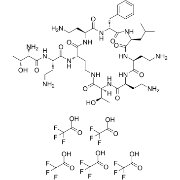
| Sequence | H-Thr-Dab-Dab(1)-Dab-D-Phe-Leu-Dab-Dab-Thr-(1).5TFA |
| Appearance | White to off-white solid powder |
| Hydrogen Bond Donor Count | 17 |
| Hydrogen Bond Acceptor Count | 21 |
| Rotatable Bond Count | 18 |
| Heavy Atom Count | 75 |
| Complexity | 1770 |
| Defined Atom Stereocenter Count | 11 |
| SMILES | FC(C(=O)O)(F)F.O=C1[C@H](CC(C)C)NC([C@@H](CC2C=CC=CC=2)NC([C@H](CCN)NC([C@H](CCNC([C@H]([C@@H](C)O)NC([C@H](CCN)NC([C@H](CCN)N1)=O)=O)=O)NC([C@H](CCN)NC([C@H]([C@@H](C)O)N)=O)=O)=O)=O)=O |
| InChi Key | XEIKWJLAIULVMY-YNQICIJSSA-N |
| InChi Code | InChI=1S/C43H74N14O11.5C2HF3O2/c1-22(2)20-31-40(65)52-26(10-15-44)35(60)51-29(13-18-47)39(64)57-34(24(4)59)43(68)49-19-14-30(53-36(61)28(12-17-46)54-42(67)33(48)23(3)58)38(63)50-27(11-16-45)37(62)56-32(41(66)55-31)21-25-8-6-5-7-9-25;5*3-2(4,5)1(6)7/h5-9,22-24,26-34,58-59H,10-21,44-48H2,1-4H3,(H,49,68)(H,50,63)(H,51,60)(H,52,65)(H,53,61)(H,54,67)(H,55,66)(H,56,62)(H,57,64);5*(H,6,7)/t23-,24-,26+,27+,28+,29+,30+,31+,32-,33+,34+;;;;;/m1...../s1 |
| Chemical Name | (2S,3R)-2-amino-N-[(2S)-4-amino-1-oxo-1-[[(3S,6S,9S,12S,15R,18S,21S)-6,9,18-tris(2-aminoethyl)-15-benzyl-3-[(1R)-1-hydroxyethyl]-12-(2-methylpropyl)-2,5,8,11,14,17,20-heptaoxo-1,4,7,10,13,16,19-heptazacyclotricos-21-yl]amino]butan-2-yl]-3-hydroxybutanamide;2,2,2-trifluoroacetic acid |
| Synonyms | Polymyxin B nonapeptide TFA; 2220175-42-6; (2S,3R)-2-Amino-N-((S)-4-amino-1-oxo-1-(((3S,6S,9S,12S,15R,18S,21S)-6,9,18-tris(2-aminoethyl)-15-benzyl-3-((R)-1-hydroxyethyl)-12-isobutyl-2,5,8,11,14,17,20-heptaoxo-1,4,7,10,13,16,19-heptaazacyclotricosan-21-yl)amino)butan-2-yl)-3-hydroxybutanamide2,2,2; HY-106783A; DA-66797; CS-0128232; G17443; (2S,3R)-2-amino-N-[(2S)-4-amino-1-oxo-1-[[(3S,6S,9S,12S,15R,18S,21S)-6,9,18-tris(2-aminoethyl)-15-benzyl-3-[(1R)-1-hydroxyethyl]-12-(2-methylpropyl)-2,5,8,11,14,17,20-heptaoxo-1,4,7,10,13,16,19-heptazacyclotricos-21-yl]amino]butan-2-yl]-3-hydroxybutanamide;2,2,2-trifluoroacetic acid |
| HS Tariff Code | 2934.99.9001 |
| Storage |
Powder-20°C 3 years 4°C 2 years In solvent -80°C 6 months -20°C 1 month Note: Please store this product in a sealed and protected environment, avoid exposure to moisture. |
| Shipping Condition | Room temperature (This product is stable at ambient temperature for a few days during ordinary shipping and time spent in Customs) |
Biological Activity
| Targets | Polypeptide antibacterial |
| ln Vitro |
By binding to the bacterial lipopolysaccharide (LPS), Polymyxin B nonapeptide, a cationic cyclic peptide produced by enzymatic processing of the naturally occurring peptide Polymyxin B, can increase the permeability of Gram-negative bacteria's outer membrane toward hydrophobic antibiotics[1]. Polymyxin B nonapeptide (PMBN), a cationic cyclic peptide derived by enzymatic processing from the naturally occurring peptide polymyxin B, is able to increase the permeability of the outer membrane of Gram-negative bacteria toward hydrophobic antibiotics probably by binding to the bacterial lipopolysaccharide (LPS). We have synthesized 11 cyclic analogues of PMBN and evaluated their activities compared to that of PMBN. The synthetic peptides were much less potent than PMBN in their capacity to sensitize Escherichia coli and Klebsiella pneumoniae toward novobiocin and to displace dansyl-PMBN from Escherichia coli LPS. Moreover, unlike PMBN, none of the analogues were able to inhibit the growth of Pseudomonas aeruginosa. The structural-functional features of PMBN were characterized and identified with regard to the ring size, the distance between positive charges and peptide backbone, the chirality of the DPhe-Leu domain, and the nature of the charged groups. Apparently, the structure of PMBN is highly specific for efficient perturbation of the outer membrane of Gram-negative bacteria as well as for LPS binding. The present study further increases our understanding of the complex PMBN-LPS and may, potentially, enable the design of compounds having enhanced permeabilization potency of the Gram-negative outer membrane. [1] |
| ln Vivo | Polymyxin B nonapeptide, derived by cleavage of the fatty acyl diaminobutyric acid from polymyxin B, is considerably less toxic, lacks bactericidal activity, and retains its ability to render gram-negative bacteria susceptible to several antibiotics by permeabilizing their outer membranes. The peptide rendered all 53 polymyxin-susceptible strains tested more susceptible to novobiocin, lowering the MIC of novobiocin eightfold or more. The combination of polymyxin B nonapeptide with novobiocin or with erythromycin administered intraperitoneally in multiple doses synergistically protected mice infected with gram-negative bacteria. This combination may be clinically useful because of the apparent rarity of the acquisition of resistance. [2] |
| Enzyme Assay |
Binding of Peptides to LPS. [1] The fluorescence of dansyl-PMBN bound to E. coli LPS was measured using a MC200 monochromator set at an excitation wavelength of 340 nm and an emission wavelength of 485 nm. 20 To a quartz cuvette containing LPS solution (2 mL, 3 μg/mL) in N-[2-hydroxyethyl]piperazine-N‘-[2-ethanesulfonic acid] buffer (HEPES; 5 mM, pH 7.2) was added 5 or 10 μL of dansyl-PMBN solution (1 × 10⁻⁶−1 × 10⁻³ M) at 5-min intervals for up to 1 h until a plateau in the fluorescence intensity was reached (i.e., saturation of dansyl-PMBN binding to LPS). The amount of dansyl-PMBN bound to LPS at saturation was calculated as described elsewhere.12 Briefly, a binding curve of dansyl-PMBN to excess LPS (400 μg/mL) was plotted and referred to as maximal binding (Fmax). The amount of bound dansyl-PMBN was calculated from: [dansyl-PMBN] = (Fexp/Fmax) × [dansyl-PMBNtotal], where Fexp is the fluorescence obtained with 3 μg/mL LPS. To determine the binding of a given peptide to LPS, a displacement assay was performed in which 5 or 10 μL (1 × 10⁻⁵−1 × 10⁻³ M) of the tested peptide at desired concentrations were added at 5-min intervals to a preequilibrated mixture of LPS solution (2 mL, 3 μg/mL, ∼2 × 10⁻⁷ M) in HEPES buffer (5 mM, pH 7.2) and dansyl-PMBN (0.55 μM). The fluorescence intensity was recorded after each time interval of adding the tested peptide. Each experiment was repeated 2−3 times. The percent inhibition of fluorescence intensity was plotted as a function of the peptide concentration from which the concentration required for maximal (Imax) and 50% (IC50) displacement of the dansyl-PMBN from LPS was derived. Circular Dichroism (CD) Studies. [1] CD spectra were recorded on an Aviv-202 circular dichroism spectrometer. Duplicate scans over a wavelength range of 190−250 nm were taken at a chart speed of 12 nm/min in a 0.1-cm path length quartz cell at room temperature. Peptides were dissolved in 5 mM phosphate buffer (PB), pH 7.2, at a final concentration of 0.1 mM. The CD of pPMBN and sPMBN were evaluated, as well, in a mixture of TFE/aqueous buffer (1:1, v/v) or in bulk TFE. A baseline was recorded and subtracted after each spectrum. Ellipticity is reported as the mean residue ellipticity [ϑ] in deg cm2 dmol-1. [ϑ] = [ϑ]obs × (MRW/10lc), where [ϑ]obs is the ellipticity measured in mdeg, MRW is the mean residue molecular weight of the peptide (molecular weight divided by the number of peptide bonds), c is the concentration of the sample in mg/mL, and l is the optical path length of the cell in cm. |
| Cell Assay |
Determination of Minimal Inhibitory Concentration (MIC). [1] Clinical isolates of E. coli, K. pneumoniae and P. aeruginosa, obtained as described elsewhere, were employed.18 The Gram-negative bacteria were grown on nutrient agar plates and kept at 4 °C. An overnight culture in isotonic sensitest broth was adjusted to 1 × 10⁵ CFU/mL and inoculated into microtiter plate wells containing each 100 μL of a serial 2-fold dilution (1000−0.5 μg/mL) of the tested antibiotics in ISB. MIC was defined as the lowest concentration at which there was no visible bacterial growth after incubation for 20 h at 37 °C. The results are reported for 4−8 separate tests that varied by no more than one dilution. Sensitizing Activity. [1] Bacterial suspension (10 μL, 1 × 10⁵ CFU) was inoculated into microtiter plate wells containing 100 μL of a serial 2-fold dilution (1000−0.5 μg/mL) of novobiocin in ISB. To each well was added 10 μL of the test peptide to a final concentration of 50 μg/mL. The fold decrease in MIC for novobiocin comparing wells with and without test peptides was calculated and designated as sensitizing activity. The relative sensitizing activity of the test peptide was calculated as percent of the sensitizing activity of 50 μg/mL pPMBN. |
| References |
[1]. Structure-function studies of polymyxin B nonapeptide: implications to sensitization of gram-negative bacteria. J Med Chem. 2000 Aug 10;43(16):3085-92. [2]. Antibacterial synergism of polymyxin B nonapeptide and hydrophobic antibiotics in experimental gram-negative infections in mice. Antimicrob Agents Chemother. 1994 Feb;38(2):374-7. |
| Additional Infomation | Antimicrobial resistance is a global public health threat. Antibiotic development pipeline has few new drugs; therefore, using antibiotic adjuvants has been envisioned as a successful method to preserve existing medications to fight multidrug-resistant (MDR) pathogens. In this study, we investigated the synergistic effect of a polymyxin derivative known as polymyxin B nonapeptide (PMBN) with azithromycin (AZT). A total of 54 Escherichia coli strains were first characterized for macrolide resistance genes, and susceptibility to different antibiotics, including AZT. A subset of 24 strains was then selected for synergy testing by the checkerboard assay. PMBN was able to re-sensitize the bacteria to AZT, even in strains with high minimum inhibitory concentrations (MIC: 32 to ≥128 μg/ml) for AZT, and in strains resistant to the last resort drugs such as colistin and meropenem. The fractional inhibitory concentration index was lower than 0.5, demonstrating that PMBN and AZT combinations had a synergistic effect. The combinations worked efficiently in strains carrying mphA gene encoding macrolide phosphotransferase which can cause macrolide inactivation. However, the combinations were inactive in strains having an additional ermB gene encoding macrolide methylase which causes ribosomal drug target alteration. Killing kinetics study showed a significant reduction of bacterial growth after 6 h of treatment with complete killing achieved after 24 h. Transmission electron microscopy showed morphological alterations in the bacteria treated with PMBN alone or in combination with AZT, with evidence of damage to the outer membrane. These results suggested that PMBN acted by increasing the permeability of bacterial outer membrane to AZT, which was also evident using a fluorometric assay. Using multiple antimicrobial agents could therefore be a promising strategy in the eradication of MDR bacteria. PMBN is a good candidate for use with other antibiotics to potentiate their activity, but further studies are required in vivo. This will significantly contribute to resolving antimicrobial resistance crisis.https://pubmed.ncbi.nlm.nih.gov/36212888/ |
Solubility Data
| Solubility (In Vitro) |
DMSO : ~100 mg/mL (~65.22 mM) H2O : ~50 mg/mL (~32.61 mM) |
| Solubility (In Vivo) |
Solubility in Formulation 1: 100 mg/mL (65.22 mM) in PBS (add these co-solvents sequentially from left to right, and one by one), clear solution; with sonication. (Please use freshly prepared in vivo formulations for optimal results.) |
| Preparing Stock Solutions | 1 mg | 5 mg | 10 mg | |
| 1 mM | 0.9284 mL | 4.6418 mL | 9.2837 mL | |
| 5 mM | 0.1857 mL | 0.9284 mL | 1.8567 mL | |
| 10 mM | 0.0928 mL | 0.4642 mL | 0.9284 mL |