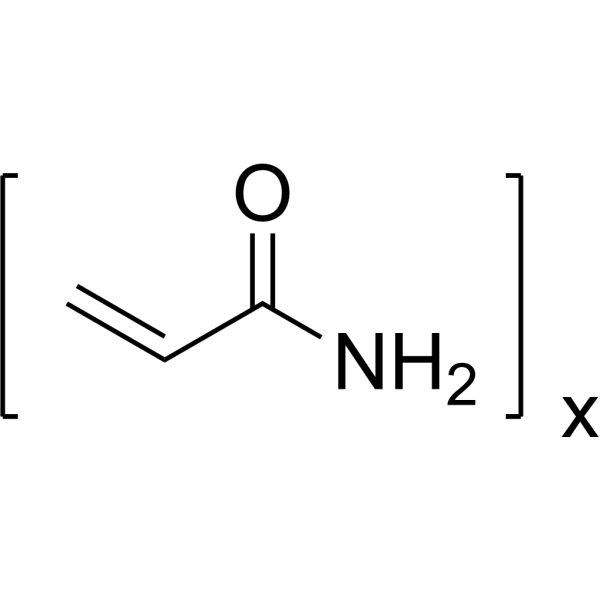
Physicochemical Properties
| Molecular Formula | C3H5NO)X |
| Exact Mass | 71.037 |
| CAS # | 9003-05-8 |
| Related CAS # | 9003-05-8 |
| PubChem CID | 6579 |
| Appearance |
Flake-like crystals from benzene White crystalline ... solid WHITE SOLID |
| Density | 1.3 |
| Boiling Point | 231.7±0.0 °C at 760 mmHg |
| Melting Point | >300 °C |
| Flash Point | 79.0±19.8 °C |
| Vapour Pressure | 0.1±0.4 mmHg at 25°C |
| Index of Refraction | 1.433 |
| LogP | -0.78 |
| Hydrogen Bond Donor Count | 1 |
| Hydrogen Bond Acceptor Count | 1 |
| Rotatable Bond Count | 1 |
| Heavy Atom Count | 5 |
| Complexity | 57.9 |
| Defined Atom Stereocenter Count | 0 |
| InChi Key | HRPVXLWXLXDGHG-UHFFFAOYSA-N |
| InChi Code | InChI=1S/C3H5NO/c1-2-3(4)5/h2H,1H2,(H2,4,5) |
| Chemical Name | prop-2-enamide |
| Synonyms | PAM,average Mn 40000 |
| HS Tariff Code | 2934.99.9001 |
| Storage |
Powder-20°C 3 years 4°C 2 years In solvent -80°C 6 months -20°C 1 month |
| Shipping Condition | Room temperature (This product is stable at ambient temperature for a few days during ordinary shipping and time spent in Customs) |
Biological Activity
| ADME/Pharmacokinetics |
Absorption, Distribution and Excretion Acrylamide is well absorbed after oral, dermal, inhalational, and parenteral exposure, including through intact skin and mucous membranes. Efficient absorption of this compound is demonstrated by the observation that peak blood concentrations occur at approximately 1 hour after exposure. It is estimated that human elimination rates of acrylamide are only one-fifth that seen in rats. Acrylamide is primarily (90 to 95%) excreted in the urine as conjugated metabolite with less then 2% parent compound appearing in the urine. Smaller amounts of metabolites are also present in feces, bile, and other biological matrices, still with only small amounts being eliminated as unchanged parent. Acrylamide elimination is biphasic with an alpha half-life of less than 5 hours and a beta half-life of 6 to 8 days. In rats given 0.5-100 mg/kg bw of either (1-14(C))- or (2,3-14(C))acrylamide intravenously or orally, radioactivity was distributed rapidly throughout the body, with no selective accumulation in any tissue. Radioactivity was also distributed evenly among tissues of beagle dogs and miniature pigs ... Can be absorbed through ... mucous membranes and lungs as well as the GI tract. For more Absorption, Distribution and Excretion (Complete) data for ACRYLAMIDE (14 total), please visit the HSDB record page. MICROSPHERES OF (14)C-LABELED POLYACRYLAMIDE WERE MAINLY (APPROX 80%) FOUND IN LIVER & SPLEEN BOTH AFTER IV & IP INJECTION IN MOUSE & RAT, ALSO DETECTED EARLY (1 HR AFTER IV INJECTION) IN BONE MARROW, & PARTICLE AGGREGATES WERE ALSO INITIALLY FOUND IN LUNGS. Metabolism / Metabolites Acrylamide is primarily (90 to 95%) excreted in the urine as conjugated metabolite with less then 2% parent compound appearing in the urine. Smaller amounts of metabolites are also present in feces, bile, and other biological matrices, still with only small amounts being eliminated as unchanged parent. Acrylamide elimination is biphasic with an alpha half-life of less than 5 hours and a beta half-life of 6 to 8 days. Urinary metabolites among acrylamide-exposed animals were identified as N-acetyl-S- (3-amino-3-oxopropyl) cysteine (the N-acetyl-cysteine conjugate of acrylamide, following glutathione conjugation accounting for 67% of the total urinary metabolites found in rats, 41% of the total found in mice), N-acetyl-S- (3-amino-2-hydroxy-3-oxopropyl) cysteine (16% in rats, 21% in mice), N-acetyl-S- (1-carbamoyl-2-hydroxyethyl) cysteine (9% in rats, 12% in mice), glycidamide (6% in rats, 17% in mice), 2,3-dihydroxy-propionamide (2% in rats, 5% in mice), and a small amount of the parent compound (which was not possible to quantify). ... In the present study, a low-dose of acrylamide (ACR; 18 mg/kg) was administered to male Wistar rats for 40 days. Ultra performance liquid chromatography/time of flight mass spectrometry (UPLC-Q-TOF MS) was used to examine urine samples from ACR-dosed and control animals. Multiple statistical analyses with principal component analysis (PCA) were used to investigate metabolite profile changes in urine samples, and to screen for potential neurotoxicity biomarkers. PCA showed differences between the ACR-dosed and control groups 20 days after the start of dosing; a bigger separation between the two groups was seen after dosing for 40 days. Levels of 4-guanidinobutanoic acid and 2-oxoarginine were significantly higher in urine from the ACR-dosed group than in urine from the control group after 10 days (p<0.05). Receiver operator characteristic (ROC) curve analysis suggested that 4-guanidinobutanoic acid and 2-oxoarginine were the major metabolites. Our results suggest that high levels of 4-guanidinobutanoic acid and 2-oxoarginine may be related to ACR neurotoxicity. These metabolites could, therefore, act as sensitive biomarkers for ACR exposure and be useful for investigating toxic mechanisms. They may also provide a scientific foundation for assessing the effects of chronic low-dose ACR exposure on human health. To study the toxic effect of chronic exposure to acrylamide (AA) at low-dose levels, we applied a metabolomics approach based on ultra-performance liquid chromatography/mass spectrometry (UPLC-MS). A total of 40 male Wistar rats were randomly assigned to different groups: control, low-dose AA (0.2 mg/kg bw), middle-dose AA (1 mg/kg bw) and high-dose AA (5 mg/kg bw). The rats continuously received AA via drinking water for 16 weeks. Rat urine samples were collected at different time points for measurement of metabolomic profiles. Thirteen metabolites, including the biomarkers of AA exposure (AAMA, GAMA and iso-GAMA), were identified from the metabolomic profiles of rat urine. Compared with the control group, the treated groups showed significantly increased intensities of GAMA, AAMA, iso-GAMA, vinylacetylglycine, 1-salicylate glucuronide, PE (20:1(11Z)/14:0), cysteic acid, L-cysteine, p-cresol sulfate and 7-ketodeoxycholic acid, as well as decreased intensities of 3-acetamidobutanal, 2-indolecarboxylic acid and kynurenic acid in rat urine. Notably, three new candidate biomarkers (p-cresol sulfate, 7-ketodeoxycholic acid and 1-salicylate glucuronide) in rat urine exposed to AA have been found in this study. The results indicate exposure to AA disrupts the metabolism of lipids and amino acids, induces oxidative stress. For more Metabolism/Metabolites (Complete) data for ACRYLAMIDE (8 total), please visit the HSDB record page. Acrylamide is absorbed following oral, inhalation, and dermal exposure and is widely distributed, tending to accumulate in the red blood cells. In the proposed major metabolic pathway acrylamide reacts with glutathione to form S-beta-propionamide glutathione conjugate which is excreted in the urine as cysteine or N-acetylcysteine derivatives. The major urinary metabolite (accounting for 48% of the excreted dose) is N-acetylcysteine-S-beta-propionamide. Alternately, acrylamide may be oxidized to glycidamide by CYP2E1. Glycidamide then goes on to form similar glutathione conjugates or undergos hydrolysis, leading to the formation of 2,3-dihydroxypropionamide and 2,3-dihydroxypropionicacid. (A635, A324, L1887) Biological Half-Life Plasma (animal studies): 2 days; whole body (animal studies): 6-18 days; [TDR, p. 40] Acrylamide elimination is biphasic with an alpha half-life of less than 5 hours and a beta half-life of 6 to 8 days. The distribution and metabolism of 2,3-(14)C-labeled acrylamide were studied in male rats. Three dose levels of acrylamide (1.0, 10, or 100 mg/kg) were administered orally. ... Elimination of the radiolabel from most tissues was biphasic with a terminal half-life of approx 8 days. The amount of (14)C in blood remained constant at 12% of the dose for about 7 days. However, (14)C in plasma was eliminated readily. The concn-time curve of parent acrylamide in tissues and blood fit a monoexponential curve with a half-life of approx 2 hr. Elimination from the tissues occurs in two phases: in the first phase its half-life is 5 hr, and in the second (delayed) phase, 8 days or less. The excretion half-life of parent acrylamide in rat urine was 7.8 hr. Using [1-14C]-acrylamide, ... /it was reported/ that approx 6% of the dose was exhaled as /carbon dioxide/. In an extensive study of the kinetics of both orally and iv admin [2,3-14C]-acrylamide, it was shown that the rate of elimination of the radiolabel in urine was independent of the route of admin. Within 24 hr, about 2/3 of the dose was excreted in the urine and 3/4 in 7 days. Fecal excretion was small (4.8% in 24 hr and 6% by 7 days). Since 15% of the dose appeared in the bile within 6 hr, acrylamide or its derivatives must undergo enterohepatic circulation. Thus, approx 80% of the radiolabel was excreted within 7 days and, of this, a very large proportion (90%) was in the form of metabolites. MICROSPHERES OF (14)C-LABELED POLYACRYLAMIDE WERE USED TO FOLLOW DISTRIBUTION & FATE IN MOUSE & RAT AFTER IV & IP INJECTION. PARTICLES WERE RAPIDLY CLEARED FROM CIRCULATION (T/2 IN RAT APPROX 40 MIN) BY MACROPHAGES OF RETICULOENDOTHELIAL SYSTEM. |
| Toxicity/Toxicokinetics |
Toxicity Summary IDENTIFICATION AND USE: Acrylamide is a white crystalline solid. Acrylamide is mainly used in the production of polymers and copolymers for various purposes. All acrylamide in the environment is man-made, the main source being the release of the monomer residues from polyacrylamide used in water treatment or in industry. HUMAN EXPOSURE AND TOXICITY: Acrylamide is toxic and an irritant. Cases of acrylamide poisoning show signs and symptoms of local effects due to irritation of the skin and mucous membranes and systemic effects due to the involvement of the central, peripheral, and autonomic nervous systems. Local irritation of the skin or mucous membranes is characterized by blistering and desquamation of the skin of the hands (palms) and feet (soles) combined with blueness of the hand and feet. Effects on the central nervous system are characterized by abnormal fatigue, sleepiness, memory difficulties, and dizziness. With severe poisoning, confusion, disorientation, and hallucinations occur. Truncal ataxia is a characteristic feature, sometimes combined with nystagmus and slurred speech. Excessive sweating in the limb extremities is a common observation. Sign of central nervous system and local skin involvement may precede peripheral neuropathy by as much as several weeks. Peripheral neuropathy can involve loss of tendon reflexes, impairment of vibration sense, loss of other sensation, and muscular wasting in peripheral parts of the extremities. Nerve biopsy shows loss of large diameter nerve fibers as well as regenerating fibers. Autonomic nervous system involvement is indicated by excessive sweating, peripheral vasodilation, and difficulties in micturition and defecation. After cessation of exposure to acrylamide, most cases recover, although the course of improvement is prolonged and can extend over months to years. There are no epidemiological data available on cancer due to exposure to acrylamide. There is no evidence in man of any teratogenic effects resulting from acrylamide exposure. ANIMAL STUDIES: In rats, biotransformation of acrylamide occurs through glutathione conjugation and through decarboxylation. At least 4 urinary metabolites have been found in rat urine, of which mercapturic acid and cysteine- S-propionamide have been identified. Acrylamide and its metabolites are accumulated (protein-bound) in both nervous system tissue and blood (hemoglobin-bound). Accumulation in the liver and kidney as well as the male reproductive system has also been demonstrated. In animal studies, early changes in visual-evoked potentials (VEP), preceding clinical signs, as well as changes in somatosensory-evoked potentials (SEP), have been seen. Degenerative changes have been described in peripheral nerve axons, with less severe changes in the longer fibers of the CNS. Degeneration of Purkinje cells has been observed in chronically-intoxicated animals. The changes are most pronounced in the nerve endings of myelinated sensory fibers. The nerve endings show enlarged "boutons terminaux" and a widespread enlargement of nerve terminals from the accumulation of neurofilaments. This occurs in both the peripheral and central nervous systems. Impairment of axonal transport has been found in sensory fibers, and interference with glycolysis and protein synthesis has been observed in biochemical studies. Studies of neurotransmitter distribution and receptor binding in the brains of rats have revealed changes induced by acrylamide. In rats, changes in the concentration of neurotransmitters and in striatal dopamine receptor binding have been related to behavioral changes. Degenerative changes in renal convoluted tubular epithelium and glomeruli and fatty generation and necrosis of the liver have been seen in monkeys given large doses of acrylamide. In rats, acrylamide disrupted the metabolism of lipids and amino acids, induced oxidative stress, impaired hepatic porphyrin metabolism. Acrylamide was not mutagenic in Salmonella typhimurium with or without metabolic activation. Acrylamide induced chromosomal aberrations in the spermatocytes of male mice and increased cell transformation frequency in Balb 3T3 cells with a metabolic activation. Acrylamide was shown to be an initiator for skin tumors in mice. It increased the incidence of lung tumors in mice-screening assays. Absorption of acrylamide by the fetus has been demonstrated in animal (pig, dog, rabbit, and rat) studies. Oral administration of acrylamide, between the 7-16th days of gestation in rats, decreased the binding of dopamine receptors in the striatal membranes in 2-week-old pups. Degeneration of seminiferous tubules and chromosome aberrations in spermatocytes has been seen in acrylamide-treated male mice. Depressed plasma levels of testosterone and prolactin have also been observed. A statistically-significant increase in the incidence of mesothelioma of the scrotal cavity was observed in rats after long-term (2-year) administration of acrylamide in the drinking-water. Administration over 2 years of acrylamide not only increased the incidence of a variety of tumor types (both benign and malignant) but also decreased the life expectancy in both male and female rats. ECOTOXICITY STUDIES: Acrylamide was genotoxic in C. auratus peripheral blood cells. The fish exposure also produced a dose-dependent increase in total DNA strand breakage, the formation of erythrocytic nuclear abnormalities and in the levels of hepatic cytochrome P4501A (CYP1A) and glutathione S-transferase (GST) activity. Acrylamide may induce gonadotoxicity in mussels. Acrylamide produces a central-peripheral distal axonopathy when administered chronically. This is characterized functionally by decreases in the monosynaptic reflex and dorsal root potential and alterations in the characteristics of the dorsal root reflex. Acrylamide's neurotoxic effects may be caused by the disruption of fast axonal transport. Acrylamide is thought to bind to kinesin, which leads to impairment of the fast axonal transport system responsible for the distal delivery of macromolecules. This results in deficiencies in proteins responsible for maintaining axonal structure and function. Acrylamide may also disrupt nitric oxide signaling at nerve terminals by forming adducts with soft nucleophilic sulfhydryl groups on cysteine residues. In terms of reproductive toxicity, data suggest that acrylamide-induced male dominant lethal mutations may involve clastogenic events from binding of acrylamide and/or glycidamide to spermatid protamines or spindle fiber proteins and/or direct alkylation of DNA by glycidamide. Adverse effects on mounting, sperm motility, and intromission could also be related to distal axonopathy resulting from binding of acrylamide to motor proteins. Acrylamide's mechanism of carcinogenicity is likely mutagenic, as the metabolite glycidamide is believed to react with proteins and DNA, causing mutations that persist in viable somatic cells and resulting in tumor formation. In addition, acrylamide's affinity for binding sulfhydryl groups on proteins could inactive proteins/enzymes involved in DNA repair and other critical cell functions. (A322, L1887, A2877) Toxicity Data 0.83 microg/kg/day based on reproductive effects, 1.2 microg/kg/day based on neurotoxicity and 1.5 microg/kg/day based on cancer (A15337) Interactions The primary objective of this investigation was to assess the neuroprotective efficacy of lithium in an acrylamide (ACR)-induced neuropathy model in mice. In this study, Kunming male mice were administered ACR (25 mg/kg bw, i.p. once a day) with or without lithium (25 mg/kg bw, i.p. once a day) for 2 weeks. All ACR-administered mice exhibited severe symptoms of neuropathy. We found that treatment with lithium effectively alleviated behavioral deficits in animals elicited by acrylamide. Interestingly, the reduction of hippocampal neurogenesis resulting from ACR injection was promoted by administration of lithium. Further, lithium treatment significantly offset ACR-induced depletion in p-GSK-3beta (Ser9) levels in hippocampus. Collectively our findings suggest the propensity of lithium to attenuate ACR-induced neuropathy. Further studies are necessary to understand the precise molecular mechanism by which the lithium attenuates neuropathy. Nevertheless, our data clearly demonstrate the beneficial effects of lithium on ACR-induced neuropathy in mice and suggest its possible therapeutic application as an adjuvant in the management of other forms of neuropathy in humans. ... The present study investigated the efficacy of geraniol (GE, a natural monoterpene) to mitigate acrylamide (ACR)-induced oxidative stress, mitochondrial dysfunction and neurotoxicity in a rat model and compared its efficacy to that of curcumin (CU, a spice active principle with multiple biological activities). ACR administration (50 mg/kg bw, i.p. 3 times/week) for 4 weeks to growing rats caused typical symptoms of neuropathy. ACR rats provided with daily oral supplements of phytoconstituents (GE: 100 mg/kg bw/d; CU: 50 mg/kg bw/d, 4 weeks) exhibited marked improvement in behavioral tests. Both phytoconstituents markedly attenuated ACR-induced oxidative stress as evidenced by the diminished levels of reactive oxygen species, malondialdehyde and nitric oxide and restored the reduced glutathione levels in sciatic nerve (SN) and brain regions (cortex - Ct, cerebellum - Cb). Further, both phytoconstituents effectively diminished ACR-induced elevation in cytosolic calcium levels in SN and Cb. Furthermore, diminution in the levels of oxidative markers in the mitochondria was associated with elevation in the activities of antioxidant enzymes. While ACR mediated elevation in the acetylcholinesterase activity was reduced by both actives, the depletion in dopamine levels was restored only by CU in brain regions. Taken together our findings ... demonstrate that the neuromodulatory propensity of GE is indeed comparable to that of CU and may be exploited as a therapeutic adjuvant in the management of varied human neuropathy conditions. ... The current study assessed mixture effects of the three known genotoxic chemicals, 2,4-dichlorophenoxyacetic acid (2,4-D), acrylamide (AA), and maleic hydrazide (MH), in an experiment with a fixed ratio design setup. The genotoxic effects were assessed with the single-cell gel electrophoresis assay (comet assay) for both single chemicals and the ternary mixture. The concentration ranges used were 0-1.4, 0-20, and 0-37.7 mM for 2,4-D, AA, and MH, respectively. Mixture toxicity was tested with a fixed ratio design at a 10:23:77% ratio for 2.4-D:AA:MH. Results indicated that the three chemicals yielded a synergistic mixture effect. It is not clear which mechanisms are responsible for this interaction. The aim of this study was to investigate the protective effects of N-acetylcysteine NAC) against acrylamide toxicity in liver and small and large intestine tissues in rats. The rats were divided into four groups. Acrylamide administration increased malondialdehyde (MDA) levels in all tissues significantly (p<0.05). Butacrylamide+NAC administration decreased MDA levels significantly as compared to the acrylamide group, and lowered it to a level close to the control group values (p<0.05). Glitathione (GSH) levels in liver and small intestine tissues reduced significantly in the acrylamide group (p<0.05). Butacrylamide+NAC administration increased GSH levels significantly in all tissues. Whereas GST activity decreased significantly in the acrylamide group in liver and small intestine tissues as compared to the other groups (p<0.05), the GST activity increased significantly in the acrylamide+NAC group in all tissues as compared to the acrylamide group (p<0.05). Liver histopathology showed that the liver epithelial cells were damaged significantly in the acrylamide group. Small intestine histopathology showed that the intestinal villous epithelial cells were damaged significantly in the acrylamide group. Our results indicate that a high level of acrylamide causes oxidative damage in liver and small and large intestine tissues, while N-acetylcysteine administration in a pharmacological dose shows to have an antioxidant effect in preventing this damage. For more Interactions (Complete) data for ACRYLAMIDE (33 total), please visit the HSDB record page. Non-Human Toxicity Values LD50 Mouse ip 170 mg/kg LD50 Rat dermal 1.68 ml/kg LD50 Rat oral 124 mg/kg LD50 Rat skin 400 mg/kg For more Non-Human Toxicity Values (Complete) data for ACRYLAMIDE (9 total), please visit the HSDB record page. |
| References |
[1]. Biochemical reagents[M]//Methods of Enzymatic Analysis. Academic Press, 1965: 967-1037. |
| Additional Infomation |
Acrylamide is a colorless, odorless, crystalline solid that can react violently when melted. When it is heated, sharp fumes may be released.Acrylamide is used to make polyacrylamide, which is mainly used in treating waste water discharge from water treatment plants and industrial processes.In addition, acrylamide and polyacrylamides are used in the production of dyes and organic chemicals, contact lenses, cosmetics and toiletries, permanent-press fabrics, paper and textile production, pulp and paper production, ore processing, sugar refining, and as a chemical grouting agent and soil stabilizer for the construction of tunnels, sewers, wells and reservoirs.Acrylamide is formed in foods that are rich in carbohydrates when they are fried, grilled, or baked. Acrylamide can cause cancer according to The World Health Organization's International Agency for Research on Cancer (IARC) and The Environmental Protection Agency (EPA). It can cause developmental toxicity and male reproductive toxicity according to The National Toxicology Program's Center for the Evaluation of Risks to Human Reproduction. Acrylamide appears as white crystalline solid shipped either as a solid or in solution. A confirmed carcinogen. Toxic by skin absorption. Less dense than water and soluble in water. May be toxic by ingestion. Used for sewage and waste treatment, to make dyes, adhesives. The solid is stable at room temperature, but upon melting may violently polymerize. Toxic, irritating to skin, eyes, etc. Acrylamide solution, [aqueous] appears as a colorless aqueous solution of a solid. Often at a concentration of 40% (w/v). Spills can easily penetrate the soil and contaminate groundwater and nearby streams. Used for sewage and waste treatment and to make dyes and adhesives. Toxic, irritating to skin, eyes, etc. Produce toxic oxides of nitrogen when burned. Acrylamide solution, [flammable liquid label] appears as a solution of a colorless crystalline solid. Flash point depends on the solvent but below 141 °F. Less dense than water. Vapors heavier than air. Toxic oxides of nitrogen produced during combustion. Used for sewage and waste treatment, to make dyes and adhesives. Acrylamide is a member of the class of acrylamides that results from the formal condensation of acrylic acid with ammonia. It has a role as a carcinogenic agent, a neurotoxin, a mutagen, an alkylating agent and a Maillard reaction product. It is a N-acylammonia, a primary carboxamide and a member of acrylamides. It is functionally related to an acrylic acid. The largest use for acrylamide is as an intermediate in the production of organic chemicals and in the synthesis of polyacrylamides. Acute (short-term) and chronic (long-term) oral exposures to acrylamide have resulted in damage to the nervous system in humans and animals. Human data are inadequate on acrylamide and cancer risk. In rats orally exposed to acrylamide, significantly increased incidences of tumors at multiple sites have been observed. EPA has classified acrylamide as a Group B2, probable human carcinogen. Acrylamide is a colorless, odorless, crystalline amide that polymerizes rapidly and can form as a byproduct during the heating of starch-rich foods to high temperatures. Acrylamide is used in the production of polymers mainly in the water treatment industry, pulp and paper industry and textile treatment industry and is used as a laboratory reagent. The polymer is nontoxic, but exposure to the monomer can cause central and peripheral nervous system damage resulting in hallucinations, drowsiness and numbness in the hands and legs. Acrylamide is reasonably anticipated to be a human carcinogen. (NCI05) Acrylamide (ACR) is a chemical used in many industries around the world and more recently was found to form naturally in foods cooked at high temperatures. Acrylamide is a neurotoxicant, reproductive toxicant, and carcinogen in animal species. Only the neurotoxic effects have been observed in humans and only at high levels of exposure in occupational settings. The mechanism underlying neurotoxic effects of ACR may be basic to the other toxic effects seen in animals. This mechanism involves interference with the kinesin-related motor proteins in nerve cells or with fusion proteins in the formation of vesicles at the nerve terminus and eventual cell death. Neurotoxicity and resulting behavioral changes can affect reproductive performance of ACR-exposed laboratory animals with resulting decreased reproductive performance. Further, the kinesin motor proteins are important in sperm motility, which could alter reproduction parameters. Effects on kinesin proteins could also explain some of the genotoxic effects on ACR. These proteins form the spindle fibers in the nucleus that function in the separation of chromosomes during cell division. This could explain the clastogenic effects of the chemical noted in a number of tests for genotoxicity and assays for germ cell damage. Other mechanisms underlying ACR-induced carcinogenesis or nerve toxicity are likely related to an affinity for sulfhydryl groups on proteins. Binding of the sulfhydryl groups could inactive proteins/enzymes involved in DNA repair and other critical cell functions. Direct interaction with DNA may or may not be a major mechanism for cancer induction in animals. The DNA adducts that form do not correlate with tumor sites and ACR is mostly negative in gene mutation assays except at high doses that may not be achievable in the diet. All epidemiologic studies fail to show any increased risk of cancer from either high-level occupational exposure or the low levels found in the diet. In fact, two of the epidemiologic studies show a decrease in cancer of the large bowel. A number of risk assessment studies were performed to estimate increased cancer risk. The results of these studies are highly variable depending on the model. There is universal consensus among international food safety groups in all countries that examined the issue of ACR in the diet that not enough information is available at this time to make informed decisions on which to base any regulatory action. Too little is known about levels of this chemical in different foods and the potential risk from dietary exposure. Avoidance of foods containing ACR would result in worse health issues from an unbalanced diet or pathogens from under cooked foods. There is some consensus that low levels of ACR in the diet are not a concern for neurotoxicity or reproductive toxicity in humans, although further research is need to study the long-term, low-level cumulative effects on the nervous system. Any relationship to cancer risk from dietary exposure is hypothetical at this point and awaits more definitive studies. (A2877). A colorless, odorless, highly water soluble vinyl monomer formed from the hydration of acrylonitrile. It is primarily used in research laboratories for electrophoresis, chromatography, and electron microscopy and in the sewage and wastewater treatment industries. See also: Polyquaternium-53 (monomer of); Polyacrylamide (1500 MW) (monomer of); Polyacrylamide (1300000 MW) (monomer of) ... View More ... Mechanism of Action ... In this study, we first investigated the effects of acrylamide (ACR) on slow axonal transport of neurofilaments in cultured rat dorsal root ganglia (DRG) neurons through live-cell imaging approach. Then for the underlying mechanisms exploration, the protein level of neurofilament subunits, motor proteins kinesin and dynein, and dynamitin subunit of dynactin in DRG neurons were assessed by western blotting and the concentrations of ATP was detected using the ATP Assay Kit. The results showed that ACR treatment results in a dose-dependent decrease of slow axonal transport of neurofilaments. Furthermore, ACR intoxication significantly increases the protein levels of the three neurofilament subunits (NF-L, NF-M, NF-H), kinesin, dynein, and dynamitin subunit of dynactin in DRG neurons. In addition, ATP level decreased significantly in ACR-treated DRG neurons. Our findings indicate that ACR exposure retards slow axonal transport of NF-M, and suggest that the increase of neurofilament cargoes, motor proteins, dynamitin of dynactin, and the inadequate ATP supply contribute to the ACR-induced retardation of slow axonal transport. Acrylamide produces a central-peripheral distal axonopathy when administered chronically. This is characterized functionally by decreases in the monosynaptic reflex and dorsal root potential and alterations in the characteristics of the dorsal root reflex. Acute administration of acrylamide inhibits the oxidative enzyme complex nicotinamide adenine dinucleotide (reduced form)-tetrazolium reductase and slows retrograde axoplasmic transport. This study was carried out to determine if the spinal cord reflexes are also affected following acute administration of acrylamide. Dose response studies revealed a dose-dependent increase in both the monosynaptic reflex and dorsal root reflex. A single injection of 50 mg/kg acrylamide caused an increase in both the monosynaptic reflex and dorsal root reflex within 15 min and continued for over 3 hr. These data are paradoxical since chronic administration of acrylamide results in decreased function. Two possible mechanisms are proposed. First, calcium ion regulation may be involved in both the acute and chronic effects of acrylamide on spinal cord reflexes. Second, a depolarization of the neurons is occurring just prior to cell injury or death. |
Solubility Data
| Solubility (In Vitro) | May dissolve in DMSO (in most cases), if not, try other solvents such as H2O, Ethanol, or DMF with a minute amount of products to avoid loss of samples |
| Solubility (In Vivo) |
Note: Listed below are some common formulations that may be used to formulate products with low water solubility (e.g. < 1 mg/mL), you may test these formulations using a minute amount of products to avoid loss of samples. Injection Formulations (e.g. IP/IV/IM/SC) Injection Formulation 1: DMSO : Tween 80: Saline = 10 : 5 : 85 (i.e. 100 μL DMSO stock solution → 50 μL Tween 80 → 850 μL Saline) *Preparation of saline: Dissolve 0.9 g of sodium chloride in 100 mL ddH ₂ O to obtain a clear solution. Injection Formulation 2: DMSO : PEG300 :Tween 80 : Saline = 10 : 40 : 5 : 45 (i.e. 100 μL DMSO → 400 μLPEG300 → 50 μL Tween 80 → 450 μL Saline) Injection Formulation 3: DMSO : Corn oil = 10 : 90 (i.e. 100 μL DMSO → 900 μL Corn oil) Example: Take the Injection Formulation 3 (DMSO : Corn oil = 10 : 90) as an example, if 1 mL of 2.5 mg/mL working solution is to be prepared, you can take 100 μL 25 mg/mL DMSO stock solution and add to 900 μL corn oil, mix well to obtain a clear or suspension solution (2.5 mg/mL, ready for use in animals). Injection Formulation 4: DMSO : 20% SBE-β-CD in saline = 10 : 90 [i.e. 100 μL DMSO → 900 μL (20% SBE-β-CD in saline)] *Preparation of 20% SBE-β-CD in Saline (4°C,1 week): Dissolve 2 g SBE-β-CD in 10 mL saline to obtain a clear solution. Injection Formulation 5: 2-Hydroxypropyl-β-cyclodextrin : Saline = 50 : 50 (i.e. 500 μL 2-Hydroxypropyl-β-cyclodextrin → 500 μL Saline) Injection Formulation 6: DMSO : PEG300 : castor oil : Saline = 5 : 10 : 20 : 65 (i.e. 50 μL DMSO → 100 μLPEG300 → 200 μL castor oil → 650 μL Saline) Injection Formulation 7: Ethanol : Cremophor : Saline = 10: 10 : 80 (i.e. 100 μL Ethanol → 100 μL Cremophor → 800 μL Saline) Injection Formulation 8: Dissolve in Cremophor/Ethanol (50 : 50), then diluted by Saline Injection Formulation 9: EtOH : Corn oil = 10 : 90 (i.e. 100 μL EtOH → 900 μL Corn oil) Injection Formulation 10: EtOH : PEG300:Tween 80 : Saline = 10 : 40 : 5 : 45 (i.e. 100 μL EtOH → 400 μLPEG300 → 50 μL Tween 80 → 450 μL Saline) Oral Formulations Oral Formulation 1: Suspend in 0.5% CMC Na (carboxymethylcellulose sodium) Oral Formulation 2: Suspend in 0.5% Carboxymethyl cellulose Example: Take the Oral Formulation 1 (Suspend in 0.5% CMC Na) as an example, if 100 mL of 2.5 mg/mL working solution is to be prepared, you can first prepare 0.5% CMC Na solution by measuring 0.5 g CMC Na and dissolve it in 100 mL ddH2O to obtain a clear solution; then add 250 mg of the product to 100 mL 0.5% CMC Na solution, to make the suspension solution (2.5 mg/mL, ready for use in animals). Oral Formulation 3: Dissolved in PEG400 Oral Formulation 4: Suspend in 0.2% Carboxymethyl cellulose Oral Formulation 5: Dissolve in 0.25% Tween 80 and 0.5% Carboxymethyl cellulose Oral Formulation 6: Mixing with food powders Note: Please be aware that the above formulations are for reference only. InvivoChem strongly recommends customers to read literature methods/protocols carefully before determining which formulation you should use for in vivo studies, as different compounds have different solubility properties and have to be formulated differently. (Please use freshly prepared in vivo formulations for optimal results.) |