Physicochemical Properties
| Molecular Formula | C23H28O13 |
| Molecular Weight | 512.4606 |
| Exact Mass | 512.153 |
| CAS # | 39012-20-9 |
| PubChem CID | 11944602 |
| Appearance | White to yellow solid powder |
| Density | 1.7±0.1 g/cm3 |
| Boiling Point | 780.8±60.0 °C at 760 mmHg |
| Flash Point | 267.9±26.4 °C |
| Vapour Pressure | 0.0±2.8 mmHg at 25°C |
| Index of Refraction | 1.681 |
| LogP | -2.5 |
| Hydrogen Bond Donor Count | 6 |
| Hydrogen Bond Acceptor Count | 13 |
| Rotatable Bond Count | 8 |
| Heavy Atom Count | 36 |
| Complexity | 839 |
| Defined Atom Stereocenter Count | 11 |
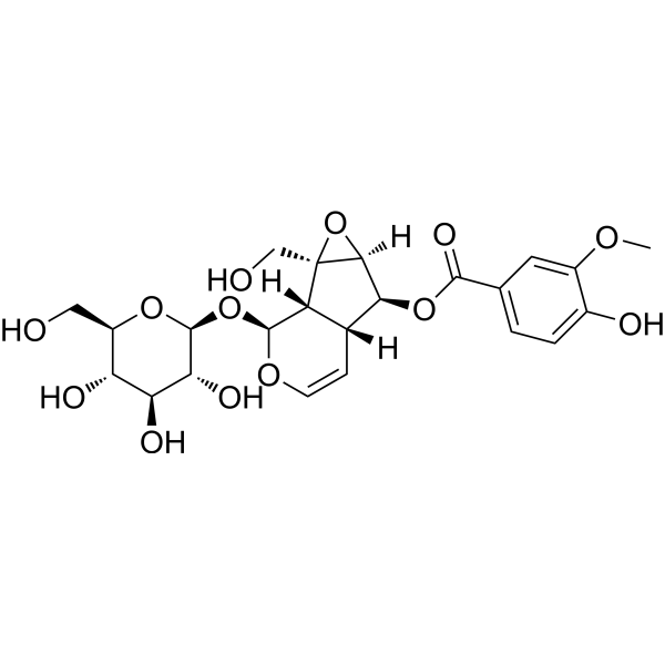
| SMILES | COC1=C(C=CC(=C1)C(=O)O[C@H]2[C@@H]3C=CO[C@H]([C@@H]3[C@@]4([C@H]2O4)CO)O[C@H]5[C@@H]([C@H]([C@@H]([C@H](O5)CO)O)O)O)O |
| InChi Key | AKNILCMFRRDTEY-NUGKWEEESA-N |
| InChi Code | InChI=1S/C23H28O13/c1-31-12-6-9(2-3-11(12)26)20(30)34-18-10-4-5-32-21(14(10)23(8-25)19(18)36-23)35-22-17(29)16(28)15(27)13(7-24)33-22/h2-6,10,13-19,21-22,24-29H,7-8H2,1H3/t10-,13-,14-,15-,16+,17-,18+,19+,21+,22+,23-/m1/s1 |
| Chemical Name | [(1S,2S,4S,5S,6R,10S)-2-(hydroxymethyl)-10-[(2S,3R,4S,5S,6R)-3,4,5-trihydroxy-6-(hydroxymethyl)oxan-2-yl]oxy-3,9-dioxatricyclo[4.4.0.02,4]dec-7-en-5-yl] 4-hydroxy-3-methoxybenzoate |
| HS Tariff Code | 2934.99.9001 |
| Storage |
Powder-20°C 3 years 4°C 2 years In solvent -80°C 6 months -20°C 1 month Note: This product requires protection from light (avoid light exposure) during transportation and storage. |
| Shipping Condition | Room temperature (This product is stable at ambient temperature for a few days during ordinary shipping and time spent in Customs) |
Biological Activity
| Targets |
- Target of Picroside II in sepsis model: Nuclear factor-kappa B (NF-κB) signaling pathway- Target of Picroside II in cerebral ischemia-reperfusion model: Oxidative stress-related pathways (Nrf2/HO-1, MAPK) and tight junction (TJ) protein-related signaling[2] |
| ln Vitro |
1. Anti-inflammatory activity in RAW264.7 macrophages (sepsis-related): RAW264.7 cells were treated with lipopolysaccharide (LPS, 1 μg/mL) and Picroside II (5 μM, 10 μM, 20 μM) for 24 hours. ELISA results showed Picroside II reduced LPS-induced secretion of tumor necrosis factor-α (TNF-α) and interleukin-6 (IL-6) in a concentration-dependent manner: 20 μM Picroside II decreased TNF-α and IL-6 levels by ~65% and ~60%, respectively. Western blot showed Picroside II inhibited LPS-induced phosphorylation of NF-κB p65 and IκBα, and reduced nuclear translocation of p65 [1] 2. Protective effect on brain microvascular endothelial cells (BMECs) (cerebral ischemia-related): BMECs were subjected to oxygen-glucose deprivation/reoxygenation (OGD/R) and treated with Picroside II (2.5 μM, 5 μM, 10 μM). Transendothelial electrical resistance (TEER) measurement showed Picroside II reversed OGD/R-induced decrease in TEER (10 μM Picroside II restored TEER to ~85% of normal level). DCFH-DA staining showed Picroside II reduced OGD/R-induced reactive oxygen species (ROS) production (10 μM reduced ROS by ~55%). Western blot showed Picroside II upregulated OGD/R-downregulated TJ proteins (ZO-1, occludin) and Nrf2/HO-1 pathway proteins, while downregulating phosphorylated p38 MAPK [2] |
| ln Vivo |
1. Protective effect in mouse sepsis model (CLP-induced): Male C57BL/6 mice were subjected to cecal ligation and puncture (CLP) to induce sepsis, and divided into sham group, CLP model group, Picroside II 20 mg/kg group, Picroside II 40 mg/kg group. Picroside II was administered via intraperitoneal injection 1 hour before CLP and once daily for 3 days. Survival rate analysis showed Picroside II increased 7-day survival rate (40 mg/kg group: 65% vs. 20% in model group). Histopathology showed Picroside II alleviated CLP-induced lung, liver, and kidney damage (reduced inflammatory cell infiltration and tissue necrosis). ELISA showed Picroside II decreased serum TNF-α, IL-6, and IL-1β levels (40 mg/kg reduced TNF-α by ~70%), and Western blot showed reduced NF-κB activation in lung tissue [1] 2. Protective effect in rat cerebral ischemia-reperfusion model (MCAO-induced): Male Sprague-Dawley rats were subjected to middle cerebral artery occlusion (MCAO) for 2 hours followed by 24 hours of reperfusion. Rats were divided into sham group, MCAO model group, Picroside II 10 mg/kg group, Picroside II 20 mg/kg group. Picroside II was administered via tail vein injection immediately after reperfusion. Neurological deficit score showed Picroside II reduced scores (20 mg/kg group: 1.5 vs. 3.2 in model group). TTC staining showed Picroside II decreased cerebral infarction volume (20 mg/kg reduced volume by ~45%). Evans blue (EB) leakage assay showed Picroside II reduced blood-brain barrier permeability (20 mg/kg reduced EB leakage by ~50%). Biochemical analysis showed Picroside II increased superoxide dismutase (SOD) activity and decreased malondialdehyde (MDA) content in brain tissue [2] |
| Cell Assay |
1. RAW264.7 macrophage experiment (sepsis-related): RAW264.7 cells were seeded in 6-well plates (5×10^5 cells/well) and cultured in DMEM containing 10% fetal bovine serum. When cells reached 80% confluence, they were divided into control group, LPS group (1 μg/mL), and Picroside II groups (5 μM, 10 μM, 20 μM + 1 μg/mL LPS). After 24-hour incubation at 37°C in 5% CO2, cell supernatant was collected for ELISA (TNF-α, IL-6 detection), and cells were lysed for Western blot (p-p65, p-IκBα, nuclear p65 detection). For nuclear protein extraction, cells were treated with nuclear extraction kit, and nuclear fractions were used for Western blot [1] 2. BMEC experiment (cerebral ischemia-related): BMECs were seeded in Transwell inserts (for TEER measurement) or 6-well plates (5×10^5 cells/well) and cultured in endothelial cell medium. When cells formed a confluent monolayer, OGD was induced by replacing medium with glucose-free DMEM and incubating in 1% O2 for 4 hours; reoxygenation was performed by replacing with normal medium and incubating in 5% CO2 for 24 hours. Picroside II (2.5 μM, 5 μM, 10 μM) was added at the start of reoxygenation. TEER was measured at 0, 6, 12, 24 hours of reoxygenation. For ROS detection, cells were loaded with DCFH-DA (10 μM) for 30 minutes, and fluorescence intensity was measured by flow cytometry. For Western blot, cells were lysed to detect ZO-1, occludin, Nrf2, HO-1, and p-p38 [2] |
| Animal Protocol |
1. Mouse sepsis model (CLP) protocol: Male C57BL/6 mice (20-25 g) were anesthetized with isoflurane. A midline abdominal incision was made, cecum was ligated (70% of cecum), and punctured twice with a 22-gauge needle. Cecum was returned to abdomen, and incision was closed. Sham group only had abdominal incision without ligation/puncture. Mice were divided into 4 groups (n=15/group): sham, CLP model, Picroside II 20 mg/kg, Picroside II 40 mg/kg. Picroside II was dissolved in normal saline (0.9% NaCl) and administered via intraperitoneal injection (0.1 mL/10 g body weight) 1 hour before CLP, then once daily on days 1-3 after CLP. On day 4, mice were sacrificed; serum was collected for ELISA, and lung, liver, kidney tissues were collected for histopathology and Western blot. Survival rate was recorded for 7 days [1] 2. Rat cerebral ischemia-reperfusion model (MCAO) protocol: Male Sprague-Dawley rats (250-300 g) were anesthetized with chloral hydrate. MCAO was performed by inserting a nylon suture into the internal carotid artery to block the middle cerebral artery for 2 hours; suture was removed for 24-hour reperfusion. Sham group only had artery dissection without suture insertion. Rats were divided into 4 groups (n=8/group): sham, MCAO model, Picroside II 10 mg/kg, Picroside II 20 mg/kg. Picroside II was dissolved in normal saline and administered via tail vein injection (0.1 mL/10 g body weight) immediately after reperfusion. After 24-hour reperfusion, neurological deficit score was evaluated (0-5 scale). Rats were then sacrificed; brain was collected for TTC staining (infarction volume) and EB leakage assay (blood-brain barrier permeability). Brain tissue homogenate was prepared to detect SOD activity and MDA content [2] |
| Toxicity/Toxicokinetics |
1. In mouse sepsis model: After 4-day treatment with Picroside II (20-40 mg/kg, intraperitoneal injection), serum levels of alanine transaminase (ALT), aspartate transaminase (AST), blood urea nitrogen (BUN), and creatinine (Cr) were measured. No significant differences were found between Picroside II-treated groups and sham group, indicating no hepatotoxicity or nephrotoxicity. Body weight of Picroside II-treated mice was stable, with no obvious weight loss [1] 2. In rat cerebral ischemia model: After 24-hour treatment with Picroside II (10-20 mg/kg, tail vein injection), serum ALT, AST, BUN, Cr levels were within normal ranges, and no abnormal changes in organ weight (brain, liver, kidney) were observed, suggesting no acute toxicity [2] |
| References |
[1]. Picroside II protects against sepsis via suppressing inflammation in mice. Am J Transl Res. 2016 Dec 15;8(12):5519-5531. eCollection 2016. [2]. Picroside II protects the blood-brain barrier by inhibiting the oxidative signaling pathway in cerebral ischemia-reperfusion injury. PLoS One. 2017 Apr 7;12(4):e0174414. |
| Additional Infomation |
Picroside II has been reported in Veronica anagallis, Catalpa ovata, and other organisms with data available. 1. Picroside II is a major active component isolated from the roots of Picrorhiza scrophulariiflora , a traditional Chinese herb used for anti-inflammatory and hepatoprotective purposes [1][2] 2. Mechanism in sepsis: Picroside II exerts anti-sepsis effects by inhibiting the NF-κB signaling pathway, reducing the production of pro-inflammatory cytokines, and alleviating organ damage caused by excessive inflammation [1] 3. Mechanism in cerebral ischemia: Picroside II protects the blood-brain barrier by inhibiting oxidative stress (activating Nrf2/HO-1 pathway, reducing ROS/MDA) and preserving TJ protein expression, while inhibiting p38 MAPK-mediated cell damage [2] |
Solubility Data
| Solubility (In Vitro) | DMSO : ~100 mg/mL (~195.14 mM) |
| Solubility (In Vivo) |
Solubility in Formulation 1: ≥ 2.5 mg/mL (4.88 mM) (saturation unknown) in 10% DMSO + 40% PEG300 + 5% Tween80 + 45% Saline (add these co-solvents sequentially from left to right, and one by one), clear solution. For example, if 1 mL of working solution is to be prepared, you can add 100 μL of 25.0 mg/mL clear DMSO stock solution to 400 μL PEG300 and mix evenly; then add 50 μL Tween-80 to the above solution and mix evenly; then add 450 μL normal saline to adjust the volume to 1 mL. Preparation of saline: Dissolve 0.9 g of sodium chloride in 100 mL ddH₂ O to obtain a clear solution. Solubility in Formulation 2: ≥ 2.5 mg/mL (4.88 mM) (saturation unknown) in 10% DMSO + 90% (20% SBE-β-CD in Saline) (add these co-solvents sequentially from left to right, and one by one), clear solution. For example, if 1 mL of working solution is to be prepared, you can add 100 μL of 25.0 mg/mL clear DMSO stock solution to 900 μL of 20% SBE-β-CD physiological saline solution and mix evenly. Preparation of 20% SBE-β-CD in Saline (4°C,1 week): Dissolve 2 g SBE-β-CD in 10 mL saline to obtain a clear solution. Solubility in Formulation 3: ≥ 2.5 mg/mL (4.88 mM) (saturation unknown) in 10% DMSO + 90% Corn Oil (add these co-solvents sequentially from left to right, and one by one), clear solution. For example, if 1 mL of working solution is to be prepared, you can add 100 μL of 25.0 mg/mL clear DMSO stock solution to 900 μL of corn oil and mix evenly. (Please use freshly prepared in vivo formulations for optimal results.) |
| Preparing Stock Solutions | 1 mg | 5 mg | 10 mg | |
| 1 mM | 1.9514 mL | 9.7569 mL | 19.5137 mL | |
| 5 mM | 0.3903 mL | 1.9514 mL | 3.9027 mL | |
| 10 mM | 0.1951 mL | 0.9757 mL | 1.9514 mL |