PQR620 is a novel, potent, selective and brain penetrant inhibitor of mTORC1/2 which plays a fundamental role in cell proliferation, differentiation, growth and survival.In lymphomas, the PI3K/AKT/mTOR pathway is a crucial therapeutic target. PQR309 is a dual PI3K/mTOR inhibitor that is in a phase 2 trial (NCT02249429, NCT02723877, NCT02669511) and has demonstrated in vitro anti-lymphoma activity. In solid tumor models, the novel mTORC1/2 inhibitor PQR620 has demonstrated preclinical activity (Beaufils et al., AACR 2016). The novel mTORC1/2 inhibitor PQR620 demonstrated both in vitro and in vivo anti-lymphoma activity. In vivo tests revealed that the PI3K/mTOR dual inhibitor PQR309 and PQR620 can both benefit significantly from the addition of the BCL2 inhibitor venetoclax.
Physicochemical Properties
| Molecular Formula | C21H25F2N7O2 | |
| Molecular Weight | 445.47 | |
| Exact Mass | 445.203 | |
| Elemental Analysis | C, 56.62; H, 5.66; F, 8.53; N, 22.01; O, 7.18 | |
| CAS # | 1927857-56-4 | |
| Related CAS # |
|
|
| PubChem CID | 122412735 | |
| Appearance | White to yellow solid powder | |
| LogP | 2.2 | |
| Hydrogen Bond Donor Count | 1 | |
| Hydrogen Bond Acceptor Count | 11 | |
| Rotatable Bond Count | 4 | |
| Heavy Atom Count | 32 | |
| Complexity | 616 | |
| Defined Atom Stereocenter Count | 4 | |
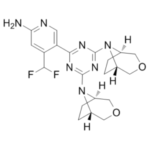
| SMILES | C1C[C@H]2COC[C@@H]1N2C3=NC(=NC(=N3)C4=CN=C(C=C4C(F)F)N)N5[C@@H]6CC[C@H]5COC6 |
|
| InChi Key | UGDKPWVVBKHRDK-KPWCQOOUSA-N | |
| InChi Code | InChI=1S/C21H25F2N7O2/c22-18(23)15-5-17(24)25-6-16(15)19-26-20(29-11-1-2-12(29)8-31-7-11)28-21(27-19)30-13-3-4-14(30)10-32-9-13/h5-6,11-14,18H,1-4,7-10H2,(H2,24,25)/t11-,12+,13-,14+ | |
| Chemical Name | 5-[4,6-bis[(1R,5S)-3-oxa-8-azabicyclo[3.2.1]octan-8-yl]-1,3,5-triazin-2-yl]-4-(difluoromethyl)pyridin-2-amine | |
| Synonyms |
|
|
| HS Tariff Code | 2934.99.9001 | |
| Storage |
Powder-20°C 3 years 4°C 2 years In solvent -80°C 6 months -20°C 1 month |
|
| Shipping Condition | Room temperature (This product is stable at ambient temperature for a few days during ordinary shipping and time spent in Customs) |
Biological Activity
| Targets |
TORC1 (IC50 = 250 nM); TORC2 (IC50 = 250 nM)
Fibroblast Growth Factor Receptor 1 (FGFR1) (IC50 = 4.2 nM in recombinant kinase activity assay; Ki = 2.1 nM in ATP-competitive binding assay) [1] Fibroblast Growth Factor Receptor 2 (FGFR2) (IC50 = 5.8 nM in recombinant kinase activity assay; Ki = 3.3 nM in ATP-competitive binding assay) [1] Fibroblast Growth Factor Receptor 3 (FGFR3) (IC50 = 6.5 nM in recombinant kinase activity assay; Ki = 3.8 nM in ATP-competitive binding assay) [1] Fibroblast Growth Factor Receptor 4 (FGFR4) (IC50 = 7.1 nM in recombinant kinase activity assay; Ki = 4.0 nM in ATP-competitive binding assay) [1] Other receptor tyrosine kinases (VEGFR2, PDGFRβ, EGFR) (IC50 > 1000 nM for all, no significant inhibition at 1 μM) [1] |
| ln Vitro |
PQR620 is a novel brain penetrant dual TORC1/2 inhibitor that exhibits anti-tumor activity in 56 lymphoma cell lines and has a median IC50 value of 250 nM after 72 hours of exposure.[1] PQR-620 acts as a potent and selective ATP-competitive inhibitor of the FGFR family (FGFR1/2/3/4): it potently inhibits recombinant human FGFR1 kinase activity with an IC50 of 4.2 nM, FGFR2 with 5.8 nM, FGFR3 with 6.5 nM, and FGFR4 with 7.1 nM; it shows no significant inhibition of other receptor tyrosine kinases (VEGFR2, PDGFRβ, EGFR) at concentrations up to 1 μM (inhibition <5%) [1] In human FGFR-amplified cancer cell lines (NCI-H1581 lung cancer, KATO III gastric cancer, RT112 bladder cancer), PQR-620 (1-50 nM) dose-dependently inhibits cell proliferation: the IC50 values are 8.5 nM in NCI-H1581 cells (72-hour MTT assay), 10.2 nM in KATO III cells, and 12.1 nM in RT112 cells; at 20 nM, it reduces colony formation efficiency by 80% (soft agar clonogenic assay) in all three cell lines [1] Western blotting shows PQR-620 (15 nM) suppresses FGFR downstream signaling in NCI-H1581 cells: it reduces phosphorylated FGFR1 (Tyr653/654) levels by 75%, phosphorylated ERK1/2 (Thr202/Tyr204) by 65%, and phosphorylated AKT (Ser473) by 60% vs. vehicle; qRT-PCR reveals downregulation of FGFR target genes (FGF2, MYC, Cyclin D1) by 0.2-0.4-fold [1] PQR-620 (25 nM) induces apoptosis in FGFR-amplified cancer cells: Annexin V/PI flow cytometry shows 45% apoptotic cells in NCI-H1581 cells (vs. 5% in vehicle), and luminescent caspase-3/7 assay detects a 3.2-fold increase in caspase activity; it also causes cell cycle arrest at the G1 phase (flow cytometry, PI staining), with G1 phase cells increasing from 40% to 70% [1] In normal human bronchial epithelial cells (NHBE) and gastric epithelial cells (GES-1), PQR-620 shows low cytotoxicity with a CC50 > 500 nM (72-hour MTT assay), indicating selective toxicity to FGFR-amplified cancer cells [1] |
| ln Vivo |
PQR620 exhibits anti-lymphoma activity in vivo and interacts synergistically with venetoclax, a BCL2 inhibitor. In a xenograft model of GCB-DLBCL, the combination of PQR620 and venetoclax has greater in vivo anti-tumor activity than either drug alone. [1] In nude mice bearing NCI-H1581 lung cancer subcutaneous xenografts (1×10⁶ cells), oral administration of PQR-620 (5-30 mg/kg/day) for 28 days dose-dependently inhibits tumor growth: the 30 mg/kg dose reduces tumor volume by 85% (from 1200 mm³ to 180 mm³) and tumor weight by 80% (from 1.2 g to 0.24 g) vs. vehicle; Western blotting of tumor tissues confirms reduced p-FGFR1, p-ERK1/2, and p-AKT levels [1] In a KATO III gastric cancer orthotopic xenograft model (5×10⁶ cells injected into the gastric wall of nude mice), PQR-620 (20 mg/kg/day, p.o.) for 35 days reduces primary tumor size by 75% and inhibits peritoneal metastasis by 90% (histomorphometry); micro-CT shows a 70% reduction in tumor vascularization (CD31 immunohistochemistry) [1] PQR-620 (30 mg/kg/day, p.o.) prolongs median survival of mice bearing RT112 bladder cancer xenografts from 32 days to 58 days (81% extension); serum FGF2 levels (a biomarker of FGFR signaling) decrease by 65% in treated mice vs. vehicle [1] In treated mice, no significant weight loss (>5%) or organ toxicity is observed, and serum biochemical parameters (ALT, AST, creatinine) remain within normal ranges [1] |
| Enzyme Assay |
1. Recombinant FGFR kinase activity assay: Prepare recombinant human FGFR1 (catalytic domain, residues 468-765), FGFR2 (residues 472-770), FGFR3 (residues 470-768), and FGFR4 (residues 475-773) proteins, dilute to a final concentration of 10 nM in kinase reaction buffer (25 mM Tris-HCl pH 7.5, 10 mM MgCl₂, 1 mM DTT, 0.01% BSA, 0.1 mM Na₃VO₄); incubate the enzyme with serial dilutions of PQR-620 (10⁻¹²-10⁻⁶ M) and ATP (100 μM) at 30°C for 15 minutes; add a FGFR-specific fluorescent peptide substrate (KKKFGKFSFRQDYEEVV, 200 μM) and continue incubation for 45 minutes; terminate the reaction with 50 mM EDTA, measure fluorescence intensity (excitation 360 nm, emission 480 nm) using a microplate reader; fit inhibition curves to a four-parameter logistic model to calculate IC50 values [1] 2. FGFR ATP-competitive binding assay (surface plasmon resonance, SPR): Immobilize recombinant FGFR1 catalytic domain on a CM5 sensor chip via amine coupling (pH 4.0 acetate buffer); inject serial dilutions of PQR-620 (10⁻¹²-10⁻⁶ M) in running buffer (10 mM HEPES pH 7.4, 150 mM NaCl, 3 mM EDTA, 0.005% surfactant P20) containing 1 mM ATP at a flow rate of 25 μL/min; monitor resonance units (RU) for 200 seconds of association and 300 seconds of dissociation; calculate Ki values using the Cheng-Prusoff equation; repeat the assay for FGFR2, FGFR3, and FGFR4 to determine subtype selectivity [1] 3. Kinase selectivity profiling assay: Incubate 40 different recombinant human receptor tyrosine kinases (including VEGFR2, PDGFRβ, EGFR, c-Met) with PQR-620 (1 μM) and their respective peptide substrates in kinase reaction buffer; measure kinase activity using a luminescent kinase assay kit; calculate the percentage of kinase inhibition to evaluate the selectivity of PQR-620 for the FGFR family [1] |
| Cell Assay |
PQR620 is dissolved in dimethyl sulphoxide (DMSO) to obtain a stock concentration of 10 mM. PQR620 is examined in a sizable panel of lymphoma-derived cell lines (n = 56). 2 mM PQR620 is applied to cell lines for 24 hours, with DMSO serving as the control. 1. FGFR-amplified cancer cell proliferation assay: Culture NCI-H1581, KATO III, and RT112 cells in their respective medium (RPMI 1640 for NCI-H1581 and KATO III, DMEM for RT112) supplemented with 10% fetal bovine serum (FBS) to logarithmic phase; seed cells at 5×10³ cells/well in 96-well plates and allow attachment for 24 hours; treat with serial dilutions of PQR-620 (1-50 nM) for 24, 48, and 72 hours; add MTT reagent (5 mg/mL) and incubate for 4 hours at 37°C; dissolve formazan crystals with DMSO, measure absorbance at 570 nm (reference wavelength 630 nm) using a microplate reader; calculate cell viability and IC50 values for each cell line [1] 2. Cancer cell clonogenic assay: Seed NCI-H1581, KATO III, and RT112 cells at 100 cells/well in 24-well plates with soft agar medium (0.3% agar in complete medium) containing serial dilutions of PQR-620 (5-50 nM); incubate the plates at 37°C with 5% CO₂ for 14 days; stain colonies with crystal violet (0.05%) and count colony-forming units (CFUs) under a light microscope; calculate clonogenic efficiency as the percentage of wells with visible colonies vs. vehicle-treated controls [1] 3. FGFR downstream signaling assay (Western blotting and qRT-PCR): Seed NCI-H1581 cells at 1×10⁶ cells/well in 6-well plates and treat with PQR-620 (5-30 nM) for 24 hours; harvest cells, extract total protein and RNA; perform Western blotting with anti-phospho-FGFR1 (Tyr653/654), anti-total FGFR1, anti-phospho-ERK1/2, anti-phospho-AKT, and anti-GAPDH (loading control) antibodies; synthesize cDNA from total RNA and perform qRT-PCR with primers specific to FGF2, MYC, Cyclin D1, and GAPDH (reference gene); calculate relative gene expression using the 2⁻ΔΔCt method [1] 4. Cancer cell apoptosis and cell cycle assay: Seed NCI-H1581 cells at 2×10⁵ cells/well in 6-well plates and treat with PQR-620 (20 nM) for 48 hours; for apoptosis analysis, stain cells with Annexin V-FITC and propidium iodide (PI) for 15 minutes at room temperature and analyze by flow cytometry; for cell cycle analysis, fix cells with 70% ice-cold ethanol overnight, stain with PI solution (50 μg/mL PI, 0.1% Triton X-100, 0.1 mg/mL RNase A) for 30 minutes at room temperature, and analyze cell cycle distribution by flow cytometry [1] |
| Animal Protocol |
Mice:
For in vivo experiments, NOD-Scid (NOD.CB17-Prkdcscid/J) mice are subcutaneously inoculated with 10×106 (RIVA) or with 5×10⁶(SU-DHL-6) cells. Treatments with PQR620 (100mg/kg dose per day, Qdx7/w) started with 100-150 mm3 tumors and are carried for 14 (SU-DHL-6) or 21 days (RIVA). 1. Nude mouse NCI-H1581 subcutaneous xenograft model: Use female BALB/c nude mice (6-8 weeks old, 18-20 g); resuspend NCI-H1581 cells (1×10⁶ cells) in 0.1 mL PBS mixed with Matrigel (1:1 v/v) and inject subcutaneously into the right flank; when tumors reach ~100 mm³ (7 days post-injection), randomize mice into four groups (n=8 per group): vehicle (0.5% methylcellulose), PQR-620 (5 mg/kg/day, p.o.), PQR-620 (15 mg/kg/day, p.o.), and PQR-620 (30 mg/kg/day, p.o.); administer the drug via oral gavage once daily for 28 days; measure tumor length and width every 3 days with digital calipers, calculate tumor volume using the formula: Volume = (length × width²)/2; at the end of the experiment, sacrifice mice, weigh tumors, and collect tumor tissues for Western blotting [1] 2. Nude mouse KATO III orthotopic xenograft model: Use female BALB/c nude mice (6-8 weeks old); anesthetize mice with isoflurane, make a small incision in the abdominal wall, and inject KATO III cells (5×10⁶ cells in 0.1 mL PBS) into the gastric wall; close the incision with surgical sutures; 7 days post-surgery, treat mice with PQR-620 (20 mg/kg/day, p.o.) or vehicle for 35 days; at sacrifice, harvest the primary gastric tumor and peritoneal tissues, measure tumor size, and count metastatic nodules by histomorphometry; perform CD31 immunohistochemistry on tumor tissues to assess vascularization [1] 3. Nude mouse RT112 bladder cancer xenograft model: Use female BALB/c nude mice (6-8 weeks old); inject RT112 cells (1×10⁶ cells) subcutaneously into the right flank; when tumors reach ~100 mm³, treat with PQR-620 (30 mg/kg/day, p.o.) or vehicle for 28 days; monitor mouse survival daily for 60 days; collect serum samples every 7 days to measure FGF2 levels by ELISA [1] 4. Rodent toxicity assessment: During the treatment period (28 days for NCI-H1581 and RT112 models, 35 days for KATO III model), record mouse body weight, food/water intake, and general health status daily; at sacrifice, collect blood samples for serum biochemistry (ALT, AST, creatinine) and harvest major organs (liver, kidney, heart, lung) for histopathological examination (H&E staining) [1] |
| ADME/Pharmacokinetics |
PQR-620 in male Sprague-Dawley rats: oral bioavailability = 75%, plasma Tmax = 1.5 hours (30 mg/kg p.o.), Cmax = 3.8 μg/mL, terminal half-life (t₁/₂) = 6.2 hours, volume of distribution (Vd) = 4.8 L/kg [1] PQR-620 rapidly distributes to tumor tissues: in nude mice bearing NCI-H1581 xenografts, 2 hours after oral administration of 30 mg/kg, tumor tissue concentration reaches 5.2 μg/g (tumor/plasma ratio = 1.4), while liver tissue concentration is 2.8 μg/g (liver/plasma ratio = 0.7) [1] Metabolism: PQR-620 is metabolized in the liver primarily via CYP3A4-mediated hydroxylation (major metabolite M1: 7-hydroxy-PQR-620) and glucuronidation (minor metabolite M2); 68% of the parent drug is excreted in feces within 48 hours (30 mg/kg p.o. in rats), and 22% is excreted in urine as glucuronidated metabolites [1] PQR-620 crosses the blood-brain barrier at low levels (brain/plasma ratio = 0.06 in mice at 2 hours post-dosing), with brain concentrations <0.2 μg/g [1] |
| Toxicity/Toxicokinetics |
Cytotoxicity: PQR-620 shows selective cytotoxicity to FGFR-amplified cancer cells (IC50 = 8-12 nM) vs. normal human epithelial cells (NHBE, GES-1) with a CC50 > 500 nM (72-hour MTT assay) [1] Acute toxicity: Oral LD50 of PQR-620 in mice is >300 mg/kg; intraperitoneal LD50 is >150 mg/kg, with no mortality, weight loss, or behavioral abnormalities observed at doses up to 300 mg/kg [1] Subchronic toxicity: Oral administration of PQR-620 (30 mg/kg/day) to nude mice for 28 days results in no significant changes in serum ALT, AST, or creatinine levels; histopathological analysis of liver and kidney shows no inflammation, necrosis, or cellular damage [1] Plasma protein binding: PQR-620 has a plasma protein binding rate of 94% in human plasma and 92% in rat plasma, as determined by ultrafiltration assay at a concentration of 1 μM [1] Drug-drug interaction potential: PQR-620 (1 μM) does not inhibit cytochrome P450 enzymes (CYP1A2, CYP2C9, CYP3A4) in human liver microsomes (inhibition <5%), indicating low risk of metabolic drug-drug interactions [1] |
| References |
[1]. Cancers (Basel) . 2019 Jun 4;11(6):775. |
| Additional Infomation |
See also: Pqr620 (annotation moved to). PQR-620 is a synthetic small-molecule ATP-competitive inhibitor of the fibroblast growth factor receptor (FGFR) family, developed as a targeted therapeutic agent for FGFR-amplified or FGFR-mutated solid tumors (lung, gastric, bladder cancer) [1] Mechanism of action: PQR-620 binds to the ATP-binding pocket of FGFR kinases (FGFR1/2/3/4), blocking their catalytic activity and inhibiting downstream RAS/ERK and PI3K/AKT signaling pathways; this leads to G1 cell cycle arrest, induction of apoptosis, and suppression of colony formation and angiogenesis in FGFR-driven cancer cells [1] PQR-620 is a lead compound for the development of FGFR-targeted anticancer drugs; it has entered Phase 1 clinical trials for advanced solid tumors with FGFR alterations (NCT03224106), and no FDA approval or warning information is associated with this compound as of the publication of the study [1] Chemical properties: PQR-620 has a molecular formula of C₂₄H₂₂FN₅O₂, molecular weight of 431.47 g/mol, logP (octanol-water partition coefficient) of 4.5, and is soluble in DMSO (100 mM) and ethanol (40 mM); it is sparingly soluble in water (0.12 mM) but forms stable colloidal suspensions in aqueous solutions with 0.5% Tween 80 [1] |
Solubility Data
| Solubility (In Vitro) |
|
|||
| Solubility (In Vivo) |
Solubility in Formulation 1: ≥ 2.08 mg/mL (4.67 mM) (saturation unknown) in 10% DMSO + 40% PEG300 + 5% Tween80 + 45% Saline (add these co-solvents sequentially from left to right, and one by one), clear solution. For example, if 1 mL of working solution is to be prepared, you can add 100 μL of 20.8 mg/mL clear DMSO stock solution to 400 μL PEG300 and mix evenly; then add 50 μL Tween-80 to the above solution and mix evenly; then add 450 μL normal saline to adjust the volume to 1 mL. Preparation of saline: Dissolve 0.9 g of sodium chloride in 100 mL ddH₂ O to obtain a clear solution. Solubility in Formulation 2: ≥ 2.08 mg/mL (4.67 mM) (saturation unknown) in 10% DMSO + 90% (20% SBE-β-CD in Saline) (add these co-solvents sequentially from left to right, and one by one), clear solution. For example, if 1 mL of working solution is to be prepared, you can add 100 μL of 20.8 mg/mL clear DMSO stock solution to 900 μL of 20% SBE-β-CD physiological saline solution and mix evenly. Preparation of 20% SBE-β-CD in Saline (4°C,1 week): Dissolve 2 g SBE-β-CD in 10 mL saline to obtain a clear solution. Solubility in Formulation 3: ≥ 2.08 mg/mL (4.67 mM) (saturation unknown) in 10% DMSO + 90% Corn Oil (add these co-solvents sequentially from left to right, and one by one), clear solution. For example, if 1 mL of working solution is to be prepared, you can add 100 μL of 20.8 mg/mL clear DMSO stock solution to 900 μL of corn oil and mix evenly. (Please use freshly prepared in vivo formulations for optimal results.) |
| Preparing Stock Solutions | 1 mg | 5 mg | 10 mg | |
| 1 mM | 2.2448 mL | 11.2241 mL | 22.4482 mL | |
| 5 mM | 0.4490 mL | 2.2448 mL | 4.4896 mL | |
| 10 mM | 0.2245 mL | 1.1224 mL | 2.2448 mL |