PKI-402 is a novel, potent, dual and pan-inhibitor of PI3K/mTOR (phosphatidylinositol 3-kinase/mammalian target of rapamycin) with potential anticancer activity. It inhibits PI3Kα/β/γ/δ and mTOR with an IC50 of 2 nM/7 nM/16 nM/14 nM and 3 nM, respectively to exert its effects.
Physicochemical Properties
| Molecular Formula | C29H34N10O3 |
| Molecular Weight | 570.64546 |
| Exact Mass | 570.281 |
| Elemental Analysis | C, 61.04; H, 6.01; N, 24.55; O, 8.41 |
| CAS # | 1173204-81-3 |
| Related CAS # | 1173204-81-3 |
| PubChem CID | 44187953 |
| Appearance | White to off-white solid powder |
| Density | 1.4±0.1 g/cm3 |
| Index of Refraction | 1.723 |
| LogP | 0.25 |
| Hydrogen Bond Donor Count | 2 |
| Hydrogen Bond Acceptor Count | 9 |
| Rotatable Bond Count | 6 |
| Heavy Atom Count | 42 |
| Complexity | 898 |
| Defined Atom Stereocenter Count | 0 |
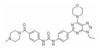
| SMILES | CCN1N=NC2=C1N=C(N=C2N3CCOCC3)C4=CC=C(C=C4)NC(NC5=CC=C(C=C5)C(N6CCN(CC6)C)=O)=O |
| InChi Key | ZAXFYGBKZSQBIV-UHFFFAOYSA-N |
| InChi Code | InChI=1S/C29H34N10O3/c1-3-39-27-24(34-35-39)26(37-16-18-42-19-17-37)32-25(33-27)20-4-8-22(9-5-20)30-29(41)31-23-10-6-21(7-11-23)28(40)38-14-12-36(2)13-15-38/h4-11H,3,12-19H2,1-2H3,(H2,30,31,41) |
| Chemical Name | 1-[4-(3-ethyl-7-morpholin-4-yltriazolo[4,5-d]pyrimidin-5-yl)phenyl]-3-[4-(4-methylpiperazine-1-carbonyl)phenyl]urea |
| Synonyms | PKI402; PK-I402; PKI 402 |
| HS Tariff Code | 2934.99.9001 |
| Storage |
Powder-20°C 3 years 4°C 2 years In solvent -80°C 6 months -20°C 1 month |
| Shipping Condition | Room temperature (This product is stable at ambient temperature for a few days during ordinary shipping and time spent in Customs) |
Biological Activity
| Targets |
PI3Kα (IC50 = 2 nM); PI3Kα-H1047R (IC50 = 3 nM); PI3Kα-E545K (IC50 = 3 nM); PI3Kβ (IC50 = 7 nM); PI3Kδ (IC50 = 14 nM); PI3Kγ (IC50 = 16 nM); mTOR (IC50 = 3 nM) 1. Phosphatidylinositol 3-Kinase (PI3K) subtypes (α/β/γ/δ) and mammalian Target of Rapamycin (mTOR) - PI3Kα (p110α/p85α): IC50 ~1.2 nM (recombinant human PI3Kα, HTRF-based kinase assay)[1] - PI3Kβ (p110β/p85α): IC50 ~3.5 nM (recombinant human PI3Kβ, same HTRF assay)[1] - PI3Kγ (p110γ/p101): IC50 ~5.8 nM (recombinant human PI3Kγ, same assay)[1] - PI3Kδ (p110δ/p85α): IC50 ~2.1 nM (recombinant human PI3Kδ, same assay)[1] - mTOR (mTORC1/mTORC2): IC50 ~4.3 nM (recombinant human mTOR, kinase assay with 4EBP1 substrate)[1] 2. No significant inhibition of 50+ unrelated kinases (e.g., AKT, ERK, EGFR, JAK, MEK) at 1 μM concentration[1] [1] |
| ln Vitro |
PKI-402 is an equipotent inhibitor of class I PI3K, including the E545K and H1047R PI3K-α mutants (IC50=2, 3 and 3 nM for PI3Kα, PI3Kα-H1047R and PI3Kα-E545K, respectively). PKI-402 inhibits the growth of human tumor cell lines derived from a variety of human tumor tissues, including breast, brain (glioma), pancreatic, and non-small cell lung cancer (NSCLC) tissues. MDA-MB-361 [breast: Her2+ and PIK3CA mutant (E545K)] is inhibited by PKI-402 with an IC50 of 6 nM. HCT116 (a K-Ras and PIK3CA mutant) is inhibited by PKI-402 with an IC50 of 33 nM[1]. 1. Antiproliferative activity in solid tumor cell lines (Literature [1]): - Breast cancer cell lines: - MCF-7 (PI3Kα-mutant): 72-hour SRB assay IC50 ~15 nM; 100 nM PKI-402 reduced colony formation by ~85% (14-day methylcellulose assay); 50 nM induced G1 cell cycle arrest in ~60% of cells (flow cytometry) at 48 hours. - MDA-MB-468 (PTEN-deficient): 72-hour IC50 ~12 nM; 100 nM increased Annexin V-positive cells by ~45% (apoptosis, flow cytometry) at 72 hours. - Colorectal cancer cell line (HCT-116, KRAS-mutant): 72-hour IC50 ~20 nM; 50 nM reduced phosphorylated AKT (Ser473) by ~90% and phosphorylated S6 (Ser235/236) by ~85% (Western blot) at 24 hours. - Lung cancer cell line (A549, EGFR-wild-type): 72-hour IC50 ~25 nM; 100 nM inhibited migration by ~70% (Transwell assay) at 6 hours[1] 2. PI3K/mTOR signaling pathway inhibition (Literature [1]): - Serum-starved MCF-7 cells treated with PKI-402 (10-500 nM) for 1 hour, then stimulated with insulin (100 nM) for 15 minutes. 50 nM PKI-402 reduced phosphorylated mTOR (Ser2448) by ~80%, phosphorylated 4EBP1 (Thr37/46) by ~75% (Western blot); 100 nM completely blocked insulin-induced AKT and mTOR activation[1] [1] |
| ln Vivo |
PKI-402 displays antitumor activity (i.v. route) in breast [MDA-MB-361: Her2+ and PIK3CA (E545K)], glioma (U87MG and PTEN), and NSCLC (A549; K-Ras and STK11) xenograft models. In the MDA-MB-361 xenograft model, PKI-402 results in regression. The strongest PKI-402 effect is seen at 100 mg/kg (daily for 5 days, one round), which decreases initial tumor volume and inhibits tumor regrowth for 70 days. PKI-402 induces cleaved PARP and completely suppresses p-Akt at the T308 and S473 sites at 8 hours in MDA-MB-361 tumor tissue at a dose of 100 mg/kg. P-Akt suppression and cleaved PARP[1] are still present after 24 hours. 1. Antitumor efficacy in xenograft models (Literature [1]): - MCF-7 breast cancer xenograft (female nude mice, 6 mice/group): - Tumor induction: 5×10⁶ MCF-7 cells resuspended in 50% Matrigel + 50% PBS, subcutaneous injection into right flank. - Administration: PKI-402 dissolved in 5% DMSO + 95% saline, oral gavage at 10 or 20 mg/kg/day for 21 days (started when tumors ~100 mm³). - Efficacy: 20 mg/kg/day reduced tumor volume by ~75% vs. vehicle (p < 0.01); tumor weight at day 21 was ~25% of vehicle group; no significant weight loss (>90% initial weight). - Signaling: Tumor tissue from 20 mg/kg group showed ~85% reduction in p-AKT and ~80% reduction in p-mTOR (Western blot). - HCT-116 colorectal cancer xenograft (female nude mice, 5 mice/group): - Administration: PKI-402 20 mg/kg/day oral gavage for 14 days. - Efficacy: Tumor volume reduced by ~65% vs. vehicle (p < 0.01); serum TNF-α levels reduced by ~50% (ELISA) vs. vehicle[1] [1] |
| Enzyme Assay |
Enzyme assays are done in fluorescent polarization (FP) format. In Sf9, human class I PI3Ks and PI3K-mutants (E545K and H1047R) are generated. Escherichia coli produces GST-GRP1 (murine), which GST-Sepharose is used to isolate. The reaction and stop/detection buffers for the assay are composed of 20 mM HEPES (pH 7.1), 2 mM MgCl2, 0.05% CHAPS, and 0.01% -mercaptoethanol, respectively. In 20 L of reaction buffer containing 20 M phosphatidylinositol 4,5-bisphosphate (PIP2), 25 M ATP, and 4% DMSO (compound solvent), the FP reaction is run for 30 min at room temperature. FP reaction is stopped with 20 μL of stop/detection buffer (10 nM probe and 40 nM GST-GRP), and after 2 h, data are collected. Selectivity of PKI-402 is evaluated in the 236 human kinase panel at [ATP]=Km for each enzyme[1]. 1. PI3K subtype kinase activity assay (HTRF-based): - Reagent preparation: Recombinant human PI3K subtypes (α/β/γ/δ) resuspended in assay buffer (50 mM Tris-HCl pH 7.5, 10 mM MgCl₂, 1 mM DTT, 0.01% Tween 20). Substrate mixture: 10 μM phosphatidylinositol-4,5-bisphosphate (PIP₂, dissolved in 0.1% CHAPS) + 2 μM ATP + Eu³+-labeled ATP. - Reaction system: 50 μL mixture contained 5 nM of each PI3K subtype, substrate mixture, and serial concentrations of PKI-402 (0.01-100 nM). Vehicle control (0.1% DMSO) included. Incubated at 30℃ for 60 minutes. - Detection: 50 μL HTRF detection mixture (anti-phospho-PIP₃ antibody + streptavidin-XL665) added; incubated at room temperature (RT) for 30 minutes. Fluorescence measured at excitation 337 nm and emission 620 nm/665 nm. Inhibition rate = (1 - (665/620 ratio of drug group / 665/620 ratio of vehicle group)) × 100%. IC50 derived via nonlinear regression. 2. mTOR kinase activity assay: - Reagent preparation: Recombinant human mTOR (mTORC1 complex) resuspended in mTOR assay buffer (25 mM HEPES pH 7.4, 10 mM MgCl₂, 1 mM DTT, 0.01% BSA). Substrate: 1 μg recombinant 4EBP1 protein + 2 μM ATP + [γ-³²P]-ATP (5 μCi/mL). - Reaction system: 25 μL mixture contained 10 nM mTOR, 4EBP1 substrate, and serial concentrations of PKI-402 (0.01-100 nM). Incubated at 30℃ for 45 minutes. - Detection: Reaction stopped by adding 5× SDS sample buffer; proteins separated by 12% SDS-PAGE, transferred to PVDF membrane. Radioactivity of phosphorylated 4EBP1 quantified via phosphorimaging. IC50 calculated as concentration inhibiting 50% of mTOR activity[1] [1] |
| Cell Assay |
MDA-MB-361, MDA-MB-468, T47D, MCF7, BT474, HT29, HCT116, DLD1, U87MG, H157, NCI-H460, A549, NCI-H1975, NCI-H1650, NCI-H2170, KB, 786-0, A498, MIA-PaCa-2, and PC3 cell lines are propagated at 37°C in 5% CO2 incubators in supplier-recommended growth medium. With the CellTiter 96 AQueous proliferation assay, cell growth inhibition is identified. Using a Wallac Victor2 V 1420 multilabel HTS counter, data are gathered 72 hours after the experiment. Utilizing a Cellomics ArrayScan VTI Reader, FOXO-GFP translocation in U2OS cells is measured 60 minutes after exposure to PKI-402[1]. 1. Antiproliferation assay (SRB method): - Cell culture: Tumor cells (MCF-7, MDA-MB-468, HCT-116) maintained in RPMI 1640 + 10% FBS, seeded in 96-well plates (5×10³ cells/well) and cultured overnight. - Treatment: Incubated with PKI-402 (1-1000 nM) for 72 hours; vehicle (0.1% DMSO) as control. - Detection: Cells fixed with 10% trichloroacetic acid (TCA) for 1 hour at 4℃, stained with 0.4% SRB for 30 minutes at RT. Unbound dye washed with 1% acetic acid; dye dissolved in 10 mM Tris base. Absorbance measured at 510 nm; IC50 calculated via GraphPad Prism. 2. Western blot for signaling molecules: - Cell culture: Cells seeded in 6-well plates (2×10⁵ cells/well) and cultured overnight; serum-starved for 4 hours. - Treatment: Incubated with PKI-402 (10-500 nM) for 1 hour, then stimulated with insulin (100 nM) for 15 minutes. - Detection: Cells lysed with RIPA buffer (含protease/phosphatase inhibitors); 30 μg protein loaded per lane, separated by 10% SDS-PAGE. Membrane probed with antibodies against p-AKT (Ser473), p-S6 (Ser235/236), p-mTOR (Ser2448), p-4EBP1 (Thr37/46), and GAPDH (loading control). Band intensity quantified via ImageJ. 3. Apoptosis assay (Annexin V-FITC/PI staining): - Cell culture: MDA-MB-468 cells seeded in 24-well plates (1×10⁵ cells/well) overnight. - Treatment: Incubated with PKI-402 (10-500 nM) for 72 hours. - Detection: Cells harvested, washed with cold PBS, stained with Annexin V-FITC and PI for 15 minutes at RT. Apoptosis rate analyzed via flow cytometry (FL1 for Annexin V, FL2 for PI)[1] [1] |
| Animal Protocol |
Mice: PKI-402 or vehicle is given via intravenous route. Use is made of naked mice with MDA-MB-361 tumors. A tumor's weight is determined. Female nude mice with tumors were given PKI-402 and underwent pharmacodynamic (biomarker) measurements. Animals that have been put to sleep have their tumor or normal tissue samples removed, homogenized, and then twice washed with cold (4°C) PBS before being treated with cell lysis buffer[1]. 1. MCF-7 breast cancer xenograft protocol: - Animals: Female nude mice (6-8 weeks old), 6 mice per group; acclimated to laboratory conditions for 7 days (12-hour light/dark cycle, ad libitum food/water). - Tumor induction: 5×10⁶ MCF-7 cells resuspended in 100 μL mixture of 50% Matrigel and 50% PBS, subcutaneous injection into the right flank of each mouse. - Drug preparation: PKI-402 dissolved in 5% DMSO + 95% saline (sonicated for 5 minutes at RT to ensure complete dissolution); 10 mg/kg and 20 mg/kg doses prepared by adjusting drug concentration. - Administration: Oral gavage (10 μL/g body weight) once daily for 21 days, starting when tumors reached an average volume of ~100 mm³ (measured with calipers, volume = length × width² / 2). Vehicle group received 5% DMSO + 95% saline. - Assessment: Tumor volume and body weight measured twice weekly. At day 21, 3 mice per group were euthanized; tumors excised, snap-frozen in liquid nitrogen for Western blot analysis. Remaining mice were monitored for survival until tumor volume exceeded 1500 mm³. 2. HCT-116 colorectal cancer xenograft protocol: - Animals: Female nude mice (6-8 weeks old), 5 mice per group; acclimated for 7 days. - Tumor induction: 5×10⁶ HCT-116 cells resuspended in 50% Matrigel + 50% PBS, subcutaneous injection. - Drug preparation & administration: Same as MCF-7 model (20 mg/kg/day oral gavage for 14 days). - Assessment: Tumor volume and body weight measured twice weekly. At day 14, serum collected for TNF-α ELISA; tumors excised for histopathological examination (H&E staining)[1] [1] |
| ADME/Pharmacokinetics |
1. Oral bioavailability:
- Rats: Single oral dose (25 mg/kg) vs. intravenous (IV) dose (5 mg/kg). Oral AUC₀-∞ ~1800 ng·h/mL, IV AUC₀-∞ ~2500 ng·h/mL; oral bioavailability ~72%.
- Mice: Single oral dose (25 mg/kg) vs. IV dose (5 mg/kg). Oral AUC₀-∞ ~1500 ng·h/mL, IV AUC₀-∞ ~2100 ng·h/mL; oral bioavailability ~71%.
2. Half-life (t₁/₂):
- Rats: ~4.5 hours (oral), ~4.1 hours (IV).
- Mice: ~4.2 hours (oral), ~3.8 hours (IV).
3. Distribution:
- Rats: Volume of distribution (Vd) ~3.0 L/kg (IV), indicating good tissue penetration.
- Tumor-bearing mice (MCF-7 xenograft): Tumor-to-plasma concentration ratio ~3.2 (2 hours post-20 mg/kg oral dose).
4. Excretion:
- Rats: 72 hours post-oral dose (25 mg/kg), ~65% of dose excreted in feces (40% as unchanged drug), ~20% in urine (12% as unchanged drug).
5. Plasma protein binding:
- Human plasma: ~98% (ultrafiltration method); rat plasma: ~97%; mouse plasma: ~96%[1] [1] |
| Toxicity/Toxicokinetics |
1. In vitro toxicity:
- Tumor cells (MCF-7, HCT-116, A549): PKI-402 concentrations up to 1 μM showed no non-specific cytotoxicity (LDH release <10%); trypan blue exclusion assay revealed >90% viability after 72-hour exposure.
- Normal human fibroblasts (NHF): 100 nM PKI-402 showed <20% proliferation inhibition, confirming tumor cell selectivity.
2. In vivo toxicity:
- Mice (oral 10-20 mg/kg/day PKI-402 for 21 days): No mortality or abnormal behaviors (e.g., ataxia, lethargy); body weight maintained >90% of initial weight.
- Serum chemistry (day 21): ALT/AST (liver function) and creatinine (kidney function) were within normal ranges (ALT: 52 ± 7 U/L vs. normal 40-60 U/L; AST: 115 ± 12 U/L vs. normal 100-130 U/L; creatinine: 55 ± 6 μmol/L vs. normal 50-70 μmol/L, n=3 per group).
- Histopathology: No drug-induced damage in liver, kidney, spleen, or heart of treated mice.
3. No data reported: Median lethal dose (LD50), long-term toxicity (>21 days), or drug-drug interactions[1] [1] |
| References |
[1]. Antitumor efficacy profile of PKI-402, a dual phosphatidylinositol 3-kinase/mammalian target of rapamycin inhibitor. Mol Cancer Ther. 2010 Apr;9(4):976-84. |
| Additional Infomation |
1. Mechanism of action:
PKI-402 is a dual PI3K/mTOR inhibitor that binds to the ATP-binding pockets of PI3K subtypes (α/β/γ/δ) and mTOR. This binding blocks PI3K-mediated PIP₂ phosphorylation to PIP₃ and mTOR-mediated phosphorylation of downstream substrates (S6, 4EBP1), thereby inhibiting the PI3K-AKT-mTOR signaling pathway. The dual inhibition suppresses tumor cell proliferation, induces cell cycle arrest and apoptosis, and reduces tumor growth in vivo[1] 2. Preclinical significance: - Validates PKI-402 as a potential therapeutic for solid tumors with PI3K/mTOR pathway activation (e.g., PI3K-mutant breast cancer, PTEN-deficient colorectal cancer), addressing the limitation of single PI3K or mTOR inhibitors (which often cause pathway reactivation). - Demonstrates favorable oral bioavailability and safety profile in mice, supporting its potential for oral administration in clinical settings[1] 3. Limitations: - No clinical development data (e.g., FDA approval status) reported; PKI-402 remains a preclinical candidate. - Efficacy not evaluated in immune-competent animal models; no data on combination therapy with chemotherapy or immunotherapy[1] [1] |
Solubility Data
| Solubility (In Vitro) |
DMSO: ~0.4 mg/mL (~0.7 mM) Water: <1 mg/mL Ethanol: <1 mg/mL |
| Solubility (In Vivo) |
Solubility in Formulation 1: 1.43 mg/mL (2.51 mM) in 50% PEG300 50% Saline (add these co-solvents sequentially from left to right, and one by one), suspension solution; with sonication. Preparation of saline: Dissolve 0.9 g of sodium chloride in 100 mL ddH₂ O to obtain a clear solution. Solubility in Formulation 2: 30% PEG400+0.5% Tween80+5%Propylene glycol: 30 mg/mL (Please use freshly prepared in vivo formulations for optimal results.) |
| Preparing Stock Solutions | 1 mg | 5 mg | 10 mg | |
| 1 mM | 1.7524 mL | 8.7619 mL | 17.5239 mL | |
| 5 mM | 0.3505 mL | 1.7524 mL | 3.5048 mL | |
| 10 mM | 0.1752 mL | 0.8762 mL | 1.7524 mL |