PIK-75 HCl, the hydrochloride salt of PIK-75, is a novel, potent, selective and imidazopyridine-based p110α inhibitor with potential anticancer activity and with IC50 of 5.8 nM, which is 200-fold more potent than p110β. Additionally, in cell-free assays, it strongly inhibits DNA-PK with an IC50 of 2 nM. In a variety of cell types, PIK-75, which was created as part of a PI 3-kinase drug discovery program, can attenuate insulin stimulation of Akt/PKB at a concentration of 100 nM. With an IC50 value in the range of 50 nM, PIK-75 has been shown to inhibit the growth of several different cell lines. The growth of HeLa cell xenografts in mouse models was inhibited by PIK-75 (at 50 mg/kg), according to in vivo studies. The p110 isoform of PI3K was the target of PIK-75 in acute myeloid leukemia (AML) cells, which resulted in the breakage of the link between Bcl-xL and Bak.
Physicochemical Properties
| Molecular Formula | C16H14BRN5O4S.HCL |
| Molecular Weight | 488.74 |
| Exact Mass | 486.971 |
| Elemental Analysis | C, 39.32; H, 3.09; Br, 16.35; Cl, 7.25; N, 14.33; O, 13.09; S, 6.56 |
| CAS # | 372196-77-5 |
| Related CAS # | PIK-75;372196-67-3 |
| PubChem CID | 45265864 |
| Appearance | White to off-white solid powder |
| Density | 1.7±0.1 g/cm3 |
| Index of Refraction | 1.701 |
| LogP | 3.84 |
| Hydrogen Bond Donor Count | 1 |
| Hydrogen Bond Acceptor Count | 7 |
| Rotatable Bond Count | 4 |
| Heavy Atom Count | 28 |
| Complexity | 679 |
| Defined Atom Stereocenter Count | 0 |
| SMILES | BrC1C([H])=C([H])C2=NC([H])=C(/C(/[H])=N/N(C([H])([H])[H])S(C3C([H])=C(C([H])=C([H])C=3C([H])([H])[H])[N+](=O)[O-])(=O)=O)N2C=1[H].Cl[H] |
| InChi Key | VOUDEIAYNKZQKM-MYHMWQFYSA-N |
| InChi Code | InChI=1S/C16H14BrN5O4S.ClH/c1-11-3-5-13(22(23)24)7-15(11)27(25,26)20(2)19-9-14-8-18-16-6-4-12(17)10-21(14)16;/h3-10H,1-2H3;1H/b19-9+; |
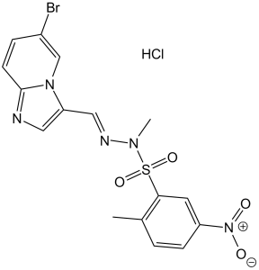
| Chemical Name | N-[(E)-(6-bromoimidazo[1,2-a]pyridin-3-yl)methylideneamino]-N,2-dimethyl-5-nitrobenzenesulfonamide;hydrochloride |
| Synonyms | PIK-75 hydrochloride; PIK-75 HCl; PIK75 HCl; PIK 75 HCl |
| HS Tariff Code | 2934.99.9001 |
| Storage |
Powder-20°C 3 years 4°C 2 years In solvent -80°C 6 months -20°C 1 month Note: Please store this product in a sealed and protected environment, avoid exposure to moisture. |
| Shipping Condition | Room temperature (This product is stable at ambient temperature for a few days during ordinary shipping and time spent in Customs) |
Biological Activity
| Targets |
DNA-PK (IC50 = 2 nM); p110α (IC50 = 5.8 nM); p110γ (IC50 = 76 nM); p110δ (IC50 = 510 nM); p110β (IC50 = 1.3 μM); hsVPS34 (IC50 = 2.6 μM); PI3KC2β (IC50 = 1 μM); PI3KC2α (IC50 = 10 μM); mTORC1 (IC50 = 1 μM); mTORC2 (IC50 = 10 μM); ATM (IC50 = 2.3 μM); ATR (IC50 = 21 μM); PI4KIIIβ (IC50 = 50 μM) 1. Phosphatidylinositol 3-Kinase α (PI3Kα) - IC50 ~5 nM (recombinant human PI3Kα, HTRF kinase activity assay); - Ki ~3.2 nM (recombinant human PI3Kα, ATP-competitive binding assay)[1] 2. High selectivity over other PI3K subtypes: - IC50 > 1000 nM (PI3Kβ), >800 nM (PI3Kγ), >1500 nM (PI3Kδ) (same HTRF assay as PI3Kα)[1] 3. Weak inhibition of mTOR (off-target): IC50 ~200 nM (recombinant human mTOR, radioactive kinase assay)[5] |
| ln Vitro |
PIK-75 shows the impressive potency and isoform selectivity at p110α while the corresponding IC50 values are 1300 nM, 76 nM and 510 nM for other PI3K isoforms, p110β, -γ, and -δ, respectively. Furthermore, when binding to purified p110α, Additionally, PIK-75 is a competitive inhibitor of the substrate PI with a Ki of 2.3 nM and a noncompetitive inhibitor of ATP when binding to purified p110. [1] DNA-PK is effectively inhibited by PIK-75 as well. PIK-75 (1 μM) reduces cell survival by significantly decreasing mitochondrial activity in unstimulated nonasthmatic airway smooth muscle (ASM) cells, asthmatic ASM cells, and lung fibroblasts. While in TGFβ stimulated ASM cells, PIK75 has no effects on non-asthmatic cells and only reduces mitochondrial activity in asthmatic cells. [2] According to a recent study, PIK-75 (10 nM) significantly reduces TNF-α-induced ADP-ribosyl cyclase activity and TNF-α-induced CD38 mRNA expression in human airway smooth muscle cells.[3] 1. Tumor cell antiproliferation and signaling inhibition (Literature [1]): - MCF-7 (breast cancer, PI3Kα-activated): PIK-75 HCl (1-100 nM) dose-dependently inhibited proliferation. 72-hour MTT IC50 ~12 nM; 50 nM reduced colony formation by ~85% (14-day assay). - Western blot: 20 nM PIK-75 HCl reduced p-AKT (Ser473) by ~90%, p-S6 (Ser235/236) by ~85% at 24 hours; no effect on p-ERK (MAPK pathway). - Primary human breast cancer cells (PIK3CA-mutant): 50 nM inhibited proliferation by ~70% (³H-thymidine incorporation)[1] 2. Cardiomyocyte survival regulation (Literature [2]): - Rat neonatal cardiomyocytes: Hypoxia-induced cell death (48 hours) was reduced by PIK-75 HCl (10-50 nM). 50 nM increased viability by ~60% (MTT); reduced caspase-3 activity by ~55% (luminescent assay). - Signaling: 20 nM PIK-75 HCl reduced hypoxia-induced p-AKT (Thr308) by ~70% (Western blot), blocking pro-survival AKT activation[2] 3. Airway epithelial cell inflammation modulation (Literature [3]): - Human bronchial epithelial cells (HBECs): PIK-75 HCl (5-50 nM) inhibited TNF-α-induced IL-8 secretion. 50 nM reduced IL-8 by ~70% (ELISA) at 24 hours; reduced NF-κB p65 nuclear translocation by ~65% (immunofluorescence). - Cell viability: >90% survival at 100 nM (trypan blue exclusion)[3] 4. Glioblastoma (GBM) cell inhibition (Literature [4]): - U87MG (GBM, PTEN-deficient): 72-hour SRB IC50 ~15 nM; 50 nM induced apoptosis in ~45% of cells (Annexin V-FITC staining). - Orthotopic GBM cell migration: 20 nM PIK-75 HCl reduced migration by ~60% (transwell assay); reduced MMP-9 expression by ~50% (qPCR)[4] 5. PI3Kα-mTOR signaling crosstalk (Literature [5]): - HEK293 cells overexpressing PI3Kα: 10 nM PIK-75 HCl reduced p-AKT by ~85%, 100 nM reduced p-mTOR (Ser2448) by ~40% (Western blot). - Recombinant mTOR inhibition: 200 nM PIK-75 HCl inhibited mTOR kinase activity by ~50% (radioactive assay)[5] [1][2][3][4][5] |
| ln Vivo |
In the ErbB3WT tumor model, PIK-75 lowers pAkt levels by 40% and lessens the in vitro chemotactic response to HRGβ1. Additionally, PIK-75 significantly lowers in vivo invasion and tumor cell motility in ErbB3WT primary tumors. [4] PIK-75 significantly impairs the insulin tolerance test (ITT), glucose tolerance test (GTT), and increases glucose production during the pyruvate tolerance test (PTT) in CD1 male mice.
PIK-75 potentiates anticancer activity of gemcitabine in vivo [3] The effect of PIK-75/gemcitabine combination was further demonstrated by in vivo mouse xenograft model. Mice bearing tumors of MIA PaCa-2 were administered with gemcitabine (20 mg/kg), PIK-75 (2 mg/kg), or combination of both drugs. Since PIK-75 is a reversible inhibitor, PIK-75 was administered 5 times per week to ensure maintaining sufficient inhibitory effects. Gemcitabine was administered twice per week. As shown in Fig. 7A, gemcitabine or PIK-75 reduced the tumor growth to similar degree. Beneficial effect of PIK-75/gemcitabine was evident as this combination markedly reduced the tumor growth in vivo without affecting the body weights of mice (Fig. 7B). 1. U87MG GBM xenograft model (Literature [4]): - Animals: Female nude mice (6-8 weeks old) with subcutaneous U87MG tumors (~100 mm³). - Administration: PIK-75 HCl dissolved in 10% DMSO + 90% PEG400, intraperitoneal (i.p.) injection 10, 20 mg/kg every other day for 21 days. - Efficacy: 20 mg/kg reduced tumor volume by ~75% (vs. vehicle); tumor weight reduced by ~70% at day 21; median survival extended from 38 days (vehicle) to 56 days (p < 0.01). Tumor p-AKT reduced by ~65% (IHC)[4] 2. Mouse allergic asthma model (Literature [3]): - Animals: Male BALB/c mice (8-10 weeks old) sensitized with OVA (ovalbumin) to induce asthma. - Administration: PIK-75 HCl dissolved in 0.5% methylcellulose + 0.1% Tween 80, oral gavage 5, 10 mg/kg/day for 7 days (during OVA challenge). - Efficacy: 10 mg/kg reduced BALF (bronchoalveolar lavage fluid) eosinophil count by ~60%; reduced lung IL-5/IL-13 levels by ~55% (ELISA); lung inflammation score improved by ~40% (histology)[3] |
| Enzyme Assay |
The PI3K inhibitor PIK-75 is dissolved at 10 mM in dimethyl sulfoxide and stored at −20°C until use. The PI3K enzyme's activity is assessed in 50 μL of 20 mM HEPES, pH 7.5, 5 mM MgCl2, 180 μM phosphatidyl inositol, and 2.5 μCi of [γ-32P]ATP. The reaction is sparked by the addition of 100 μM ATP. The enzyme reaction is stopped by adding 50 μL of 1 M HCl after it has been incubated at room temperature for 30 minutes. Then, phospholipids are extracted using 250 μL of 2 M KCl and 100 ml of a 1:1 chloroform/methanol mixture to extract the phospholipids for liquid scintillation counting. Using Prism version 5.00 for Windows, the concentration versus inhibition of enzyme activity curve is created by diluting inhibitors in 20% (v/v) dimethyl sulfoxide. The IC50 is then calculated using this analysis. An assay that detects ATP consumption is used for kinetic analysis. PI and ATP are used at various concentrations to measure the activity of the PI3K enzyme in 50 L of 20 mM HEPES, pH 7.5, and 5 mM MgCl2. After a 60-minute incubation at room temperature, the reaction is stopped by adding 50 μL of Kinase-Glo, followed by an additional 15 minutes of incubation. The Fluostar plate reader is then used to read the luminescence. Prism is used to analyze the outcomes. 1. PI3Kα kinase activity assay (HTRF, Literature [1]): - Reagent preparation: Recombinant human PI3Kα (catalytic subunit p110α + regulatory subunit p85α) resuspended in assay buffer (50 mM Tris-HCl pH 7.5, 10 mM MgCl₂, 1 mM DTT, 0.01% Tween 20). - Reaction system: 50 μL mixture contained 5 nM PI3Kα, 10 μM PIP₂ (substrate), 2 μM ATP, and serial PIK-75 HCl (0.1-100 nM). Incubated at 30℃ for 60 minutes. - Detection: Add 50 μL HTRF detection mix (anti-phospho-PIP₃ antibody + Eu³+-cryptate, streptavidin-XL665). Incubate 30 minutes at RT. Measure fluorescence (excitation 337 nm, emission 620 nm/665 nm). Inhibition rate = (1 - (665/620 ratio)drug/(665/620 ratio)vehicle) × 100%. IC50 derived via nonlinear regression[1] 2. mTOR kinase activity assay (radioactive, Literature [5]): - Reagent preparation: Recombinant human mTOR (full-length) resuspended in assay buffer (25 mM HEPES pH 7.4, 10 mM MgCl₂, 1 mM EGTA, 1 mM DTT). - Reaction system: 25 μL mixture contained 10 nM mTOR, 1 μg 4E-BP1 (substrate), 1 μCi [γ-³²P]-ATP, and serial PIK-75 HCl (10-500 nM). Incubated at 37℃ for 45 minutes. - Detection: Reaction terminated by 5×SDS loading buffer. Proteins separated by SDS-PAGE, transferred to PVDF membrane. Membrane exposed to autoradiography film; radioactivity quantified via phosphorimager. IC50 calculated via dose-response curve[5] [1][5] |
| Cell Assay |
Mitochondrial activity is measured using the 3-(4,5-dimethylthiazol-2-yl)-2,5-diphenyltetrazolium (MTT) assay after stimulation with TGF with or without inhibitors for 48 hours. Before serial dilution (1:2) in duplicates, harvested washed cells are resuspended in DMEM-lO% FCS and aliquoted (500 μL) into 24-well cluster plates. Immediately after dilution, 100 μL of an appropriate MTT concentration (dissolved in PBS and filtered through a 0.2 μm filter before use to remove any blue formazan product) is added to each well. The cells are then incubated for 3.5 hours at 37 °C. 500 μL of 10% sodium dodecyl sulfate (SDS) in 0.01 M HCl are added to each well to dissolve the resulting blue formazan product over the course of 16 hours at 37°C. In a 96-well microplate, a sample (150 μL) from each duplicate well is transferred, and the optical density is calculated using automated spectrophotometry in comparison to a reagent blank (no cells).A reference wavelength of 690 nm and a test wavelength of 570 nm are used to measure absorbance. Results from three to six wells from each treatment are averaged for each primary cell culture, and data are expressed as absorbance 570 to 690 nm. 1. Tumor cell proliferation assay (MTT/SRB, Literatures [1], [4]): - Literature [1] (MCF-7): - Cell culture: MCF-7 cells maintained in RPMI 1640 + 10% FBS, seeded in 96-well plates (5×10³ cells/well) overnight. - Treatment: Incubated with PIK-75 HCl (0.1-100 nM) for 72 hours; vehicle (0.1% DMSO) as control. - Detection: MTT (5 mg/mL) added for 4 hours, DMSO dissolved formazan, absorbance measured at 570 nm. IC50 calculated via nonlinear regression[1] - Literature [4] (U87MG): - Cell culture: U87MG cells seeded in 96-well plates (4×10³ cells/well) overnight. - Treatment: Incubated with PIK-75 HCl (0.1-100 nM) for 72 hours. - Detection: Cells fixed with 10% trichloroacetic acid, stained with 0.4% SRB. SRB dissolved in 10 mM Tris base; absorbance measured at 540 nm[4] 2. Asthma-related epithelial cell assay (Literature [3]): - Cell culture: HBECs cultured in bronchial epithelial growth medium, seeded in 24-well plates (1×10⁵ cells/well) overnight. - Treatment: Pre-incubated with PIK-75 HCl (5-50 nM) for 1 hour, then stimulated with TNF-α (10 ng/mL) for 24 hours. - Detection: Supernatant collected, IL-8 concentration measured via ELISA; NF-κB p65 localization detected by immunofluorescence (primary antibody against p65, DAPI nuclear staining)[3] 3. Signaling Western blot assay (Literatures [1], [5]): - Cell culture: MCF-7 (Literature [1])/HEK293-PI3Kα (Literature [5]) seeded in 6-well plates (2×10⁵ cells/well) overnight. - Treatment: Incubated with PIK-75 HCl (1-100 nM) for 24 hours; MCF-7 cells stimulated with insulin (100 nM) for 30 minutes before lysis. - Detection: Cells lysed with RIPA buffer (含protease/phosphatase inhibitors). Proteins separated by SDS-PAGE, transferred to PVDF membrane, probed with antibodies against p-AKT (Ser473), total AKT, p-S6, and GAPDH (loading control)[1] [5][1][3][4][5] |
| Animal Protocol |
MTLn3 cells are injected into the right fourth mammary fat pad from the head of female severe-combined immunodeficient/NCr mice. ≤1 μM Administered via i.p. Tumor xenograft study [3] MIA PaCa-2 cells (∼1.7×10⁶ cells/mouse) mixed with Matrigel were injected subcutaneously into the flank of male athymic nude (Foxn1nu) mice aged 6-weeks. Gemcitabine (50 mg/ml) was dissolved in PBS and PIK-75 (20 mg/ml) was dissolved in DMSO. Injection solution was made as 10% of Cremophor® EL and 3% of poly(ethylene glycol) 400 in sterile water. Before administration of compounds, gemcitabine was further diluted in PBS and DMSO or PIK-75 was further diluted in the injection solution and sterilized by 0.2 μm filter unit. These diluents were mixed with 1:1 ratio and administered into peritoneal cavity of the mouse. Gemcitabine (20 mg/kg) or gemcitabine (20 mg/kg)/PIK-75 (2 mg/kg) combination was administered twice per week and vehicle control and PIK-75 (2 mg/kg) were administered 5 times per week. The body weights and tumor sizes were measured 3 times per week. Tumor volumes were calculated as width (mm) × length (mm) × height (mm)/2. 1. U87MG GBM xenograft protocol (Literature [4]): - Animals: Female nude mice (6-8 weeks old), 5 mice/group; acclimated 7 days (12h light/dark, ad libitum food/water). - Tumor induction: 5×10⁶ U87MG cells injected subcutaneously (right flank). - Drug preparation: PIK-75 HCl dissolved in 10% DMSO + 90% PEG400 (sonicated 5 minutes for dissolution). - Administration: Intraperitoneal injection 10/20 mg/kg every other day (10 μL/g body weight), starting when tumors reached ~100 mm³ (volume = length×width²/2). - Assessment: Tumor volume measured twice weekly; body weight weekly; mice euthanized at day 21, tumor lysed for p-AKT IHC; survival monitored daily[4] 2. OVA-induced asthma protocol (Literature [3]): - Animals: Male BALB/c mice (8-10 weeks old), 6 mice/group. - Sensitization: Day 0/7, intraperitoneal injection of OVA (10 μg) + aluminum hydroxide (2 mg). - Drug preparation: PIK-75 HCl dissolved in 0.5% methylcellulose + 0.1% Tween 80 (stirred 2 hours at RT). - Administration: Day 14-20 (OVA challenge period), oral gavage 5/10 mg/kg/day (10 μL/g body weight); control group received vehicle. - Assessment: Day 21, collect BALF to count eosinophils (flow cytometry); measure lung IL-5/IL-13 via ELISA; lung tissue stained with H&E for inflammation scoring[3] |
| Toxicity/Toxicokinetics |
1. In vitro toxicity:
- Tumor cells (MCF-7, U87MG), HBECs, cardiomyocytes: PIK-75 HCl concentrations up to 1 μM showed no non-specific cytotoxicity (LDH release <10%); trypan blue exclusion showed >90% viability after 72-hour exposure[1] [2][3][4] 2. In vivo toxicity (Literatures [3], [4]): - GBM xenograft mice (20 mg/kg i.p., 21 days): No mortality; body weight maintained >90% of initial; serum ALT/AST (liver) and creatinine (kidney) within normal range[4] - Asthma model mice (10 mg/kg oral, 7 days): No abnormal behavior (ataxia, lethargy); lung/kidney/liver histology showed no drug-induced damage[3] |
| References |
[1]. Mol Pharmacol. 2011 Oct;80(4):657-64. [2]. J Pharmacol Exp Ther. 2011 May;337(2):557-66. [3]. Am J Respir Cell Mol Biol. 2012 Oct;47(4):427-35. [4]. Oncogene. 2012 Feb 9;31(6):706-15. [5]. Biochem J. 2012 Feb 15;442(1):161-9. |
| Additional Infomation |
The combination of molecular modeling and X-ray crystallography has failed to yield a consensus model of the mechanism for selective binding of inhibitors to the phosphoinositide 3-kinase (PI3K) p110 α-isoform. Here we have used kinetic analysis to determine that the p110α-selective inhibitor 2-methyl-5-nitro-2-[(6-bromoimidazo[1,2-α]pyridin-3-yl)methylene]-1-methylhydrazide-benzenesulfonic acid (PIK-75) is a competitive inhibitor with respect to a substrate, phosphatidylinositol (PI) in contrast to most other PI3K inhibitors, which bind at or near the ATP site. Using sequence analysis and the existing crystal structures of inhibitor complexes with the p110γ and -δ isoforms, we have identified a new region of nonconserved amino acids (region 2) that was postulated to be involved in PIK-75 p110α selectivity. Analysis of region 2, using in vitro mutation of identified nonconserved amino acids to alanine, showed that Ser773 was a critical amino acid involved in PIK-75 binding, with an 8-fold-increase in the IC(50) compared with wild-type. Kinetic analysis showed that, with respect to PI, the PIK-75 K(i) for the isoform mutant S773D increased 64-fold compared with wild-type enzyme. In addition, a nonconserved amino acid, His855, from the previously identified region 1 of nonconserved amino acids, was found to be involved in PIK-75 binding. These results show that these two regions of nonconserved amino acids that are close to the substrate binding site could be targeted to produce p110α isoform-selective inhibitors.[1] \n\nThe phosphatidylinositol 3-kinase (PI3K) signal transduction pathway is implicated in the airway remodeling associated with asthma. The class IA PI3K isoforms are known to be activated by growth factors and cytokines. Because this pathway is a possible site of pharmacological intervention for treating the disease, it is important to know which isoforms contribute to this process. Therefore, we used a pharmacological approach to investigate the roles of the three class IA PI3K isoforms (p110α, p110β, and p110δ) in airway remodeling using airway smooth muscle (ASM) cells derived from asthmatic subjects and ASM cells and lung fibroblasts from nonasthmatic subjects. These studies used the inhibitors N'-[(E)-(6-bromoimidazo[1,2-a]pyridin-3-yl)methylidene]-N,2-dimethyl-5-nitrobenzenesulfonohydrazide (PIK75) (which selectively inhibits p110α), 7-methyl-2-(4-morpholinyl)-9-[1-(phenylamino)ethyl]-4H-pyrido[1,2-a]pyrimidin-4-one (TGX221) (which selectively inhibits p110β), and 2-[(6-amino-9H-purin-9-yl)methyl]-5-methyl-3-(2-methylphenyl)-4(3H)-quinazolinone (IC87114) (which selectively inhibits p110δ). Cells were stimulated with transforming growth factor-β (TGFβ) and/or 10% fetal bovine serum in the presence or absence of inhibitor or vehicle control (dimethyl sulfoxide). PIK75, but not TGX221 or IC87114, attenuated TGFβ-induced fibronectin deposition in all cell types tested. PIK75 and TGX221 each decreased secretion of vascular endothelial growth factor and interleukin-6 in nonasthmatic ASM cells and lung fibroblasts, whereas TGX221 was not as effective in asthmatic ASM cells. In addition, PIK75 decreased cell survival in TGFβ-stimulated asthmatic, but not nonasthmatic, ASM cells. In conclusion, specific PI3K isoforms may play a role in pathophysiological events relevant to airway wall remodeling.[2] \n\nThe ADP-ribosyl cyclase activity of CD38 generates cyclic ADP-ribose, a Ca(2+)-mobilizing agent. In human airway smooth muscle (HASM) cells, TNF-α mediates CD38 expression through mitogen-activated protein kinases and NF-κB and AP-1. The phosphatidylinositol-3 kinase/Akt (PI3K/Akt) pathway is involved in TNF-α signaling and contributes to airway hyperresponsiveness and airway remodeling. We hypothesized that PI3Ks mediate CD38 expression and are involved in the differential induction of CD38 by TNF-α in asthmatic HASM cells. HASM cells were treated with pan-PI3K inhibitors (LY294002 or wortmannin) or class I-selective (GDC0941) or isoform-selective PI3K inhibitors (p110α-PIK-75 and p110β-TGX-221) with or without TNF-α. HASM cells were transfected with a catalytically active form of PI3K or phosphatase and tensin homolog (PTEN) or nontargeting or p110 isoform-targeting siRNAs before TNF-α exposure. CD38 expression and activation of Akt, NF-κB, and AP-1 were determined. LY294002 and wortmannin inhibited TNF-α-induced Akt activation, whereas only LY294002 inhibited CD38 expression. P110 expression caused Akt activation and basal and TNF-α-induced CD38 expression, whereas PTEN expression attenuated Akt activation and CD38 expression. Expression levels of p110 isoforms α, β, and δ were comparable in nonasthmatic and asthmatic HASM cells. Silencing of p110α or -δ, but not p110β, resulted in comparable attenuation of TNF-α-induced CD38 expression in asthmatic and nonasthmatic cells. NF-κB and AP-1 activation were unaltered by the PI3K inhibitors. In HASM cells, regulation of CD38 expression occurs by specific class I PI3K isoforms, independent of NF-κB or AP-1 activation, and PI3K signaling may not be involved in the differential elevation of CD38 in asthmatic HASM cells.[3] 1. Mechanism of action: PIK-75 HCl binds to the ATP-binding pocket of PI3Kα, blocking its catalytic activity and inhibiting PIP₂ phosphorylation to PIP₃. This suppresses downstream AKT-S6 signaling, inhibiting tumor cell proliferation/migration and reducing inflammation (via NF-κB inhibition in epithelial cells). It weakly inhibits mTOR at high concentrations (>100 nM), contributing to anti-tumor effects[1] [3][4][5] 2. Preclinical significance: - Literature [1]/[4]: Establishes PIK-75 HCl as a tool for PI3Kα-driven tumors (breast cancer, GBM), especially PTEN-deficient/PIK3CA-mutant subtypes. [1][4] - Literature [3]: Demonstrates potential in inflammatory airway diseases (asthma) by targeting PI3Kα-mediated IL-8/NF-κB signaling. [3] - Literature [2]: Suggests role in regulating cardiomyocyte survival, providing insights into PI3Kα in cardiac pathophysiology. [2] |
Solubility Data
| Solubility (In Vitro) |
|
|||
| Solubility (In Vivo) |
Solubility in Formulation 1: ≥ 1.1 mg/mL (2.25 mM) (saturation unknown) in 10% DMSO + 40% PEG300 + 5% Tween80 + 45% Saline (add these co-solvents sequentially from left to right, and one by one), clear solution. For example, if 1 mL of working solution is to be prepared, you can add 100 μL of 11.0 mg/mL clear DMSO stock solution to 400 μL PEG300 and mix evenly; then add 50 μL Tween-80 to the above solution and mix evenly; then add 450 μL normal saline to adjust the volume to 1 mL. Preparation of saline: Dissolve 0.9 g of sodium chloride in 100 mL ddH₂ O to obtain a clear solution. Solubility in Formulation 2: ≥ 1.1 mg/mL (2.25 mM) (saturation unknown) in 10% DMSO + 90% (20% SBE-β-CD in Saline) (add these co-solvents sequentially from left to right, and one by one), clear solution. For example, if 1 mL of working solution is to be prepared, you can add 100 μL of 11.0 mg/mL clear DMSO stock solution to 900 μL of 20% SBE-β-CD physiological saline solution and mix evenly. Preparation of 20% SBE-β-CD in Saline (4°C,1 week): Dissolve 2 g SBE-β-CD in 10 mL saline to obtain a clear solution. Solubility in Formulation 3: ≥ 1.1 mg/mL (2.25 mM) (saturation unknown) in 10% DMSO + 90% Corn Oil (add these co-solvents sequentially from left to right, and one by one), clear solution. For example, if 1 mL of working solution is to be prepared, you can add 100 μL of 11.0 mg/mL clear DMSO stock solution to 900 μL of corn oil and mix evenly. Solubility in Formulation 4: PIK-75 HCl; PIK75 HCl; PIK 75 HCl. (Please use freshly prepared in vivo formulations for optimal results.) |
| Preparing Stock Solutions | 1 mg | 5 mg | 10 mg | |
| 1 mM | 2.0461 mL | 10.2304 mL | 20.4608 mL | |
| 5 mM | 0.4092 mL | 2.0461 mL | 4.0922 mL | |
| 10 mM | 0.2046 mL | 1.0230 mL | 2.0461 mL |