Physicochemical Properties
| Molecular Formula | C16H16F2N2O4S2 |
| Molecular Weight | 402.43604850769 |
| Exact Mass | 402.052 |
| CAS # | 141286-78-4 |
| PubChem CID | 6603828 |
| Appearance | White to off-white solid powder |
| LogP | 4.071 |
| Hydrogen Bond Donor Count | 2 |
| Hydrogen Bond Acceptor Count | 8 |
| Rotatable Bond Count | 9 |
| Heavy Atom Count | 26 |
| Complexity | 541 |
| Defined Atom Stereocenter Count | 0 |
| InChi Key | GTACSIONMHMRPD-UHFFFAOYSA-N |
| InChi Code | InChI=1S/C16H16F2N2O4S2/c17-13-8-11(9-14(18)16(13)24-10-15(19)21)25-7-6-20-26(22,23)12-4-2-1-3-5-12/h1-5,8-9,20H,6-7,10H2,(H2,19,21) |
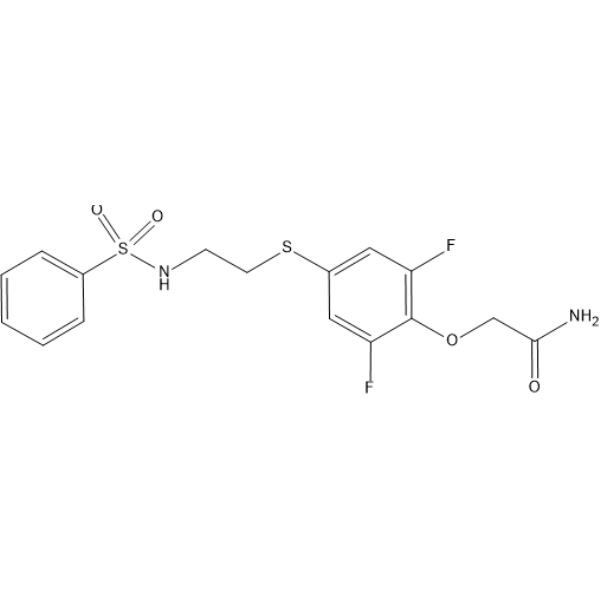
| Chemical Name | 2-[4-[2-(benzenesulfonamido)ethylsulfanyl]-2,6-difluorophenoxy]acetamide |
| HS Tariff Code | 2934.99.9001 |
| Storage |
Powder-20°C 3 years 4°C 2 years In solvent -80°C 6 months -20°C 1 month |
| Shipping Condition | Room temperature (This product is stable at ambient temperature for a few days during ordinary shipping and time spent in Customs) |
Biological Activity
| Targets |
Allosteric potentiator of AMPA receptors, preferentially acting on flop splice variants [1] |
| ln Vitro |
In rat hippocampal cultures, PEPA (100 µM) applied alone did not elicit an increase in intracellular free calcium ion concentration in any of the cells tested [1] PEPA (25 µM) potentiated the AMPA (1 µM)-induced increase in the 340/380 fluorescence ratio (indicative of increased intracellular calcium) in a small number of hippocampal cells. This potentiation was observed in a much larger number of cells when 150 µM PEPA was applied [1] In rat hippocampal cultures, PEPA (100 µM) potentiated the AMPA (10 µM)-induced increase in intracellular free calcium concentration ([Ca²⁺]i), with the potentiation factor varying between 1.0-fold (no potentiation) and 27.7-fold across different cells (n=80) [1] The ratio of potentiation by PEPA (100 µM) to that by cyclothiazide (100 µM) (P/C ratio) for the AMPA (10 µM) response in hippocampal cells varied between 0 and 2.15, revealing cell-to-cell heterogeneity. Populations were identified with low P/C ratios (<0.15; 34% of cells) and high P/C ratios (≥2.00; 1% of cells) [1] In Xenopus oocytes expressing various AMPA receptor subunit combinations, PEPA (100 µM) potentiated the AMPA (10 µM)-induced currents. The potentiation varied with subunit and splice-variant composition. For example, potentiation was 1.90-fold in oocytes expressing GluR1-flip alone, and 2.20-fold in oocytes expressing GluR1-flop alone. In oocytes expressing GluR1-flip and GluR2-flip (1:4 ratio), potentiation was 6.41-fold [1] The P/C ratio in Xenopus oocytes also varied with subunit composition. Oocytes expressing predominantly flip variants (e.g., GluR1-flip + GluR2-flip, 1:4) exhibited low P/C ratios (0.19), while those expressing predominantly flop variants (e.g., GluR1-flop alone) exhibited high P/C ratios (2.20) [1] |
| Cell Assay |
For intracellular calcium imaging in hippocampal cultures, cells (8 days in vitro) were loaded with 10 µM Fura-2 AM for 30-40 minutes at 37°C. Cells were perfused with a control medium containing tetrodotoxin. PEPA was initially dissolved in DMSO at 100 mM and then diluted into the control medium to the final working concentration. Cells were examined under epifluorescence illumination with alternating 340 nm and 380 nm excitation. The ratio of fluorescence at these wavelengths (340/380 ratio) was monitored and used as an indicator of intracellular calcium concentration. AMPA and PEPA were applied via perfusion [1] For two-electrode voltage clamp recordings in Xenopus oocytes, cRNAs encoding specific AMPA receptor subunits (e.g., GluR1, GluR2, GluR3 flip/flop variants) were injected into oocytes. After incubation for several days, oocytes were voltage-clamped at -100 mV and perfused with frog Ringer's solution. AMPA, PEPA, and cyclothiazide were applied via perfusion. The current responses were recorded, and potentiation was calculated as the ratio of the current amplitude induced by (AMPA + potentiator) to the current amplitude induced by AMPA alone [1] |
| References |
[1]. Pharmacological detection of AMPA receptor heterogeneity by use of two allosteric potentiators in rat hippocampal cultures. Br J Pharmacol. 1998 Apr;123(7):1294-303. [2]. Molecular mechanism of flop selectivity and subsite recognition for an AMPA receptor allosteric modulator: structures of GluA2 and GluA3 in complexes with PEPA. Biochemistry. 2010 Apr 6;49(13):2843-50. [3]. Facilitating actions of an AMPA receptor potentiator upon extinction of contextually conditioned fear response in stressed mice. Neurosci Lett. 2011 Jan 25;488(3):242-6. [4]. Positive modulation of AMPA receptors prevents downregulation of GluR2 expression and activates the Lyn-ERK1/2-CREB signaling in rat brain ischemia. Hippocampus. 2010 Jan;20(1):65-77. |
| Additional Infomation |
PEPA (4-[2-(phenylsulphonylamino)ethylthio]-2,6-difluoro-phenoxyacetamide) is a novel allosteric potentiator of AMPA receptors that suppresses receptor desensitization [1] PEPA acts preferentially on flop splice variants of AMPA receptors and is about 100 times more potent than aniracetam, another known potentiator with flop preference [1] PEPA is also a more potent suppressor of desensitization for receptors containing GluR3 and GluR4 subunits compared to those containing GluR1 [1] Comparing the potentiating effects of PEPA and cyclothiazide (which preferentially acts on flip variants) via the P/C ratio facilitates the detection of cell-to-cell heterogeneity in functional AMPA receptor composition in hippocampal cultures [1] |
Solubility Data
| Solubility (In Vitro) | DMSO : ~50 mg/mL (~124.24 mM) |
| Solubility (In Vivo) |
Solubility in Formulation 1: ≥ 2.5 mg/mL (6.21 mM) (saturation unknown) in 10% DMSO + 40% PEG300 + 5% Tween80 + 45% Saline (add these co-solvents sequentially from left to right, and one by one), clear solution. For example, if 1 mL of working solution is to be prepared, you can add 100 μL of 25.0 mg/mL clear DMSO stock solution to 400 μL PEG300 and mix evenly; then add 50 μL Tween-80 to the above solution and mix evenly; then add 450 μL normal saline to adjust the volume to 1 mL. Preparation of saline: Dissolve 0.9 g of sodium chloride in 100 mL ddH₂ O to obtain a clear solution. Solubility in Formulation 2: ≥ 2.5 mg/mL (6.21 mM) (saturation unknown) in 10% DMSO + 90% (20% SBE-β-CD in Saline) (add these co-solvents sequentially from left to right, and one by one), clear solution. For example, if 1 mL of working solution is to be prepared, you can add 100 μL of 25.0 mg/mL clear DMSO stock solution to 900 μL of 20% SBE-β-CD physiological saline solution and mix evenly. Preparation of 20% SBE-β-CD in Saline (4°C,1 week): Dissolve 2 g SBE-β-CD in 10 mL saline to obtain a clear solution. Solubility in Formulation 3: ≥ 2.5 mg/mL (6.21 mM) (saturation unknown) in 10% DMSO + 90% Corn Oil (add these co-solvents sequentially from left to right, and one by one), clear solution. For example, if 1 mL of working solution is to be prepared, you can add 100 μL of 25.0 mg/mL clear DMSO stock solution to 900 μL of corn oil and mix evenly. (Please use freshly prepared in vivo formulations for optimal results.) |
| Preparing Stock Solutions | 1 mg | 5 mg | 10 mg | |
| 1 mM | 2.4848 mL | 12.4242 mL | 24.8484 mL | |
| 5 mM | 0.4970 mL | 2.4848 mL | 4.9697 mL | |
| 10 mM | 0.2485 mL | 1.2424 mL | 2.4848 mL |