Physicochemical Properties
| Molecular Formula | C22H18CLF2N3O2S |
| Molecular Weight | 461.912029743195 |
| Exact Mass | 461.077 |
| Elemental Analysis | C, 57.21; H, 3.93; Cl, 7.67; F, 8.23; N, 9.10; O, 6.93; S, 6.94 |
| CAS # | 1359983-15-5 |
| PubChem CID | 66765263 |
| Appearance | Typically exists as solid at room temperature |
| LogP | 5.6 |
| Hydrogen Bond Donor Count | 1 |
| Hydrogen Bond Acceptor Count | 6 |
| Rotatable Bond Count | 6 |
| Heavy Atom Count | 31 |
| Complexity | 650 |
| Defined Atom Stereocenter Count | 0 |
| InChi Key | QNKMXCMJVQFBLM-UHFFFAOYSA-N |
| InChi Code | InChI=1S/C22H18ClF2N3O2S/c1-3-9-27-11-13(10-26-27)28-12(2)21(15-7-8-16(23)19(25)20(15)28)31-17-6-4-5-14(18(17)24)22(29)30/h4-8,10-11H,3,9H2,1-2H3,(H,29,30) |
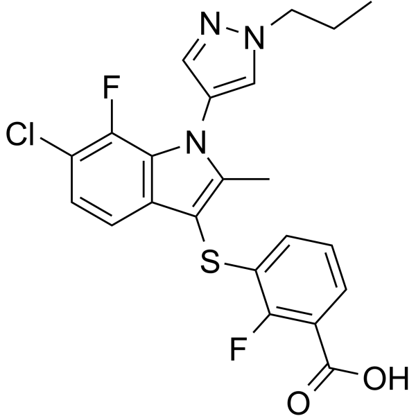
| Chemical Name | 3-[6-chloro-7-fluoro-2-methyl-1-(1-propylpyrazol-4-yl)indol-3-yl]sulfanyl-2-fluorobenzoic acid |
| Synonyms | PAT048; PAT 048; PAT-048; 1359983-15-5; 3-[6-chloro-7-fluoro-2-methyl-1-(1-propylpyrazol-4-yl)indol-3-yl]sulfanyl-2-fluorobenzoic acid; SCHEMBL671444; PAT-048 |
| HS Tariff Code | 2934.99.9001 |
| Storage |
Powder-20°C 3 years 4°C 2 years In solvent -80°C 6 months -20°C 1 month |
| Shipping Condition | Room temperature (This product is stable at ambient temperature for a few days during ordinary shipping and time spent in Customs) |
Biological Activity
| Targets | Autotaxin |
| ln Vitro | PAT-048 is a type III Autotaxin inhibitor that has inhibits autotaxin with IC50 and IC90 values of 20 nM and 200 nM, respectively. |
| Enzyme Assay |
Biochemical Assay with FS-3 Substrate[2] Starting from 20 μM highest concentration, 10 μL of a dilution series of compound, 1/5 dilution, was added to the wells. Glycosylated human ATX protein (see Supporting Information) was used at a final concentration of 0.4 or 0.64 μg/mL. The enzyme was diluted in 50 mM Tris-HCl (2-amino-2-(hydroxymethyl)-1,3-propanediol hydrochloride) pH 8.0, 250 mM NaCl, 5 mM KCl, 1 mM MgCl2, 1 mM CaCl2, and 0.1% fatty acid free BSA in a total volume of 20 μL. The enzyme mixture was added to compounds, and the resulting mixture was incubated for 30 min at room temperature under shaking. The reaction was started by the addition of 20 μL of 0.75 μM FS-3 diluted in the same buffer as described above. Fluorescence was read on an Envision apparatus after 30 min incubation at room temperature (excitation 485 nm, emission 520 nM).[2] Biochemical Assay with LPC 16:0 Substrate[2] Starting from 20 μM highest concentration, 5 μL of a dilution series of compound (1/5 dilution) was added to the wells. Glycosylated human ATX protein (see Supporting Information) was used at a final concentration of 1 or 3 μg/mL. The enzyme was diluted in 50 mM Tris-HCl pH 8.5, 500 mM NaCl, 5 mM KCl, 10 mM CaCl2, and 0.1% fatty acid free BSA in a total volume of 10 μL. The reaction was started by the addition of 10 μL of 150 μM LPC 16:0 diluted in the same buffer as described above, and the mixture was incubated at 37 °C for 30 min. The reaction was terminated and choline quantified by the addition of a 25 μL mixture containing 0.6 U/mL of choline oxidase, 0.6 U/mL of horseradish peroxidase (HRP), 1.8 mM TOOS (N-ethyl-N-(2-hydroxy-3-sulfopropyl)-3-methylaniline, sodium salt dihydrate), 1.2 mM 4-aminoantipyrine, and 20 mM EGTA (ethylene glycol-bis(2-aminoethyl ether)-N,N,N′,N′-tetraacetic acid, stop-developer solution) diluted in the buffer described above. Luminescence was read on an Envision apparatus after 30 min of incubation at room temperature (excitation 555 nm, excitation light = 70%).[2] Rat Plasma Assay[2] Rat plasma was thawed on ice and added into a plate containing a dose range of compound to be tested. After 2 h of incubation at 37 °C, plasma proteins from a 10 μL aliquot were precipitated with an excess of methanol containing LPA 17:0 as internal standard. After centrifugation, the corresponding supernatant was diluted and injected on a C18 column. Analytes were eluted out of the column under isocratic conditions. No calibration curve was prepared for LPA 18:2, and all quantifications were performed based on peak area ratios (LPA 18:2/LPA 17:0). For each concentration of compound, LPA data were expressed as percentage of reduction (% reduction) using the formula: 100 – [((LPA ratio)/(LPA ratio in control sample)) × 100]. |
| References |
[1]. An Autotaxin/Lysophosphatidic Acid/Interleukin-6 Amplification Loop Drives Scleroderma Fibrosis. Arthritis Rheumatol. 2016 Dec;68(12):2964-2974. [2]. Autotaxin activity increases locally following lung injury, but is not required for pulmonary lysophosphatidic acid production or fibrosis. FASEB J. 2016 Jun;30(6):2435-50. |
| Additional Infomation |
Autotaxin (ATX) is a secreted glycoprotein that converts lysophosphatidylcholine (LPC) to the bioactive phospholipid lysophosphatidic acid (LPA) and is the major enzyme generating circulating LPA. Inhibition of LPA signaling has profound antifibrotic effects in multiple organ systems, including lung, kidney, skin, and peritoneum. However, other LPA-generating pathways exist, and the role of ATX in localized tissue LPA production and fibrosis remains unclear and controversial. In this study, we describe the preclinical pharmacologic, pharmacokinetic, and pharmacodynamic properties of a novel small-molecule ATX inhibitor, PAT-505 [3-((6-chloro-2-cyclopropyl-1-(1-ethyl-1H-pyrazol-4-yl)-7-fluoro-1H-indol-3-yl) thio)-2-fluorobenzoic acid sodium salt]. PAT-505 is a potent, selective, noncompetitive inhibitor that displays significant inhibition of ATX activity in plasma and liver tissue after oral administration. When dosed therapeutically in a Stelic Mouse Animal Model of nonalcoholic steatohepatitis (NASH), PAT-505 treatment resulted in a small but significant improvement in fibrosis with only minor improvements in hepatocellular ballooning and hepatic inflammation. In a choline-deficient, high-fat diet model of NASH, therapeutic treatment with PAT-505 robustly reduced liver fibrosis with no significant effect on steatosis, hepatocellular ballooning, or inflammation. These data demonstrate that inhibiting autotaxin is antifibrotic and may represent a novel therapeutic approach for the treatment of multiple fibrotic liver diseases, including NASH.[1]. Autotaxin (ATX) is a secreted enzyme playing a major role in the production of lysophosphatidic acid (LPA) in blood through hydrolysis of lysophosphatidyl choline (LPC). The ATX–LPA signaling axis arouses a high interest in the drug discovery industry as it has been implicated in several diseases including cancer, fibrotic diseases, and inflammation, among others. An imidazo[1,2-a]pyridine series of ATX inhibitors was identified out of a high-throughput screening (HTS). A cocrystal structure with one of these compounds and ATX revealed a novel binding mode with occupancy of the hydrophobic pocket and channel of ATX but no interaction with zinc ions of the catalytic site. Exploration of the structure–activity relationship led to compounds displaying high activity in biochemical and plasma assays, exemplified by compound 40. Compound 40 was also able to decrease the plasma LPA levels upon oral administration to rats.[2] |
Solubility Data
| Solubility (In Vitro) | May dissolve in DMSO (in most cases), if not, try other solvents such as H2O, Ethanol, or DMF with a minute amount of products to avoid loss of samples |
| Solubility (In Vivo) |
Note: Listed below are some common formulations that may be used to formulate products with low water solubility (e.g. < 1 mg/mL), you may test these formulations using a minute amount of products to avoid loss of samples. Injection Formulations (e.g. IP/IV/IM/SC) Injection Formulation 1: DMSO : Tween 80: Saline = 10 : 5 : 85 (i.e. 100 μL DMSO stock solution → 50 μL Tween 80 → 850 μL Saline) *Preparation of saline: Dissolve 0.9 g of sodium chloride in 100 mL ddH ₂ O to obtain a clear solution. Injection Formulation 2: DMSO : PEG300 :Tween 80 : Saline = 10 : 40 : 5 : 45 (i.e. 100 μL DMSO → 400 μLPEG300 → 50 μL Tween 80 → 450 μL Saline) Injection Formulation 3: DMSO : Corn oil = 10 : 90 (i.e. 100 μL DMSO → 900 μL Corn oil) Example: Take the Injection Formulation 3 (DMSO : Corn oil = 10 : 90) as an example, if 1 mL of 2.5 mg/mL working solution is to be prepared, you can take 100 μL 25 mg/mL DMSO stock solution and add to 900 μL corn oil, mix well to obtain a clear or suspension solution (2.5 mg/mL, ready for use in animals). Injection Formulation 4: DMSO : 20% SBE-β-CD in saline = 10 : 90 [i.e. 100 μL DMSO → 900 μL (20% SBE-β-CD in saline)] *Preparation of 20% SBE-β-CD in Saline (4°C,1 week): Dissolve 2 g SBE-β-CD in 10 mL saline to obtain a clear solution. Injection Formulation 5: 2-Hydroxypropyl-β-cyclodextrin : Saline = 50 : 50 (i.e. 500 μL 2-Hydroxypropyl-β-cyclodextrin → 500 μL Saline) Injection Formulation 6: DMSO : PEG300 : castor oil : Saline = 5 : 10 : 20 : 65 (i.e. 50 μL DMSO → 100 μLPEG300 → 200 μL castor oil → 650 μL Saline) Injection Formulation 7: Ethanol : Cremophor : Saline = 10: 10 : 80 (i.e. 100 μL Ethanol → 100 μL Cremophor → 800 μL Saline) Injection Formulation 8: Dissolve in Cremophor/Ethanol (50 : 50), then diluted by Saline Injection Formulation 9: EtOH : Corn oil = 10 : 90 (i.e. 100 μL EtOH → 900 μL Corn oil) Injection Formulation 10: EtOH : PEG300:Tween 80 : Saline = 10 : 40 : 5 : 45 (i.e. 100 μL EtOH → 400 μLPEG300 → 50 μL Tween 80 → 450 μL Saline) Oral Formulations Oral Formulation 1: Suspend in 0.5% CMC Na (carboxymethylcellulose sodium) Oral Formulation 2: Suspend in 0.5% Carboxymethyl cellulose Example: Take the Oral Formulation 1 (Suspend in 0.5% CMC Na) as an example, if 100 mL of 2.5 mg/mL working solution is to be prepared, you can first prepare 0.5% CMC Na solution by measuring 0.5 g CMC Na and dissolve it in 100 mL ddH2O to obtain a clear solution; then add 250 mg of the product to 100 mL 0.5% CMC Na solution, to make the suspension solution (2.5 mg/mL, ready for use in animals). Oral Formulation 3: Dissolved in PEG400 Oral Formulation 4: Suspend in 0.2% Carboxymethyl cellulose Oral Formulation 5: Dissolve in 0.25% Tween 80 and 0.5% Carboxymethyl cellulose Oral Formulation 6: Mixing with food powders Note: Please be aware that the above formulations are for reference only. InvivoChem strongly recommends customers to read literature methods/protocols carefully before determining which formulation you should use for in vivo studies, as different compounds have different solubility properties and have to be formulated differently. (Please use freshly prepared in vivo formulations for optimal results.) |
| Preparing Stock Solutions | 1 mg | 5 mg | 10 mg | |
| 1 mM | 2.1649 mL | 10.8246 mL | 21.6492 mL | |
| 5 mM | 0.4330 mL | 2.1649 mL | 4.3298 mL | |
| 10 mM | 0.2165 mL | 1.0825 mL | 2.1649 mL |